. 2018 May 15;10(5):259.
doi: 10.3390/v10050259.

Zika Virus Induces Autophagy in Human Umbilical Vein Endothelial Cells

Affiliations

Zika Virus Induces Autophagy in Human Umbilical Vein Endothelial Cells

Haoran Peng et al. Viruses. .

Abstract

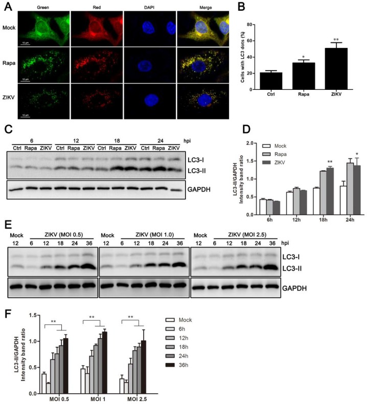
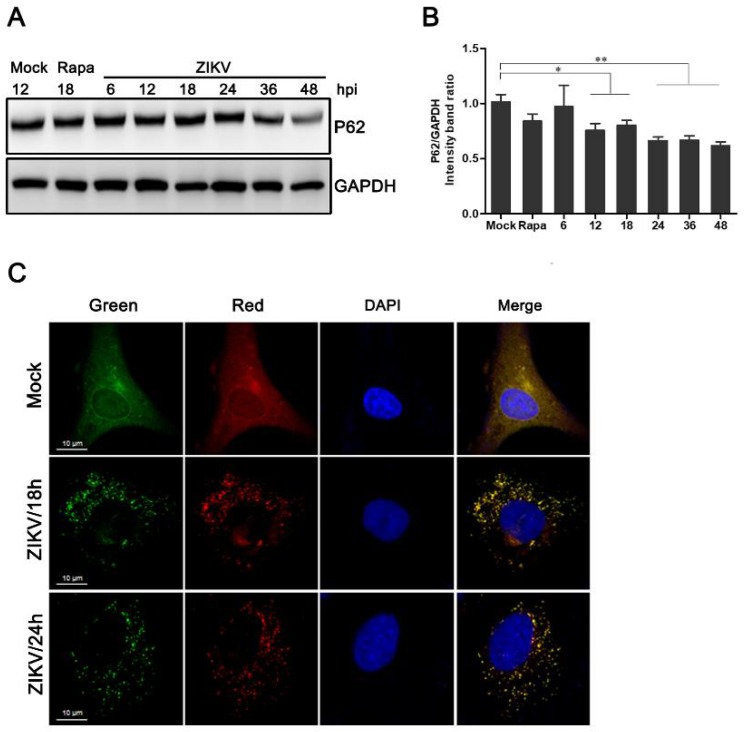
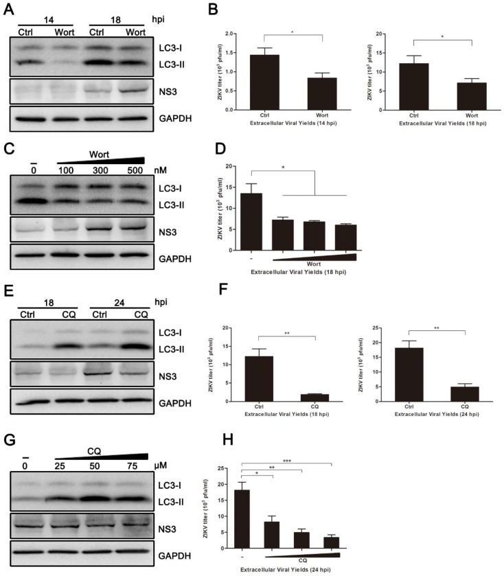
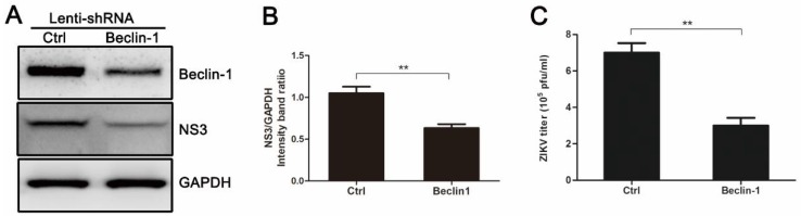
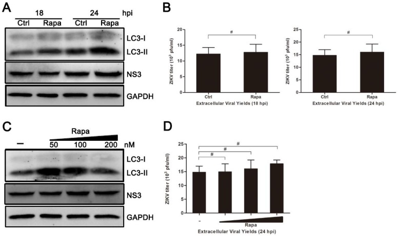
Autophagy is a common strategy for cell protection; however, some viruses can in turn adopt cellular autophagy to promote viral replication. Zika virus (ZIKV) is the pathogen that causes Zika viral disease, and it is a mosquito-borne virus. However, its pathogenesis, especially the interaction between ZIKV and target cells during the early stages of infection, is still unclear. In this study, we demonstrate that infecting human umbilical vein endothelial cells (HUVEC) with ZIKV triggers cellular autophagy. We observed both an increase in the conversion of LC3-I to LC3-II and increased accumulation of fluorescent cells with LC3 dots, which are considered to be the two key indicators of autophagy. The ratio of LC3-II/GAPDH in each group was significantly increased at different times after ZIKV infection at different MOIs, indicating that the production of lipidated LC3-II increased. Moreover, both the ratio of LC3-II/GAPDH and the expression of viral NS3 protein increased with increasing time of viral infection. The expression level of p62 decreased gradually from 12 h post-infection. Expression profile of double fluorescent protein labelling LC3 indicated that the autophagy induced by ZIKV infection was a complete process. We further investigated the role of autophagy in ZIKV replication. We demonstrated that either the treatment with inhibitors of autophagosomes formation or short hairpin RNA targeting the Beclin-1 gene, which is critical for the formation of autophagosomes, significantly reduced viral production. Taken together, our results indicate that ZIKV infection induces autophagy of HUVEC, and inhibition of ZIKV-induced autophagy restrains viral replication.

Keywords: Beclin-1; LC3; Zika virus; autophagy; viral replication; virus production.


Conflict of interest statement

The authors declare that they have no conflict of interest.

Figures

Figure 1
Figure 1
ZIKV induces autophagy in HUVEC. (A) Confocal microscopy. HUVEC were infected with lentivirus expressing mTagRFP-mWasabi-LC3 followed by treatment at 48 h post-infection with mock treatment as a negative control, rapamycin (100 nM) treatment as a positive control, or infection of ZIKV (MOI = 1) for 24 h. The cells were observed by confocal microscopy with scale bars indicating 10 µm; (B) percent of cells with LC3 dots from total LC3-expressing cells was calculated. Data were derived from at least 100 cells for each sample. * p < 0.05, ** p < 0.01; (C) western blot analysis. The turnover of LC3-I to LC3-II was detected for mock-treated cells, rapamycin-treated (100 nM) cells, or the infected cells with ZIKV at MOI 1. Cells were harvested at indicated time points and detected with anti-LC3B antibody. GAPDH was used as a loading control. Representative images are presented; (D) the intensity band ratio of LC3-II to GAPDH. Graphs represent the ratio of LC3-II to GAPDH, as measured with densitometry; (E) HUVEC were infected with ZIKV at a range of MOIs (0.5, 1, and 2.5) for the indicated time, and LC3 expression was measured by western blot. The ratios of LC3-II to GAPDH were calculated and presented (F). Data are presented as means from three independent experiments. Significance was analyzed with two-tailed Student’s t test. * p < 0.05, ** p < 0.01.
Figure 2
Figure 2
Measurement of the autophagic flux in ZIKV-infected HUVEC. (A) Western blot analysis of p62 degradation. The time course of expression of p62 in ZIKV-infected HUVEC at MOI 1 was investigated using anti-p62 antibody. Mock-infected HUVEC were used as negative controls, rapamycin (100 nM) treatment was used as a positive control, and GAPDH was used as a protein-loading control; (B) the ratios of P62 to GAPDH were calculated, and representative results are presented with graphs. Data are presented as means from three independent experiments. Compared to the control group, significance is analyzed with two-tailed Student’s t test. * p < 0.05; ** p < 0.01. (C) HUVEC transduced with lentivirus expressing mTagRFP-mWasabi-LC3 were either uninfected or infected with ZIKV at MOI 1. The cells were fixed and visualized by confocal microscopy at 18 and 24 h post-infection, respectively. Bars, 10 µm.
Figure 3
Figure 3
Inhibition of autophagy reduced ZIKV production. HUVEC were pretreated with wortmannin (Wort) (100 nM, or the indicated concentrations), chloroquine (CQ) (50 µM, or the indicated concentrations) or DMSO (control) in complete medium for 2 h and then infected with ZIKV (MOI = 1). Samples were collected at the indicated times after infection and subjected to western blot analysis or plaque assay. (A) Western blot analysis of LC3 and viral NS3 expression inhibited with Wort at 14 and 18 hpi. GAPDH was used as a protein loading control; (B) extracellular virus yields were determined by plaque assay on Vero cells and expressed as pfu/mL; (C) western blot analysis of LC3 and viral NS3 expression inhibited with Wort at the concentrations of 100, 300, and 500 nM at 18 hpi. (D) Determination of the extracellular virus yields (pfu/mL) at 18 hpi after treatment with Wort at different concentrations; (E) western blot analysis of LC3 and viral NS3 expression inhibited with CQ at 18 and 24 hpi. GAPDH was used as a protein loading control; (F) determination of the extracellular virus yields (pfu/mL); (G) western blot analysis of autophagy and viral NS3 expression inhibited with CQ at concentrations of 25, 50, and 75 µM at 24 hpi; (H) determination of the extracellular virus yields (pfu/mL) at 24 hpi after treatment with CQ at different concentrations. Data are presented as means ± SDs from three independent experiments. Significance is analyzed with two-tailed Student’s t test compared to the control group. * p < 0.05; ** p < 0.01; *** p < 0.001.
Figure 4
Figure 4
Gene silencing of Beclin-1 by pLenti-Beclin-1-shRNA (short hairpin RNA) inhibits ZIKV production. HUVEC were infected with empty lentivirus vector or pLenti-Beclin-1-shRNA. Forty-eight hours after lentivirus infection, cells were infected with ZIKV at an MOI of 1. Samples were collected at 24 h after ZIKV infection and subjected to Western blot analysis (A) or plaque assay (C). (A) Western blot analysis of Beclin-1 and viral NS3 expression. GAPDH was used as a protein loading control; (B) The ratios of NS3 to GAPDH were calculated and representative results are presented with graphs. Data are presented as means from three independent experiments. ** p < 0.01; (C) Extracellular virus yields were determined and expressed as pfu/mL. Data are presented as means ± SDs for triplicate assays. ** p < 0.01.
Figure 5
Figure 5
Treatment of rapamycin has no effect on ZIKV infection in HUVEC. (A) HUVEC were pretreated with rapamycin (Rapa) (100 nM) or DMSO (Ctrl) in complete medium and then processed as described for Figure 4A; (B) At 18 and 24 hpi, extracellular virus yields were determined by plaque assay on Vero cells and expressed as pfu/mL; (C) HUVEC were pretreated with Rapa or DMSO and then processed as described for panel A, with drug concentrations of 50, 100, 200, and 500 nM; (D) Extracellular virus yields at 24 hpi were determined and expressed as pfu/mL. Data are presented as means ± SDs from three independent experiments. Significance was analyzed with two-tailed Student’s t test and compared to the control group. # p > 0.05.

References

    1. Dick G.W.A. Zika virus (II). Pathogenicity and physical properties. Trans. R. Soc. Trop. Med. Hyg. 1952;46:521. doi: 10.1016/0035-9203(52)90043-6. - DOI - PubMed
    1. Musso D., Nilles E.J., Cao-Lormeau V.M. Rapid spread of emerging Zika virus in the Pacific area. Clin. Microbiol. Infect. Dis. 2014;20:O595–O596. doi: 10.1111/1469-0691.12707. - DOI - PubMed
    1. Chouin-Carneiro T., Vega-Rua A., Vazeille M., Yebakima A., Girod R., Goindin D., Dupont-Rouzeyrol M., Lourenco-de-Oliveira R., Failloux A.B. Differential Susceptibilities of Aedes aegypti and Aedes albopictus from the Americas to Zika Virus. PLoS Negl. Trop. Dis. 2016;10:e0004543. doi: 10.1371/journal.pntd.0004543. - DOI - PMC - PubMed
    1. Calvet G., Aguiar R.S., Melo A.S., Sampaio S.A., de Filippis I., Fabri A., Araujo E.S., de Sequeira P.C., de Mendonca M.C., de Oliveira L., et al. Detection and sequencing of Zika virus from amniotic fluid of fetuses with microcephaly in Brazil: A case study. Lancet Infect. Dis. 2016;16:653–660. doi: 10.1016/S1473-3099(16)00095-5. - DOI - PubMed
    1. Musso D., Roche C., Robin E., Nhan T., Teissier A., Cao-Lormeau V.M. Potential sexual transmission of Zika virus. Emerg. Infect. Dis. 2015;21:359–361. doi: 10.3201/eid2102.141363. - DOI - PMC - PubMed

Publication types

MeSH terms