. 2018 May 15;9(5):253.
doi: 10.3390/genes9050253.

Role of SdiA on Biofilm Formation by Atypical Enteropathogenic Escherichia coli

Affiliations

Role of SdiA on Biofilm Formation by Atypical Enteropathogenic Escherichia coli

Hebert F Culler et al. Genes (Basel). .

Abstract

Atypical enteropathogenic Escherichia coli are capable to form biofilm on biotic and abiotic surfaces, regardless of the adherence pattern displayed. Several E. coli mechanisms are regulated by Quorum sensing (QS), including virulence factors and biofilm formation. Quorum sensing is a signaling system that confers bacteria with the ability to respond to chemical molecules known as autoinducers. Suppressor of division inhibitor (SdiA) is a QS receptor present in atypical enteropathogenic E.coli (aEPEC) that detects acyl homoserine lactone (AHL) type autoinducers. However, these bacteria do not encode an AHL synthase, but they are capable of sensing AHL molecules produced by other species, establishing an inter-species bacterial communication. In this study, we performed experiments to evaluate pellicle, ring-like structure and biofilm formation on wild type, sdiA mutants and complemented strains. We also evaluated the transcription of genes involved in different stages of biofilm formation, such as bcsA, csgA, csgD, fliC and fimA. The sdiA mutants were capable of forming thicker biofilm structures and showed increased motility when compared to wild type and complemented strains. Moreover, they also showed denser pellicles and ring-like structures. Quantitative real-time PCR (qRT-PCR) analysis demonstrated increased csgA, csgD and fliC transcription on mutant strains. Biofilm formation, as well as csgD, csgA and fimA transcription decreased on wild type strains by the addition of AHL. These results indicate that SdiA participates on the regulation of these phenotypes in aEPEC and that AHL addition enhances the repressor effect of this receptor on the transcription of biofilm and motility related genes.

Keywords: acyl homoserine lactone; atypical enteropathogenic Escherichia coli; biofilm formation; confocal scanning laser microscopy; sdiA gene.


Conflict of interest statement

The authors declare no conflict of interest.

Figures
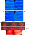
Figure 1
Figure 1
Cellulose production by ONT:H25, sdiA mutant strain (HFC01) and ONT:H25 wild type and complemented strain (HFC02), strains on LBNS agar plates supplemented with calcofluor. (A,C) 24 h of incubation at 37 °C and 26 °C, respectively; (B,D) 48 h of incubation at 37 °C and 26 °C, respectively. Curli production by ONT:H25, HFC01 and HFC02 strains on LBNS agar plates supplemented with Congo Red and Coomassie brilliant blue, after 24 h at 26 °C. (E) ONT:H25, HFC01 and HFC02; (F) rdar colonie morphotype. (G) smooth colonie morphotype.
Figure 2
Figure 2
(A) Biofilm formation by ONT:H25, HFC01 and HFC02 after 24 h of incubation at 26 °C in Luria-Bertani (LB) with dimethyl sulfoxide (DMSO), 3O-C6-DL-HSL or C8-DL-HSL. Statistic bars indicate standard deviation. (B) Biofilm formation by ONT:H25, HFC01 and HFC02 after 24 h of incubation at 37 °C in LB with DMSO, 3O-C6-DL-HSL or C8-DL-HSL. Statistic bars indicate standard deviation.
Figure 3
Figure 3
Pellicle and ring-like structure formation by ONT:H25, HFC01 and HFC02. (A) Pellicle formation on air-liquid interface in 24 h at 37 °C in LB, arrows indicate pellicle and ring-like structure formation; (B) Ring-like structure formation on air-liquid-glass interface in 72 h at 37 °C in LB, arrows indicate pellicle and ring-like structure formation; (C) Ring-like structure formation in 96-well plate after 72 h at 26 °C. 1, ONT:H25; 2, HFC01; 3, HFC02. (D) Ring-like structure formation in 96-well plate after 72 h at 37 °C in LB. 1, ONT:H25; 2, HFC01; 3, HFC02.
Figure 4
Figure 4
(A) Biofilm formation by ONT:H25, HFC01 and HFC02 after 24 h of incubation at 37 °C on LB supplemented with DMSO, 3O-C6-DL-HSL and C8-DL-HSL. (B) Biofilm formation on air-liquid-glass interface by ONT:H25, HFC01 and HFC02 after 48 h of incubation. XYxy axis, Zz axis from XY. Bacteria were stained with Propidium Iodide (red). 630× magnification.
Figure 5
Figure 5
(A) Fluorescent actin staining assay of ONT:H25, HFC01 and HFC02 in HeLa cells in Dulbecco’s Modified Eagle Medium (DMEM) supplemented with DMSO, 3O-C6-DL-HSL and C8-DL-HSL after 6 h of incubation at 37 °C. (B) Long filament-like morphology displayed by bacteria in the presence of HeLa cells. (C) Bacteria in the absence of HeLa cells. HeLa cells were stained with Alexa Fluor–Phalloidin (green) and nucleic acids were stained with Propidium Iodide (red). (A), 630× magnification; (B,C), 1000× magnification.
Figure 6
Figure 6
Transcription analysis of biofilm formation related genes (bcsA, csgD, csgA, fimA and fliC) by quantitative real-time PCR (qRT-PCR), in ONT:H25 (4157 wild type), HFC01 and HFC02 strains. Ct values were normalized by rpoA transcription. Error bars represent the standard deviation from triplicates.
Figure 7
Figure 7
Motility from ONT:H25, HFC01 and HFC02 strains, after 8 h of incubation at 37 °C.

Similar articles

Cited by

References

    1. Wardlaw T., Salama P., Brocklehurst C., Chopra M., Mason E. Diarrhoea: Why children are still dying and what can be done. Lancet. 2010;13:870–872. doi: 10.1016/S0140-6736(09)61798-0. - DOI - PubMed
    1. Ochoa T.J., Contreras C.A. Enteropathogenic Escherichia coli infection in children. Curr. Opin. Infect. Dis. 2011;24:478–483. doi: 10.1097/QCO.0b013e32834a8b8b. - DOI - PMC - PubMed
    1. Kaper J.B. Defining enteropathogenic Escherichia coli. Rev. Microbiol. 1996;27:130–133.
    1. Hernandes R.T., Elias W.P., Vieira M.A.M., Gomes T.A.T. An overview of atypical enteropathogenic Escherichia coli. FEMS Microbiol. Lett. 2009;297:137–149. doi: 10.1111/j.1574-6968.2009.01664.x. - DOI - PubMed
    1. Trabulsi L.R., Keller R., Gomes T.A.T. Typical and atypical enteropathogenic Escherichia coli. Emerg. Infect. Dis. 2002;8:508–513. doi: 10.3201/eid0805.010385. - DOI - PMC - PubMed

LinkOut - more resources