. 2018 May 15;19(5):1470.
doi: 10.3390/ijms19051470.

Silver Nanoparticles: Two-Faced Neuronal Differentiation-Inducing Material in Neuroblastoma (SH-SY5Y) Cells

Affiliations

Silver Nanoparticles: Two-Faced Neuronal Differentiation-Inducing Material in Neuroblastoma (SH-SY5Y) Cells

Ahmed Abdal Dayem et al. Int J Mol Sci. .

Abstract

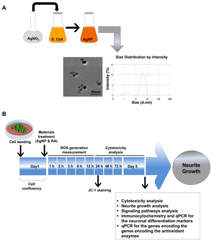
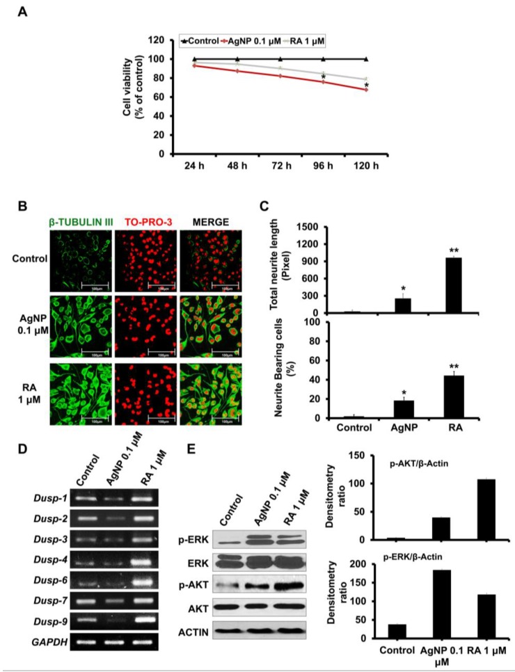
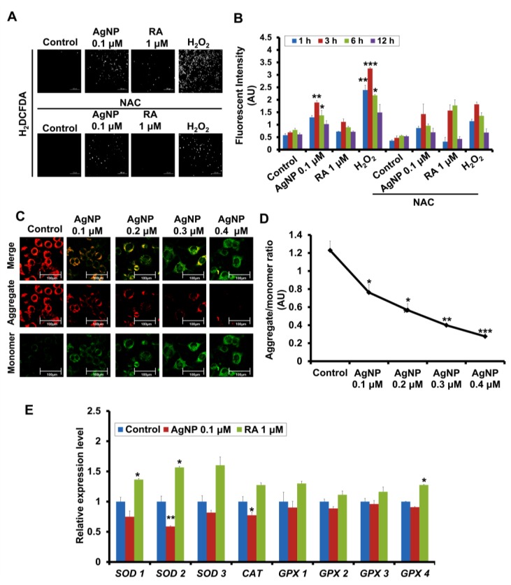
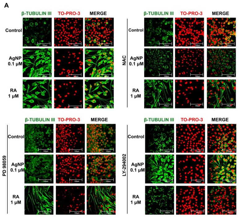
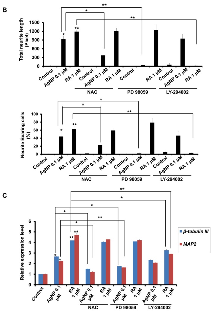
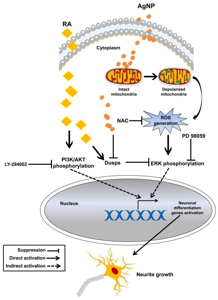
We have previously demonstrated the potential of biologically synthesized silver nanoparticles (AgNP) in the induction of neuronal differentiation of human neuroblastoma, SH-SY5Y cells; we aimed herein to unveil its molecular mechanism in comparison to the well-known neuronal differentiation-inducing agent, all-trans-retinoic acid (RA). AgNP-treated SH-SY5Y cells showed significantly higher reactive oxygen species (ROS) generation, stronger mitochondrial membrane depolarization, lower dual-specificity phosphatase expression, higher extracellular-signal-regulated kinase (ERK) phosphorylation, lower AKT phosphorylation, and lower expression of the genes encoding the antioxidant enzymes than RA-treated cells. Notably, pretreatment with N-acetyl-l-cysteine significantly abolished AgNP-induced neuronal differentiation, but not in that induced by RA. ERK inhibition, but not AKT inhibition, suppresses neurite growth that is induced by AgNP. Taken together, our results uncover the pivotal contribution of ROS in the AgNP-induced neuronal differentiation mechanism, which is different from that of RA. However, the negative consequence of AgNP-induced neurite growth may be high ROS generation and the downregulation of the expression of the genes encoding the antioxidant enzymes, which prompts the future consideration and an in-depth study of the application of AgNP-differentiated cells in neurodegenerative disease therapy.

Keywords: differentiation; kinase; mitochondria; neuroblastoma; phosphatase; reactive oxygen species; silver nanoparticles.


Conflict of interest statement

The authors declare no conflicts of interest.

Figures

Figure 1
Figure 1
Characterization and experimental scheme for the biologically synthesized silver nanoparticles (AgNP). (A) Characterization of the biologically synthesized AgNP. The biological synthesis of AgNP was performed through the reduction of the bulk material silver nitrate (AgNO3) by Escherichia coli nitrate reductase. Transmission electron microscopy (TEM) imaging of the biologically synthesized AgNP shows a spherical shape with an approximate size of 30 nm. The histogram shows the synthesized AgNP size distribution, as measured by dynamic light scattering (DLS) and presence of small population of the particles between 1 and 3 nm. Scale bar = 50 nm. (B) Schematic of the experimental procedures used to compare the neuronal differentiation processes of AgNP- and all-trans-retinoic acid (RA)-exposed neuroblastoma (SH-SY5Y) cells.
Figure 2
Figure 2
Effects of AgNP and RA on the viability, differentiation, Dual-specificity phosphatase (DUSP expression, and AKT and ERK activation status of SH-SY5Y cells. (A) SH-SY5Y cells were incubated with 0.1 μM AgNP or 1 μM RA for 24, 48, 72, 96, and 120 h and viability was analyzed using the EZ-Cytox cell viability kit. SH-SY5Y cells exposed to AgNP for 96 and 72 h showed a significant cytotoxicity. The experiment was performed in triplicate. (B) Immunocytochemistry analysis: incubation of SH-SY5Y cells with 0.1 μM AgNP or 1 μM RA for five days. Both RA-exposed and AgNP-exposed cells showed morphological changes (neurite phenotype) and high expression of β-tubulin III. Scale bars, 100 μm. (C) Neurite length and the percentage of neurite-bearing cells were measured using the neurite tracing plugin NeuriteTrace in ImageJ. Both AgNP- and RA-exposed cells significantly promoted the neurite length and increased the percentage of neurite-bearing cells. * p < 0.05; ** p < 0.01. (D) Determination of DUSP1, DUSP2, DUSP3, DUSP4, DUSP6, DUSP7, and DUSP9 expression levels in SH-SY5Y cells after 5 d of incubation with 0.1 μM AgNP or 1 μM RA. Glyceraldehyde 3-phosphate dehydrogenase (GAPDH) is a housekeeping gene. DUSPs expression level was markedly decreased and increased in AgNP- and RA-treated cells, respectively. (E) Western blot analysis was performed to determine the phosphorylation levels of extracellular-signal-regulated kinase (ERK) and AKT in 0.1 μM AgNP- or 1 μM RA-exposed SH-SY5Y cells. Western blot analysis: SH-SY5Y cells treated with 0.1 μM AgNP or 1 mM RA showed high phosphorylation of ERK and AKT signalings. AgNP-exposed cells showed higher phosphorylation of ERK than that shown in RA-exposed cells and higher AKT phosphorylation was detected in RA-exposed cells than that of AgNP-treated cells as depicted in the densitometry analysis (right panel).
Figure 3
Figure 3
Modulation of reactive oxygen species (ROS) generation, mitochondrial depolarization, and the genes encoding the antioxidant enzymes expression in AgNP- and RA-treated SH-SY5Y cells. (A,B) ROS generation in SH-SY5Y cells pretreated with NAC (1 mM), and were then treated with 0.1 μM of AgNP or 1 μM of RA or 100 μM of Hydrogen peroxide (H2O2) was measured by fluorescence microscopy (A) and a fluorometric method (B). AgNP-exposed cells showed a significant ROS generation, in particular, after 3 h and 6 h of incubation, which is inhibited by NAC pretreatment. * p < 0.05; ** p < 0.01; *** p < 0.001. (C) SH-SY5Y cells were incubated with AgNP (0.1, 0.2, 0.3, and 0.4 μM) and the mitochondrial membrane potential (ΔΨm) was measured using JC-1 staining. The qualitative analysis fluorescence intensities of the monomer (green) and an aggregate (red) form was analyzed with the fluorescence confocal microscopy. Scale bars, 100 μm. (D) The quantitative analysis of the ratio of aggregate and the monomer was determined using dual-scanning microplate spectrofluorometer. AgNP showed a significant depolarization of the mitochondrial membrane in a dose-dependent manner in SH-SY5Y cells. * p < 0.05; ** p < 0.01; *** p < 0.001. (E) Expression of genes encoding the antioxidant enzymes (SOD1, SOD2, SOD3, CAT, GPX1, GPX2, GPX3, and GPX4) by quantitative real-time PCR. AgNP-treated cells showed a significant suppression of the expression level of SOD2 and CAT. SOD: superoxide dismutase; CAT: catalase; GPX: glutathione peroxidase. * p < 0.05; ** p < 0.01.
Figure 4
Figure 4
Roles of inhibitors of ROS generation and ERK/AKT signaling in the modulation of AgNP- and RA-induced neurite growth. (A) Immunocytochemistry analysis of the expression level of the neuronal differentiation marker β-tubulin III (green florescence) of SH-SY5Y cells pretreated with the antioxidant NAC (1 mM), the ERK inhibitor PD98059 (10 μM), or the AKT inhibitor LY-294002 (10 μM) for 1 h, and then exposed to 0.1 μM AgNP or 1 μM RA for five days. Scale bars, 100 μm. (B) Neurite length and the percentage of neurite-bearing cells were measured using the neurite tracing plugin NeuriteTrace in ImageJ. The pretreatment of NAC and PD98059 significantly abolished AgNP-induced neurite growth and high neurite-bearing cells percentage, but LY-294002 pretreatment markedly suppressed the RA-induced neurite growth. * p < 0.05; ** p < 0.01. (C) The expression of the neuronal differentiation-specific markers microtubule-associated protein 2 (MAP2) and β-tubulin III was measured by quantitative real-time PCR in cells that were pretreated and AgNP- or RA-exposed, as above. The expression level of the neuronal differentiation markers in AgNP- and RA-treated cells after the pretreatment of the aforementioned inhibitors confirmed the immunocytochemistry results. * p < 0.05; ** p < 0.01.
Figure 4
Figure 4
Roles of inhibitors of ROS generation and ERK/AKT signaling in the modulation of AgNP- and RA-induced neurite growth. (A) Immunocytochemistry analysis of the expression level of the neuronal differentiation marker β-tubulin III (green florescence) of SH-SY5Y cells pretreated with the antioxidant NAC (1 mM), the ERK inhibitor PD98059 (10 μM), or the AKT inhibitor LY-294002 (10 μM) for 1 h, and then exposed to 0.1 μM AgNP or 1 μM RA for five days. Scale bars, 100 μm. (B) Neurite length and the percentage of neurite-bearing cells were measured using the neurite tracing plugin NeuriteTrace in ImageJ. The pretreatment of NAC and PD98059 significantly abolished AgNP-induced neurite growth and high neurite-bearing cells percentage, but LY-294002 pretreatment markedly suppressed the RA-induced neurite growth. * p < 0.05; ** p < 0.01. (C) The expression of the neuronal differentiation-specific markers microtubule-associated protein 2 (MAP2) and β-tubulin III was measured by quantitative real-time PCR in cells that were pretreated and AgNP- or RA-exposed, as above. The expression level of the neuronal differentiation markers in AgNP- and RA-treated cells after the pretreatment of the aforementioned inhibitors confirmed the immunocytochemistry results. * p < 0.05; ** p < 0.01.
Figure 5
Figure 5
Hypothetical model of the mechanisms of AgNP- and RA-induced neuronal differentiation of SH-SY5Y cells.

References

    1. Lopes F.M., Schröder R., da Frota Júnior M.L.C., Zanotto-Filho A., Müller C.B., Pires A.S., Meurer R.T., Colpo G.D., Gelain D.P., Kapczinski F., et al. Comparison between proliferative and neuron-like SH-SY5Y cells as an in vitro model for parkinson disease studies. Brain Res. 2010;1337:85–94. doi: 10.1016/j.brainres.2010.03.102. - DOI - PubMed
    1. Schiwy N., Brazda N., Müller H.W. Enhanced regenerative axon growth of multiple fibre populations in traumatic spinal cord injury following scar-suppressing treatment. Eur. J. Neurosci. 2009;30:1544–1553. doi: 10.1111/j.1460-9568.2009.06929.x. - DOI - PubMed
    1. Shipley M.M., Mangold C.A., Szpara M.L. Differentiation of the SH-SY5Y human neuroblastoma cell line. J. Vis. Exp. JoVE. 2016:53193. doi: 10.3791/53193. - DOI - PMC - PubMed
    1. Xicoy H., Wieringa B., Martens G.J. The SH-SY5Y cell line in Parkinson’s disease research: A systematic review. Mol. Neurodegener. 2017;12:10. doi: 10.1186/s13024-017-0149-0. - DOI - PMC - PubMed
    1. Krishna A., Biryukov M., Trefois C., Antony P.M., Hussong R., Lin J., Heinäniemi M., Glusman G., Köglsberger S., Boyd O., et al. Systems genomics evaluation of the SH-SY5Y neuroblastoma cell line as a model for parkinson’s disease. BMC Genom. 2014;15:1154. doi: 10.1186/1471-2164-15-1154. - DOI - PMC - PubMed

LinkOut - more resources