. 2018 Jun 15;13(6):1551-1559.
doi: 10.1021/acschembio.8b00154. Epub 2018 May 30.

Selective Inhibition of the Myeloid Src-Family Kinase Fgr Potently Suppresses AML Cell Growth in Vitro and in Vivo

Affiliations

Selective Inhibition of the Myeloid Src-Family Kinase Fgr Potently Suppresses AML Cell Growth in Vitro and in Vivo

Mark C Weir et al. ACS Chem Biol. .

Abstract

Acute myelogenous leukemia (AML) is the most common hematologic malignancy in adults and is often associated with constitutive tyrosine kinase signaling. These pathways involve the nonreceptor tyrosine kinases Fes, Syk, and the three Src-family kinases expressed in myeloid cells (Fgr, Hck, and Lyn). In this study, we report remarkable anti-AML efficacy of an N-phenylbenzamide kinase inhibitor, TL02-59. This compound potently suppressed the proliferation of bone marrow samples from 20 of 26 AML patients, with a striking correlation between inhibitor sensitivity and expression levels of the myeloid Src family kinases Fgr, Hck, and Lyn. No correlation was observed with Flt3 expression or mutational status, with the four most sensitive patient samples being wild-type for Flt3. Kinome-wide target specificity profiling coupled with in vitro kinase assays demonstrated a narrow overall target specificity profile for TL02-59, with picomolar potency against the myeloid Src-family member Fgr. In a mouse xenograft model of AML, oral administration of TL02-59 for 3 weeks at 10 mg/kg completely eliminated leukemic cells from the spleen and peripheral blood while significantly reducing bone marrow engraftment. These results identify Fgr as a previously unrecognized kinase inhibitor target in AML and TL02-59 as a possible lead compound for clinical development in AML cases that overexpress this kinase independent of Flt3 mutations.


Figures

Figure 1.
Figure 1.
Identification of TL02–59 as a potent inhibitor of AML cell growth. A) TL02–59 and nine N-phenylbenzamide analogs were evaluated for their growth inhibitory activity in the FLT3-ITD+ AML cell line, MV4–11, using the CellTiter Blue assay (Promega). IC50 values for each compound are shown, with the R-group unique to each analog plotted next to the corresponding data point. The structures of TL02–59 and the related TL8 scaffold are shown on the right. B) Proliferation of MV4–11, MOLM-14 and THP-1 AML cells in the presence of TL02–59 was measured using the CellTiter-Blue assay over a range of concentrations for 72 hours. For THP-1 cells, additional concentrations up to 3.0 μM did not significantly inhibit growth (not shown). C) MV4–11, MOLM-14 and THP-1 AML cells were incubated with tandutinib (100 nM), TL02–59 (100 nM), staurosporine (1 μM) or DMSO for 72 hours. Apoptosis and cell viability were independently measured and results are presented as mean ratios of caspase activity to cell viability from three independent experiments ± SD.
Figure 2.
Figure 2.
Expression of TL02–59 target kinases in primary AML bone marrow samples. Expression of the 27 putative TL02–59 target kinases identified by KINOMEscan analysis was determined in 26 primary AML bone marrow samples by qPCR. Relative expression values were calculated as the base 2 antilog of the qPCR ΔCt values relative to GAPDH for each kinase. These values were then plotted as a distribution relative to the mean value for all 27 kinases analyzed in each patient sample. All determinations were made on at least two independent RNA samples from each patient. Each patient is represented by a dot. The Flt3 genotype of each sample was determined by PCR and sequencing (Table S2) and results are grouped as Flt3-ITD+ (panel A) or Flt3 wild-type (Flt3-WT; panel B). Expression profiles for the AML samples most sensitive to TL02–59 are highlighted in green (top responder; cases 454 and 451) while the least responsive cases are highlighted in red (non-responder; cases 104 and 505).
Figure 3.
Figure 3.
A) Comparison of TL02–59 target kinase expression in primary AML samples from the TCGA database vs. primary AML bone marrow samples from this study. TL02–59 target kinase RNAseq expression data were downloaded as FPKM values from the TCGA database and normalized to the average expression across all 27 kinases within each sample. Average values for each kinase from the TCGA database (Flt3-ITD: n = 34; Flt3-WT: n =111) were plotted against the average values obtained for each kinase in this study for Flt3-ITD (n = 13; left) and Flt3-WT (n = 12; right) primary AML bone marrow samples by qPCR. Data were analyzed by linear regression and the best-fit line and 95% confidence intervals are shown; p < 0.0001 in both cases. B) Myeloid-restricted Src-family kinases and Syk correlate with TL02–59 sensitivity in patient AML bone marrow samples. Relative TL02–59 target kinase expression values from Figure 2 were correlated with TL02–59 IC50 values determined for each of the primary AML bone marrow samples (Table S3). Spearman’s correlations were calculated for each kinase across all of the AML samples, and the resulting correlation coefficients are plotted in ascending order. Blue bars represent kinases whose expression shows a significant negative correlation with TL02–59 IC50 values with p < 0.05; green bar (Lyn) represents p < 0.1. Grey bars represent kinases that did not show statistical significance.
Figure 4.
Figure 4.
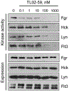
TL02–59 selectively inhibits Fgr autophosphorylation in myeloid cells. TF-1 myeloid cells were transduced with recombinant retroviruses carrying Fgr, Lyn, Hck, and Flt3-ITD. Cells were treated with TL02–59 for 6 h over the range of concentrations shown, followed by immunoprecipitation of each kinase from clarified cell protein extracts. The immunoprecipitates were resolved by SDS-PAGE, followed by immunoblotting with antibodies to the Src-family kinase activation loop tyrosine (pTyr416) or antiphosphotyrosine in the case of Flt3-ITD (upper panels). The presence of kinase protein in each lane was verified by immunoblotting with kinase-specific antibodies (lower panels). This experiment was repeated three times with equivalent results.
Figure 5.
Figure 5.
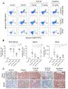
TL02–59 reduces MV4–11 cell engraftment in an AML mouse model. Human Flt3-ITD+ MV4–11 AML cells were injected into the tail vein of immunocompromised (NSG) mice and allowed to engraft for two weeks. Groups of eight mice were then treated daily by oral gavage with TL02–59 (1 and 10 mg/kg), sorafenib (10 mg/kg), or vehicle. Two mice were not engrafted and received no treatment for use as baseline controls. Following three weeks of treatment, the animals were sacrificed and the presence of MV4–11 AML cells in the bone marrow, spleen and peripheral blood were assayed by flow cytometry. A) Representative flow cytometry diagrams for a single mouse within each treatment group. For the bone marrow and spleen, the inset number represents the percentage of human CD45+/CD33+ MV4–11 leukemia cells present. For whole blood, the number of MV4–11 cells present per 25 μL of blood is shown. B) Summary of all flow cytometry results, where each dot represents a single mouse. Horizontal bars indicate the mean value in each group +/− SD. Statistical significance was determined by pairwise Student’s t-test, with p values indicated for significantly different groups; ns, not significant. C) Representative thin sections of spleen and bone marrow from all five treatment groups. MV4–11 AML cells were visualized in the sections by immunohistochemistry with an antibody specific for human CD45.

References

    1. Pratz KW; Luger SM (2014) Will FLT3 inhibitors fulfill their promise in acute meyloid leukemia? Curr. Opin. Hematol 21, 72–78. - PMC - PubMed
    1. Grunwald MR; Levis MJ (2015) FLT3 Tyrosine Kinase Inhibition as a Paradigm for Targeted Drug Development in Acute Myeloid Leukemia. Semin. Hematol 52, 193–199. - PubMed
    1. Sudhindra A; Smith CC (2014) FLT3 inhibitors in AML: are we there yet? Curr. Hematol. Malig. Rep 9, 174–185. - PubMed
    1. Smith CC; Wang Q; Chin CS; Salerno S; Damon LE; Levis MJ; Perl AE; Travers KJ; Wang S; Hunt JP; Zarrinkar PP; Schadt EE; Kasarskis A; Kuriyan J; Shah NP (2012) Validation of ITD mutations in FLT3 as a therapeutic target in human acute myeloid leukaemia. Nature , 485, 260–263. - PMC - PubMed
    1. Wander SA; Levis MJ; Fathi AT (2014) The evolving role of FLT3 inhibitors in acute myeloid leukemia: quizartinib and beyond. Ther. Adv. Hematol 5, 65–77. - PMC - PubMed

Publication types

MeSH terms