. 2018 Aug:173:91-108.
doi: 10.1016/j.exer.2018.05.009. Epub 2018 May 25.

Müller glial cells contribute to dim light vision in the spectacled caiman (Caiman crocodilus fuscus): Analysis of retinal light transmission

Affiliations

Müller glial cells contribute to dim light vision in the spectacled caiman (Caiman crocodilus fuscus): Analysis of retinal light transmission

Silke Agte et al. Exp Eye Res. 2018 Aug.

Abstract

In this study, we show the capability of Müller glial cells to transport light through the inverted retina of reptiles, specifically the retina of the spectacled caimans. Thus, confirming that Müller cells of lower vertebrates also improve retinal light transmission. Confocal imaging of freshly isolated retinal wholemounts, that preserved the refractive index landscape of the tissue, indicated that the retina of the spectacled caiman is adapted for vision under dim light conditions. For light transmission experiments, we used a setup with two axially aligned objectives imaging the retina from both sides to project the light onto the inner (vitreal) surface and to detect the transmitted light behind the retina at the receptor layer. Simultaneously, a confocal microscope obtained images of the Müller cells embedded within the vital tissue. Projections of light onto several representative Müller cell trunks within the inner plexiform layer, i.e. (i) trunks with a straight orientation, (ii) trunks which are formed by the inner processes and (iii) trunks which get split into inner processes, were associated with increases in the intensity of the transmitted light. Projections of light onto the periphery of the Müller cell endfeet resulted in a lower intensity of transmitted light. In this way, retinal glial (Müller) cells support dim light vision by improving the signal-to-noise ratio which increases the sensitivity to light. The field of illuminated photoreceptors mainly include rods reflecting the rod dominance of the of tissue. A subpopulation of Müller cells with downstreaming cone cells led to a high-intensity illumination of the cones, while the surrounding rods were illuminated by light of lower intensity. Therefore, Müller cells that lie in front of cones may adapt the intensity of the transmitted light to the different sensitivities of cones and rods, presumably allowing a simultaneous vision with both receptor types under dim light conditions.

Keywords: Caiman; Glia; Light guidance; Müller cell; Retina.


Conflict of interest statement

Conflicts of interest

None of the authors have any financial and personal relationships with other people or organizations that could inappropriately influence (bias) their work.

Figures

Fig. 1.
Fig. 1.
Double-sided imaging of the retina. A. The setup consists of two imaging units used for bidirectional imaging of retinal wholemount preparations combined with local light transmission. The wholemounts were placed with the photoreceptor side up onto the bottom of a chamber on the stage of an inverted Olympus IX81 equipped with a laser-scanning unit (FluoviewFV1000, Olympus). B, C. Example of a doublesided imaging of a wholemount preparation of the ventral peripheral retina. B. By using the LSM (10x and 40× objective), stacks of horizontal confocal images of dye-loaded Müller and photoreceptor cells were recorded within the tissue. Müller cells and rods were loaded with the vital dye Celltracker Orange and cones with FITC-coupled peanut agglutinin. The horizontal confocal images were obtained at different depths in the tissue. The images at 0-μm and 10-μm depths show the nerve fiber/ganglion cell layers with the bright Müller cell endfeet, dark neuronal perikarya and dark nerve fiber bundles. The images obtained at 20–50 μm depths within the tissue show the inner plexiform layer which shows bright Müller cell processes within the dark synaptic neuropil. The images of the photoreceptors show the thick rod segments (red) and the segments of single cones (gray, blue) and double cones (gray, blue). C. By using the 10× objective of the LSM, the wholemounts were illuminated at the inner (vitreal) side with a stationary focused laser beam (light stimulus at z = 0 μm). The red points mark the position of the green laser light (λ = 532 nm). With a 40× objective and a CCD-camera, the transmitted light (green) was recorded from the outer (scleral) surface of the retina (left: transmitted light). A wide-field illumination of the tissue with parallel light (Xenon lamp) from below allowed the recording of a transmission image of the photoreceptor segments (left: wide-field). To analyze the light transporting structures within the neuroretina and their impact on vision, the transmitted light was overlayed with the images of the photoreceptor segments (right: rods (red), cones (gray, blue)). Bars, 10 μm.
Fig. 2.
Fig. 2.
Histological structure of the caiman retina. A. Immunolocalization of the glia-specific proteins glutamine synthetase (GS; above), glial fibrillary acidic protein (GFAP; middle) and the calcium-binding protein S100β (below) in vertical vibratome sections of different retinal areas. Cell nuclei were labeled with Hoechst 33342 (blue). Note that the inner nuclear layer (INL) of the peripheral retina (above) contains fewer neuronal perikarya than the INL of the central retina. The outer nuclear layer (ONL) contains one row of photoreceptor perikarya. The arrowhead points to an inner stem process of a Müller cell that splits into two endfeet in the NFL/GCL. B. Immunolocalization of glutamine synthetase (above: purple, below: red), cone transducin (above: red, below: purple), rod transducin (below: purple) and fluorescent peanut agglutinin (PNA; left below: green). The caiman retina is rod-dominated and contains relatively few cones. Note that the INL of the peripheral retina (above) is composed of large, nonregularly formed Müller cell somata. Neuronal perikarya are located at the inner and outer margins of the INL. In the INL of the central retina (below), vertical columns of neuronal perikarya traverse the layer, which is composed of large Müller cell somata. The yellow asterisk shows a cone cell with a perikaryon that is surrounded by the outer processes of various Müller cells (yellow arrows). The blue arrows indicate a Müller cell that divides within the ONL into several processes, which surround at least two photoreceptor cell perikarya (blue asterisks). Glutamine synthetase-containing structures that extend from the outer limiting membrane (OLM) into the subretinal space are microvilli of Müller cells that run between the photoreceptor inner segments (PIS). Note that many photoreceptor nuclei penetrate the OLM. C. Double labeling of a section with antibodies against glutamine synthetase (purple) and cone transducin (red). Note the spatial alignment of Müller cell outer processes in the ONL and cone inner segments. D. Retinal sections labeled with antibodies against glutamine synthetase (left: purple, right: red) and cone transducin (left: red, right: white). The blue arrowheads point at a putative single (left) and a double cone (right), respectively. The rods have long and thick cylindrical inner and outer segments. E. Horizontal sections through the receptor segment layer in freshly isolated, vital wholemounts of different retinal areas. Rods were loaded with the vital dye Celltracker Orange (red) and cones with fluorescent peanut agglutinin (green). Large single cones and thick members of double cones (principal cells) show low fluorescence intensities, while short single cones and the thin member of double cones (accessory cells) show high fluorescence intensities. CHO choriocapillaris; IPL, inner plexiform layer; OPL, outer plexiform layer.Bars, 20 (A‒D) and 50 μm (E).
Fig. 3.
Fig. 3.
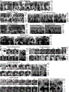
Horizontal confocal images of vital caiman retinal wholemounts. The wholemounts were loaded with Celltracker Orange that labels Müller cells and rods, and FITC-coupled peanut agglutinin that labels cones. Images of horizontal sections through the retina were recorded at depth intervals of 1 μm (every 5th image is shown), and show bright Müller cells, bright photoreceptor segments (PRS), and dark nerve fibers, synaptic neuropil and neuronal perikarya. A. Example of a recording obtained in the temporal (peripheral) retina. The image stacks were obtained from the ganglion cell layer (GCL; image 1) up to the PRS (image 25). The GCL mainly contains Müller cell endfeet that display different intensities of fluorescence as well as few dark neuronal perikarya and thin nerve fiber bundles. The records from the inner plexiform layer (IPL, images 3–9) show dark synaptic neuropil and cross-sections through bright inner stem processes of Müller cells. In the inner nuclear layer (INL, images 10–17), the dark spherical neuronal perikarya are located at the inner (images 10–13) and outer margins of the layer (images 15–17). The middle part of the INL (image 14) does not contain neuronal perikarya, but is composed of Müller cell somata. Hyperfluorescent Müller cell nuclei (arrowheads) are mainly located within the outer part of the INL (images 15–17). The image of the outer plexiform layer (OPL, image 18 and 19) shows cross-sections through bright Müller cell processes surrounded by dark neuropil. The outer nuclear layer (ONL, images 20–22) is composed of dark photoreceptor cell perikarya which are separated by bright Müller cell processes. Perikarya of double cones are not separated among each other (white arrows, image 21). The receptor segment layer (images 23–25) contains bright rod segments and gray cone segments. B. Example of a recording obtained of the dorsal peripheral retina. C. Images obtained in the nerve fiber layer (NFL, images 2 and 3) and GCL (images 4 and 5) of the dorsal central retina. The NFL contains thick nerve fiber bundles which are separated by groups of relatively thin Müller cell endfeet that are arranged in lines. Image 1 shows the innermost tissue of the retina which is solely composed of Müller cell endfeet. D. Images of the NFL (image 1), the GCL (images 2–4) and the IPL (image 5) of the ventral peripheral retina. Note that the NFL of this area contains thin nerve fiber bundles and thick Müller cell endfeet. E. Horizontal sections through the outer part of the INL. Note that each bright Müller cell process that runs between the dark neuronal perikarya contains a hyperfluorescent cell nucleus with several lobes. Bars, 10 μm.
Fig. 4.
Fig. 4.
Müller cell morphology of the vital caiman retina. Müller cells and rods were loaded with Celltracker Orange. The images show bright Müller cells, bright photoreceptor inner segments (PIS), and dark nerve fibers, synaptic neuropil, neuronal perikarya and photoreceptor cell perikarya. A. Vertical sections (z-reconstruction images) calculated by horizontal confocal images of the caiman retina. The bright cytoplasm of Müller cells traverses the whole thickness of the neuroretina. Individual Müller cells are often recognizable because of their different fluorescence intensities (above). The nerve fiber (NFL) and ganglion cell layers (GCL) are filled by the cytoplasm of Müller cell endfeet which enclose dark nerve fiber bundles (blue arrowheads). Inner stem processes of Müller cells with varying thickness run vertically or obliquely through the inner plexiform layer (IPL). Large Müller cell somata fill the inner nuclear layer (INL). Dark neuronal perikarya are located at the outer and inner margins of the INL. In the more central retinal areas, columns of stacked neuronal perikarya traverse the INL; the columns are separated by Müller cell somata. Flattened elongated hyperfluorescent Müller cell nuclei (yellow arrowheads) are present within the outer part of the INL and are often arranged along the light path. The Müller cell somata proceed into thick outer processes within the outer plexiform layer (OPL) and further into thinner processes which surround the perikarya of photoreceptor cells in the outer nuclear layer (ONL). The outer processes are the origin of microvilli that extend into the subretinal space. Note the hyperfluorescence of the photoreceptor inner segments. The morphology of Müller cells is similar in all retinal areas. The thickness of the tissue, the number of neurons in the INL and of the columns of stacked neuronal perikarya within the INL varied in dependence on the retinal topography. Arrows, Müller cells with two or several inner stem processes.*, double cone perikarya. B. Different morphologies of inner stem processes of Müller cells in the IPL, as revealed by horizontal and vertical sections obtained in wholemounts of different retinal areas. In the wholemount of the ventral midperiphery shown left above, the endfeet of three different Müller cells in the NFL/GCL are marked. Each endfoot is the origin of one thick inner stem process that traverses the IPL nearly radially. At another site of the same wholemount (middle), different numbers of inner stem processes originate in the marked endfeet (blue, red, purple). The endfoot, marked by the purple point, splits into two processes within the IPL. Then, one of these two processes assembles in one process with one of the two processes that are marked by the blue points. In the example of a wholemount of the dorsal peripheral retina (right above), one Müller cell has three endfeet. Each of the three endfeet proceeds to its individual inner process. All three processes get combined to a thick trunk within the IPL. The trunk proceeds to the Müller cell soma in the INL and further to an outer process that traverses the OPL. The NFL/GCL of the dorsal central retina (below) contains thick nerve fiber bundles (**). The nerve fiber bundles are separated by groups of Müller cell endfeet which are arranged in lines. Three endfeet are marked (red, blue, purple). Various processes, which originate in the three endfeet, assemble to one thick trunk below a nerve fiber bundle. Bars, 10 μm.
Fig. 5.
Fig. 5.
Spatial colocalization of inner stem processes of Müller cells in the inner plexiform layer (IPL) and cone photoreceptors in wholemounts of the caiman retina. Müller cells were loaded with Celltracker Orange and appear bright. A. Horizontal sections of the IPL (left) and the outer nuclear layer (ONL; middle) of a wholemount of the temporal retina. The corresponding vertical section (right) shows the retinal cross-section. The dark perikaya of many double cones in the ONL are not separated by Müller cell processes. Many inner stem processes of Müller cells (points) are located in front of the perikarya of double cones (asterisks). Most Müller cell stem processes run more or less vertically through the IPL and are often tightly combined to a larger Müller cell trunk (purple, red and blue points). B. Images of horizontal and vertical sections through the dorsal midperipheral retina. Red and yellow points mark different parts of two Müller cells (inner stem processes in the IPL, somata in the inner nuclear layer (INL), and outer processes in the ONL). Green asterisks indicate perikarya and segments of double cones (left and middle) and a single cone (right), respectively. Note that processes of Müller cells in the IPL are located in front of cone perikarya. Note also that in the example shown in the middle images, the processes of two Müller cells are assembled to one thick trunk that traverses the IPL. In the example shown in the right image, the inner stem processes of two Müller cells form one trunk in the inner part of the IPL that get split in two processes in the outer part of the IPL. GCL, ganglion cell layer; NFL, nerve fiber layer; OPL, outer plexiform layer; PIS, photoreceptor inner segments; PRS, photoreceptor segments. Bars, 5 μm.
Fig. 6.
Fig. 6.
Cell densities of the caiman retina as determined in freshly isolated retinal wholemounts. A. Mean cell densities across all retinal areas investigated. The cell densities were determined by counting the neuronal perikarya in the ganglion cell layer (GCL) and at the inner and outer margins of the inner nuclear layer (INL), by counting the Müller cell nuclei and by counting the rod and cone cells. B, C. Densities of photoreceptors (B) and Müller cell nuclei (C) in nasal, temporal and ventral retinal areas. Each bar represents mean ± SD obtained in 3–12 measurements. Significant differences obtained by Bonferroni’s multiple comparison test: *P < 0.05.
Fig. 7.
Fig. 7.
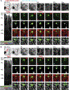
Müller glial cells improve the light transmission within the caiman retina. Stacks of horizontal confocal images of a freshly isolated wholemount of the ventral midperipheral retina which were recorded at depth intervals of 1 μm (every second is shown) within the tissue (GCL/NFL, IPL, INL, OPL, ONL) show Celltracker Orange-loaded Müller cells which appear bright and unloaded nerve fibers, synaptic neuropil, and neuronal perikarya which appear dark. The wholemount was illuminated at the inner (vitreal) side with green laser light, the positions of the light are indicated by the red and cyan points. The transmitted light was recorded at the outer (scleral) surface of the wholemount. A, B. A single straight Müller cell was investigated by a stepwise moving of the light positions along two paths (path 1 and path 2, each with seven light positions) crossing above the center of the thick Müller cell process. Shown are two z-reconstruction images (left) of the same Müller cell around the red arrows of path 1 and path 2 indicated in the horizontal confocal images at 0 μm depths above. When the incoming light is directly placed on top of the Müller cell center (indicated by the red points and the word ‘MC’), the transmitted light field shows a bright illumination of only a few receptor cells. (positions 3, 4 and 5 in A; positions 2′, 3′ and 4′ in B). For light positions within the periphery of the Müller cell endfoot (cyan points), the receptor field shows a wider distribution with a lower intensity of light. Note that the illuminated receptor fields consist of the same cone and a position dependent number of rods, whereby the cone cell always received light of higher intensity than the rod cells. GCL, ganglion cell layer; NFL, nerve fiber layer; IPL, inner plexiform layer; INL, inner nuclear layer; ONL, outer nuclear layer. Bars, 10 μm.
Fig. 8.
Fig. 8.
Propagation of light along Müller cell trunks formed by the tight assembling of Müller cell processes. Horizontal confocal images (above in A and B) were obtained at different depths in a freshly isolated wholemount of the dorsal midperipheral caiman retina. Celltracker Orange-loaded Müller cells appear bright; nerve fibers, synaptic neuropil, and neuronal and photoreceptor cell perikarya appear dark. The images left in A and B show z-reconstruction images of the investigated Müller cells around the red arrows indicated in the horizontal confocal images at 0 μm depth above. The wholemount was illuminated at the inner (vitreal) side with green laser light, the positions of the light are indicated by the red and cyan points. The images of the receptor segment layer were obtained from the outer (scleral) side of the wholemounts and show the light transmitted through the neuroretina (green) and cone segments (grey, blue). A, B. Müller cell trunks, formed by the assembling of Müller cell processes, were analyzed by the movement of light along a light path (red arrows). If the light is positioned within the trunk (positions 3–6 in A and positions 2–6 in B, indicated by the red points and the word ‘MC’) the intensity of the transmitted light increased and is distributed over some rods and individual cones (below). For positions outside of the trunk center the less intense transmitted light is distributed mainly above rod cells (positions 1, 2, 7 and 8 in A, positions 1, 7 and 8 in B, cyan points). GCL, ganglion cell layer; NFL, nerve fiber layer; IPL, inner plexiform layer; INL, inner nuclear layer; ONL, outer nuclear layer. Bars, 10 μm.
Fig. 9.
Fig. 9.
Propagation of light along individual Müller cell processes which get combined to a trunk. Horizontal confocal images (above in A and B) were obtained at different depths in a freshly isolated wholemount of the dorsal midperipheral caiman retina. Celltracker Orange-loaded Müller cells appear bright; nerve fibers, synaptic neuropil, and neuronal and photoreceptor cell perikarya appear dark. The images left in A and B show z-reconstruction images of the investigated Müller cells around the red arrows indicated in the horizontal confocal images at 0 μm depth above. The wholemount was illuminated at the inner (vitreal) side with green laser light, the positions of the light are indicated by the red and cyan points. The images of the receptor segment layer were obtained from the outer (scleral) side of the wholemounts and show the light transmitted through the neuroretina (green) and cone segments (grey). A, B. Separated inner stem processes which get combined in the outer part of the IPL were investigated by the movement of light along a light path (red arrows). The light intensity increased during the movement of the light position towards the first Müller cell process (positions 1–3 in A, cyan points). The maximum intensity is reached if the light is projected onto the first Müller cell process (positions 4, 5 and 6 in A; position 1 in B, indicated by the red points and the word ‘MC’), between the two processes (position 7 in A, position 2 in B, red points) and onto the second Müller cell process (position 8 in A; positions 3 and 4 in B, red points). Here, the light is distributed above cones and rods. If the light was positioned away from the second process center, the intensity of the transmitted light decreased (positions 9–12 in A; positions 5–7 in B, cyan points) and mainly rods are illuminated. GCL, ganglion cell layer; NFL, nerve fiber layer; IPL, inner plexiform layer; INL, inner nuclear layer; ONL, outer nuclear layer. Bars, 10 μm.
Fig. 10.
Fig. 10.
Propagation of light along a thick Müller cell trunk which split into individual processes. Horizontal confocal images (above) were obtained at different depths in a freshly isolated wholemount of the dorsal midperipheral caiman retina. Celltracker Orange-loaded Müller cells appear bright; nerve fibers, synaptic neuropil, and neuronal and photoreceptor cell perikarya appear dark. The image left shows a z-reconstruction image of the investigated Müller cell around the red arrow indicated in the horizontal confocal image at 0 μm depth above. The wholemount was illuminated at the inner (vitreal) side with green laser light, the positions of the light are indicated by the red and cyan points. The images of the receptor segment layer were obtained from the outer (scleral) side of the wholemounts and show the light transmitted through the neuroretina (green) and cone segments (grey). A large Müller cell trunk consisting of 3–4 processes which splits towards the INL, was investigated by the movement of light along a light path (red arrow). If the light was projected onto the center of the Müller cell trunk, the light intensity increased (positions 3 and 4, indicated by red points and the word ‘MC’) while projections of light beside the trunk center result in a lower intensity of light (positions 1, 2, 5, 6 and 7, indicated by cyan points). At all positions, only rods were illuminated by the transmitted light. GCL, ganglion cell layer; NFL, nerve fiber layer; IPL, inner plexiform layer; INL, inner nuclear layer; ONL, outer nuclear layer. Bars, 10 μm.

References

    1. Abelsdorff G, 1898. Physiologische Beobachtungen am Auge der Krokodile. Arch Anat Physiol Physiol Abt 155–167.
    1. Agte S, Junek S, Matthias S, Ulbricht E, Erdmann I, Wurm A, Schild D, Käs JA, Reichenbach A, 2011. Müller glial cell-provided cellular light guidance through the vital Guinea-pig retina. Biophys. J. 101, 2611–2619. - PMC - PubMed
    1. Banks MS, Sprague WW, Schmoll J, Parnell JA, Love GD, 2015. Why do animal eyes have pupils of different shapes? Sci Adv 1, e1500391. - PMC - PubMed
    1. Barer R, 1957. Refractometry and interferometry of living cells. J Opt Soc Am A Opt Image Sci Vis 47, 545–556. - PubMed
    1. Beuthan J, Minet O, Helfmann J, Herrig M, Müller G, 1996. The spatial variation of the refractive index in biological cells. Phys. Med. Biol. 41, 369–382. - PubMed

Publication types