Review
. 2018;10(5-6):502-514.
doi: 10.1159/000487818. Epub 2018 May 15.

The Understanding and Management of Organism Toxicity in Septic Shock

Affiliations
Review

The Understanding and Management of Organism Toxicity in Septic Shock

Kelly Roveran Genga et al. J Innate Immun. 2018.

Abstract

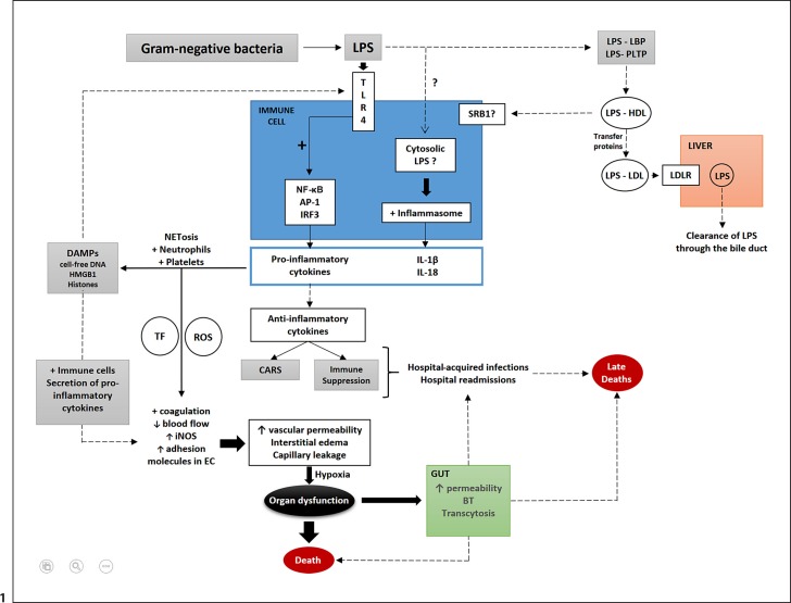
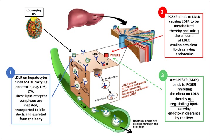
The toxicity caused by different organisms in septic shock is substantially complex and characterized by an intricate pathogenicity that involves several systems and pathways. Immune cells' pattern recognition receptors initiate the host response to pathogens after the recognition of pathogen-associated molecular patterns. In essence, the subsequent activation of downstream pathways may progress to infection resolution or to a dysregulated host response that represents the hallmark of organ injury in septic shock. Likewise, the management of organism toxicity in septic shock is complicated and comprises a multiplicity of suitable targets. In this review, the classic immune responses to pathogens are discussed as well as other factors that are relevant in the pathogenicity of septic shock, including sepsis-induced immune suppression, inflammasome activation, intestinal permeability, and the role of lipids and proprotein convertase subtilisin/kexin type 9. Current therapies aiming to eliminate the organisms causing septic shock, recent and ongoing trials in septic shock treatment, and potential new therapeutic strategies are also explored.

Keywords: Mediators; PCSK9; Sepsis; Septic shock; Therapies.


Figures

Fig. 1
Fig. 1
Schematic representation of important factors involved in the pathogenicity of septic shock caused by LPS from gram-negative bacteria. LPS from gram-negative bacteria are recognized by TLR4 on immune cells and activate the transcription of genes encoding the proinflammatory cytokines (IL-6, TNF, IFNα, and IL-8) released in the circulation. LPS may reach the cytosol of immune cells (likely mediated by an unknown endocytic pathway). LPS is then recognized by inflammatory caspases that activate the inflammasome, resulting in the release of mature IL-1β and IL-18. Proinflammatory cytokines activate neutrophils, platelets, and NET formation that generate DAMPs (histones, HMGB1, and cell-free DNA). DAMPs are recognized by TLR4 that further stimulates a proinflammatory response. Activated platelets and neutrophils lead to increases in TF and ROS that contribute to changes in endothelial cells (coagulation activation with clot formation, decreases in blood flow, and the upregulation of iNOS, ICAM, and VCAM-1 in endothelial cells. There is also upregulation of adhesion molecules in leukocytes (selectins and integrins). DAMPs also contribute to these effects on endothelial cells by the activation of immune cells, and further release of proinflammatory cytokines. Increased vascular permeability, interstitial edema, and capillary leakage cause tissue hypoxia that can progress to organ dysfunction and death. Intestinal dysfunction occurs mostly by the impairment of intestinal tight junctions and apoptosis, which lead to increases in intestinal permeability, bacterial translocation, and transcytosis. Anti-inflammatory cytokines that are also released into the circulation during sepsis may cause CARS and/or immune suppression. Both, in conjunction with the intestinal alterations in sepsis, contribute to increased rates of secondary and opportunistic hospital-acquired infections and hospital readmissions, factors associated with sepsis-associated late deaths. Alternatively, LPS can be sequestered within lipoprotein fractions in a process mediated by lipid transfer proteins such as LBP and PLTP. Initially, LPS is transferred to HDL and then from HDL to other lipoprotein fractions, such as LDL (again mediated by lipid transfer proteins such as LBP, PLTP, CETP, and possibly BPI). LPS within LDL is taken up by the LDLR and gets cleared from the body through the bile duct. Major pathways (black arrows); secondary pathways (dotted arrows); activation (+); increases (↑); decreases (↓). TLR, toll-like receptor; LPS, lipopolysaccharide; NF-κB, nuclear factor κB; AP-1, activator protein 1; IRF3, interferon regulatory transcription factor 3; IL, interleukin; NET, neutrophil extracellular trap; DAMPs, damage-associated molecular patterns; HMGB1, high-mobility group box 1; TF, tissue factor; ROS, reactive oxygen species; EC, endothelial cell; iNOS, inducible nitric oxide synthase; CARS, compensatory anti-inflammatory response syndrome; BT, bacterial translocation; SRB1, scavenger receptor class B type 1; LBP, lipopolysaccharide binding protein; PLTP, phospholipid binding protein; CETP, cholesteryl-ester transfer protein; BPI, bactericidal/permeability increasing protein; HDL, high-density lipoprotein; LDL, low-density lipoprotein; LDLR, LDL receptor; ICAM, intercellular adhesion molecule; VCAM-1, vascular cell adhesion molecule.
Fig. 2
Fig. 2
LPS clearance in the human host. (1) LPS from gram-negative bacteria carried within LDL particles binds to the LDLR on the surface of liver cells. The LPS-LDLR complex is then internalized, and while LDLR is recycled to the cell surface, LPS is cleared from the circulation through the bile duct. (2) When present, PCSK9 binds to LDLR on the cell surface and targets its lysosomal degradation. Therefore PCSK9, by preventing LDLR recycling, reduces the amount of LDLR available to clear LPS carried within LDL particles. (3) Anti-PCSK9 therapy (MAb) binds to PCSK9 and inhibits PCSK9-induced degradation of LDLR, upregulating the hepatic clearance of LPS carried within LDL. Adapted from [22]. LPS, lipopolysaccharide; LDL, low-density lipoprotein; LDLR, LDL receptor; PCSK9, proprotein convertase subtilisin/kexin type 9; Ab, monoclonal antibody.
Fig. 3
Fig. 3
We invert the usual drug discovery sequence. We start with human 'omics for drug candidate discovery (instead of animal models), confirm mechanisms in human-cell sepsis models and clinically relevant murine sepsis models, and then make go/no-go decisions for potential validated targets for clinical trials.

References

    1. Kawai T, Akira S. TLR signaling. Cell Death Differ. 2006;13:816–825. - PubMed
    1. Creagh EM, O'Neill LA. TLRs, NLRs and RLRs: a trinity of pathogen sensors that co-operate in innate immunity. Trends Immunol. 2006;27:352–357. - PubMed
    1. Kawai T, Akira S. The role of pattern-recognition receptors in innate immunity: update on Toll-like receptors. Nat Immunol. 2010;11:373–384. - PubMed
    1. Adib-Conquy M, Cavaillon JM. Compensatory anti-inflammatory response syndrome. Thromb Haemost. 2009;101:36–47. - PubMed
    1. Jin HS, Park JK, Jo EK. Toll-like receptors and NOD-like receptors in innate immune defense during pathogenic infection. J Bacteriol Virol. 2014;44:215–225.

Publication types

Substances