

Cone beam computed tomography in implant dentistry: recommendations for clinical use
- PMID: 29764458
- PMCID: PMC5952365
- DOI: 10.1186/s12903-018-0523-5
Cone beam computed tomography in implant dentistry: recommendations for clinical use
Abstract
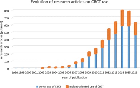
Background: In implant dentistry, three-dimensional (3D) imaging can be realised by dental cone beam computed tomography (CBCT), offering volumetric data on jaw bones and teeth with relatively low radiation doses and costs. The latter may explain why the market has been steadily growing since the first dental CBCT system appeared two decades ago. More than 85 different CBCT devices are currently available and this exponential growth has created a gap between scientific evidence and existing CBCT machines. Indeed, research for one CBCT machine cannot be automatically applied to other systems.
Methods: Supported by a narrative review, recommendations for justified and optimized CBCT imaging in oral implant dentistry are provided.
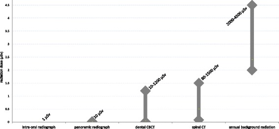
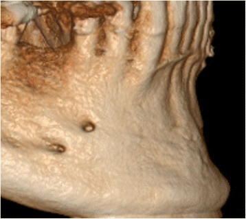
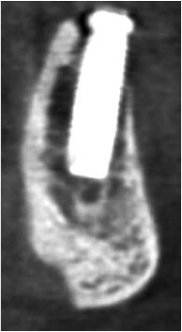
Results: The huge range in dose and diagnostic image quality requires further optimization and justification prior to clinical use. Yet, indications in implant dentistry may go beyond diagnostics. In fact, the inherent 3D datasets may further allow surgical planning and transfer to surgery via 3D printing or navigation. Nonetheless, effective radiation doses of distinct dental CBCT machines and protocols may largely vary with equivalent doses ranging between 2 to 200 panoramic radiographs, even for similar indications. Likewise, such variation is also noticed for diagnostic image quality, which reveals a massive variability amongst CBCT technologies and exposure protocols. For anatomical model making, the so-called segmentation accuracy may reach up to 200 μm, but considering wide variations in machine performance, larger inaccuracies may apply. This also holds true for linear measures, with accuracies of 200 μm being feasible, while sometimes fivefold inaccuracy levels may be reached. Diagnostic image quality may also be dramatically hampered by patient factors, such as motion and metal artefacts. Apart from radiodiagnostic possibilities, CBCT may offer a huge therapeutic potential, related to surgical guides and further prosthetic rehabilitation. Those additional opportunities may surely clarify part of the success of using CBCT for presurgical implant planning and its transfer to surgery and prosthetic solutions.
Conclusions: Hence, dental CBCT could be justified for presurgical diagnosis, preoperative planning and peroperative transfer for oral implant rehabilitation, whilst striving for optimisation of CBCT based machine-dependent, patient-specific and indication-oriented variables.
Keywords: Cone beam computed tomography; Dental implants; Guidelines; Presurgical planning; Radiation dose; Virtual patient.
Conflict of interest statement
Authors’ information
Not applicable
Ethics approval and consent to participate
Not applicable
Consent for publication
n/a: the manuscript does not contain any individual person’s data in any form.
Competing interests
Michael Bornstein is an associate editor for BMC Oral Health. Other authors declare that they have no competing interests.
Publisher’s Note
Springer Nature remains neutral with regard to jurisdictional claims in published maps and institutional affiliations.
Figures
References
-
- Jacobs R. Dental cone beam CT and its justified use in oral health care. JBR-BTR. 2011;94:254–265. - PubMed
Publication types
MeSH terms
LinkOut - more resources
Full Text Sources
Other Literature Sources
Medical
Miscellaneous
