. 2018 Apr 3;38(1):9.
doi: 10.1186/s40880-018-0276-1.

EYA4 inhibits hepatocellular carcinoma growth and invasion by suppressing NF-κB-dependent RAP1 transactivation

Affiliations

EYA4 inhibits hepatocellular carcinoma growth and invasion by suppressing NF-κB-dependent RAP1 transactivation

Shi-Jing Mo et al. Cancer Commun (Lond). .

Abstract

Background: Our previous studies demonstrated that eyes absent homolog 4 (EYA4), a member of the eye development-related EYA family in Drosophila, is frequently methylated and silenced in hepatocellular carcinoma (HCC) specimens and associated with shorter survival. The current work aimed to explore the mechanisms through which EYA4 functions as a tumor suppressor in HCC.

Methods: Stable EYA4-expressing plasmid (pEYA4) transfectants of the human HCC cell lines Huh-7 and PLC/PRF/5 (PLC) were established. Xenografts tumors were established via subcutaneous injection of the stable transfectants into BALB/c nude mice. Tissue samples were obtained from 75 pathologically diagnosed HCC patients. Quantitative real-time polymerase chain reaction, Western blotting and immunohistochemistry were performed to determine the expression of EYA4 in cell lines, xenografts and clinical specimens. The cell proliferation, colony formation, invasiveness and tumor formation of stable transfectants were studied. A gene expression microarray was utilized to screen genes regulated by EYA4 expression. The effect of EYA4 on nuclear factor-κB (NF-κB)/RAS-related protein 1 (RAP1) signaling was demonstrated through the co-transfection of pEYA4 and Flag-tagged RAS-related protein 1A gene-expressing plasmid (Flag-RAP1A), functional studies, chromatin immunoprecipitation, immunofluorescence staining and cellular ubiquitination assay.

Results: The restoration of EYA4 expression in HCC cell lines suppressed cell proliferation, inhibited clonogenic outgrowth, reduced cell invasion and restrained xenograft tumor growth, and Flag-RAP1A reversed the suppressive effects of pEYA4 in vitro. Activation of NF-κB with tumor necrosis factor-α (TNF-α) increased the binding of p65 to the RAP1A gene promoter and up-regulated RAP1 protein expression. The inhibition of NF-κB with BAY 11-7085 and p65 siRNA successfully blocked TNF-α-induced RAP1 up-regulation. EYA4 antagonized the TNF-α-induced phosphorylation and ubiquitination of inhibitor of NF-κBα (IκBα) as well as the nuclear translocation and transactivation of p65, resulting in repressed NF-κB activity and RAP1 expression. Blocking the serine/threonine phosphatase activity of EYA4 with calyculin A notably abrogated its suppressive effect on NF-κB activity. In addition, EYA4 expression was inversely correlated with IκBα/RAP1 activity in clinical HCC specimens.

Conclusion: Our findings provide a functional and mechanistic basis for identifying EYA4 as a bona fide tumor suppressor that disrupts aberrant activation of the NF-κB/RAP1 signaling pathway and thus orchestrates a physiological impediment to HCC growth and invasion.

Keywords: Eyes absent homolog 4 (EYA4); Hepatocellular carcinoma; Nuclear factor-κB (NF-κB); RAS-related protein 1 (RAP1); Transactivation.


Figures
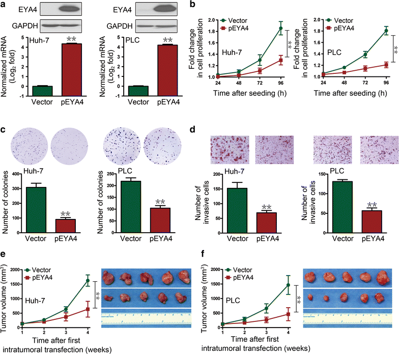
Fig. 1
Fig. 1
The overexpression of eyes absent homolog 4 (EYA4) antagonized the proliferation, clonogenicity, invasiveness and xenograft tumor growth of human hepatocellular carcinoma (HCC) cells. a Quantitative real-time polymerase chain reaction (qRT-PCR) and Western blotting analyses showed elevated EYA4 expression levels in EYA4-expressing plasmid (pEYA) transfectants. bd Suppressive effects of EYA4 overexpression on cell proliferation (b), colony formation (c) and transwell invasion (d). e, f Tumor growth curves (left panel) and images (right panel) of xenografts of Huh-7 (e) and PLC5 (f) cells after an intratumoral injection of pEYA4. **P < 0.01 between groups. EYA4 eyes absent homolog 4, GAPDH glyceraldehyde 3-phosphate dehydrogenase, HCC hepatocellular carcinoma, pEYA4 EYA4-expressing plasmid, qRT-PCR quantitative real-time polymerase chain reaction
Fig. 2
Fig. 2
EYA4 negatively regulated RAS-related protein 1 (RAP1) expression in human HCC cells. a A heat map of gene expression microarray data shows significantly repressed genes (fold change > 3) in stable pEYA4 transfectants compared with vector transfectants. b Comparison of gene expression in the indicated pEYA4 versus vector cells through microarray profiling. c Venn diagram for the selection of RAS-related protein 1A gene (RAP1A) using vector- and pEYA4-expressing Huh-7 and PLC5 cells. d qRT-PCR analyses of RAP1A and EYA4 mRNA expression in pEYA4 transfectants. **P < 0.01 and ***P < 0.001 versus the vector group. e Western blotting analyses of the RAP1 and EYA4 protein expression levels in pEYA4 transfectants. f Western blotting analyses of RAP1 protein expression levels in the indicated HCC cells after EYA4 depletion and subsequent pEYA4 transfection. g Weight of subcutaneous xenograft tumors formed by pEYA4 and vector transfectants. **P < 0.01 between groups. h Western blotting analyses of the EYA4 and RAP1 protein expression levels in the subcutaneous xenograft tumors formed by pEYA4 and vector transfectants. EYA4 eyes absent homolog 4, GAPDH glyceraldehyde 3-phosphate dehydrogenase, pEYA4 EYA4-expressing plasmid, RAP1 RAS-related protein 1, RAP1A RAS-related protein 1A gene
Fig. 3
Fig. 3
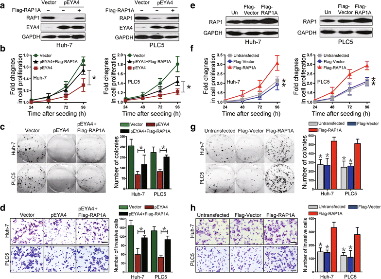
EYA4 inhibited proliferation, clonogenicity and invasiveness of HCC cells by the suppression of RAP1. a Western blotting analyses showing the RAP1 protein expression levels in pEYA4 transfectants (left: Huh-7, right: PLC5) after transfection with or without Flag-tagged human RAP1A expressing plasmid (Flag-RAP1A). bd Effects of RAP1 expression on cell proliferation (b), clonogenicity (c) and cell invasiveness (d) of EYA4-overexpressing HCC cells. pEYA4 transfectants were transfected with or without Flag-RAP1A, and the resulting cell proliferation, clonogenic outgrowth and cell invasiveness were determined by CCK-8 assays, colony formation assays and transwell invasion assays, respectively. *P < 0.05 between groups. e Western blotting analyses of the RAP1 expression levels in Huh-7 (left panel) and PLC5 (right panel) with or without Flag-RAP1A transfection. fh Effects of RAP1 expression on cell proliferation (f), clonogenicity (g) and cell invasiveness (h) of HCC cells. The effects were analyzed by CCK-8 assays, colony formation assays and transwell invasion assays, respectively. *P < 0.05 versus the Flag-RAP1A group. EYA4 eyes absent homolog 4, Flag-RAP1A Flag-tagged human RAP1A expressing plasmid, Flag-Vector Flag-tagged vector plasmid, GAPDH glyceraldehyde 3-phosphate dehydrogenase, pEYA4 EYA4-expressing plasmid, RAP1 RAS-related protein 1, RAP1A RAS-related protein 1A gene, Un untransfected control
Fig. 4
Fig. 4
Nuclear factor-κB (NF-κB) regulated RAP1 in an EYA4-dependent manner. a Western blotting analyses comparing the levels of RAP1 protein expression in Huh-7 and PLC5 cells after activation of β-catenin with epidermal growth factor (EGF, 100 ng/mL) or silencing of EYA4 with EYA4-targeted short hairpin RNA (shEYA4) in the presence or absence of pEYA4 transfection. b Chromatin immunoprecipitation (ChIP) analyses were performed to test the interactions between p65 and the RAP1A promoter in Huh-7 and PLC5 cells with or without pEYA4 transfection upon tumor necrosis factor-α (TNF-α, 10 ng/mL) stimulation. c Western blotting analyses comparing the levels of RAP1 protein expression in Huh-7 and PLC5 cells with TNF-α stimulation for the indicated times in the presence or absence of the NF-κB inhibitor BAY 11-7085 (5 µmol/L). d Western blotting analyses comparing the levels of RAP1 protein expression in Huh-7 and PLC5 cells upon TNF-α stimulation with or without p65-targeting small interfering RNA (p65 siRNA) transient transfection. e, f The expression of RAP1 protein (e) and RAP1A mRNA (f) was analyzed in Huh-7 and PLC5 cells transfected with pEYA4 alone or with pEYA4 plus shEYA4 in the presence or absence of TNF-α stimulation. **P < 0.01 vs. the pEYA4 group. g, h RAP1 and EYA4 protein expression (g) and RAP1A and EYA4 mRNA expression (h) in Huh-7 and PLC5 cells in which EYA4 was depleted by shEYA4 in the presence or absence of the NF-κB inhibitor BAY 11-7085. **P < 0.01 vs. shEYA4 group. i Western blotting analyses detecting the RAP1 protein abundance in Huh-7 and PLC5 cells in which EYA4 was silenced by shEYA4 transfection in the presence or absence of p65 siRNA transient transfection. The qRT-PCR experiments were performed five times with technical duplicates, and the real-time values were normalized to glyceraldehyde 3-phosphate dehydrogenase (GAPDH). ChIP chromatin immunoprecipitation, DMSO dimethyl sulfoxide, EGF epidermal growth factor, EYA4 eyes absent homolog 4, Flag-RAP1A Flag-tagged human RAP1A expressing plasmid, Flag-Vector Flag-tagged vector plasmid, GAPDH glyceraldehyde 3-phosphate dehydrogenase, IgG immunoglobulin G, IP immunoprecipitation, NF-κB nuclear factor-κB, P65 NF-κB p65 subunit, PCR polymerase chain reaction, pEYA4 EYA4-expressing plasmid, RAP1 RAS-related protein 1, Scr scramble short hairpin RNA, shEYA4 EYA4-targeted short hairpin RNA, siRNA small interfering RNA, TNF-α tumor necrosis factor-α
Fig. 5
Fig. 5
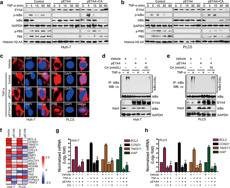
The serine/threonine-phosphatase activity of EYA4 inactivated NF-κB signaling. a, b Western blotting analyses examining the levels of IκBα phosphorylation and p65 nuclear translocation in Huh-7 (a) and PLC5 (b) cells transduced with pEYA4 in the presence or absence of the serine/threonine phosphatase inhibitor calyculin A (CA; 10 nmol/L) upon TNF-α (10 ng/mL) stimulation. Histone H2.AX served as the loading control for the nuclear fractions. c Representative immunofluorescence staining images comparing the nuclear translocation of p65 in Huh-7 and PLC5 cells transfected with pEYA4 in the presence or absence of CA treatment upon TNF-α stimulation. Scale bar = 20 µm. d, e In vivo ubiquitination assays comparing the polyubiquitination levels of IκBα in Huh-7 (d) and PLC5 (e) cells transfected with pEYA4 in the presence or absence of CA treatment upon TNF-α stimulation. f EYA4 opposed the transcriptional programs of NF-κB signaling in HCC cells. Heat map of gene expression microarray data showing the expression of NF-κB signaling-dependent genes in the indicated HCC cells. g, h qRT-PCR analyses of the NF-κB downstream target genes B cell leukemia/lymphoma 2 (BCL-2), cyclin D1 (CCND1), twist-related protein 1 (TWIST1), and X-linked inhibitor of apoptosis (XIAP) in Huh-7 (g) and PLC5 (h) cells transfected with pEYA4 in the presence or absence of CA upon TNF-α stimulation. The experiments were performed five times, each qRT-PCR assay was performed in technical duplicates, and the real-time values were normalized to GAPDH. *P < 0.05 versus the TNF-α plus pEYA4 groups. BCL-2 B cell leukemia/lymphoma 2, CA calyculin A, CCND1 cyclin D1, DAPI 4′,6-diamidino-2-phenylindole, EYA4 eyes absent homolog 4, GAPDH glyceraldehyde 3-phosphate dehydrogenase, IκBα nuclear factor of kappa light polypeptide gene enhancer in B-cells inhibitor, alpha, IP immunoprecipitation, NF-κB nuclear factor-κB, P65 NF-κB p65 subunit, p-IκBα phosphorylated IκBα, p-P65 phosphorylated P65, PCR polymerase chain reaction, pEYA4 EYA4-expressing plasmid, RAP1 RAS-related protein 1, shEYA4 EYA4-targeted short hairpin RNA, siRNA small interfering RNA, TNF-α tumor necrosis factor-α, TWIST1 twist-related protein 1, Ub ubiquitination, (Ub)n polyubiquitination, WB Western blotting, XIAP X-linked inhibitor of apoptosis
Fig. 6
Fig. 6
Clinical relevance of EYA4 expression for the IκBα/RAP1 axis in human HCC tissues. a, b Representative immunohistochemistry images (a) and bar graphs (b) showing the relevance of EYA4 expression on the phospho-IκBα-Ser32 and RAP1 expression levels in 67 clinical HCC specimens. c, d Western-blotting analyses (c) and correlation analyses (d) were used to examine whether the EYA4 protein levels are associated with the levels of phospho-IκBα-Ser32 and RAP1 in eight HCC samples. EYA4 eyes absent homolog 4, IκBα nuclear factor of kappa light polypeptide gene enhancer in B-cells inhibitor, alpha, GAPDH glyceraldehyde 3-phosphate dehydrogenase, p-IκBα phosphorylated IκBα, RAP1 RAS-related protein 1

Similar articles

Cited by

References

    1. Ferlay J, Soerjomataram I, Dikshit R, Eser S, Mathers C, Rebelo M, et al. Cancer incidence and mortality worldwide: sources, methods and major patterns in GLOBOCAN 2012. Int J Cancer. 2015;136:E359–E386. doi: 10.1002/ijc.29210. - DOI - PubMed
    1. Ferlay J, Soerjomataram I, Ervik M, Dikshit R, Eser S, Mathers C, et al. GLOBOCAN 2012 v1.0, cancer incidence and mortality worldwide: IARC CancerBase No. 11. Lyon: International Agency for Research on Cancer. 2013.
    1. Zheng R, Zeng H, Zhang S, Chen W. Estimates of cancer incidence and mortality in China, 2013. Chin J Cancer. 2017;36:66. doi: 10.1186/s40880-017-0234-3. - DOI - PMC - PubMed
    1. El-Serag HB. Hepatocellular carcinoma. N Engl J Med. 2011;365:1118–1127. doi: 10.1056/NEJMra1001683. - DOI - PubMed
    1. Kong M, Hong SE. Optimal follow-up duration for evaluating objective response to radiotherapy in patients with hepatocellular carcinoma: a retrospective study. Chin J Cancer. 2015;34:79–85. doi: 10.5732/cjc.014.10136. - DOI - PMC - PubMed

Publication types

MeSH terms