. 2018 May 15;11(530):eaao6847.
doi: 10.1126/scisignal.aao6847.

A DGKζ-FoxO-ubiquitin proteolytic axis controls fiber size during skeletal muscle remodeling

Affiliations

A DGKζ-FoxO-ubiquitin proteolytic axis controls fiber size during skeletal muscle remodeling

Jae-Sung You et al. Sci Signal. .

Abstract

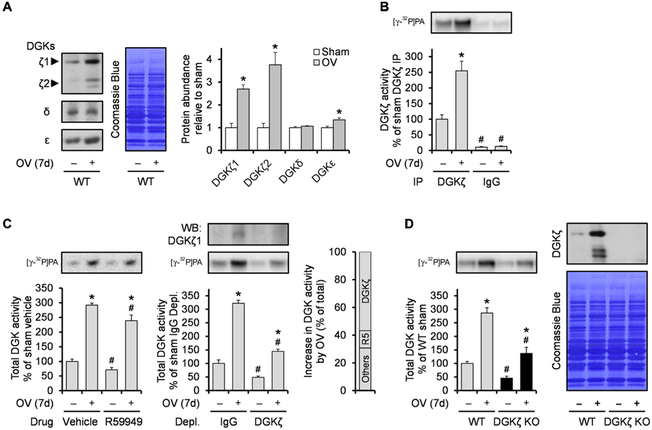
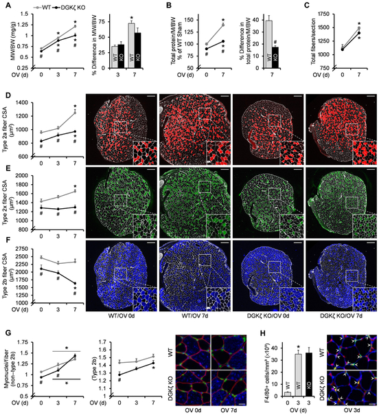
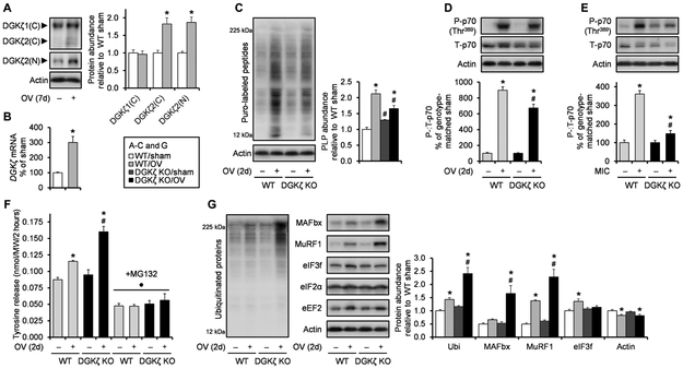
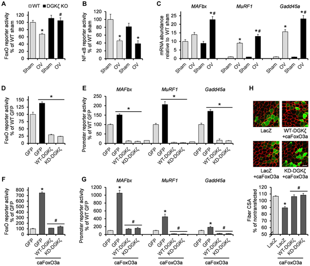
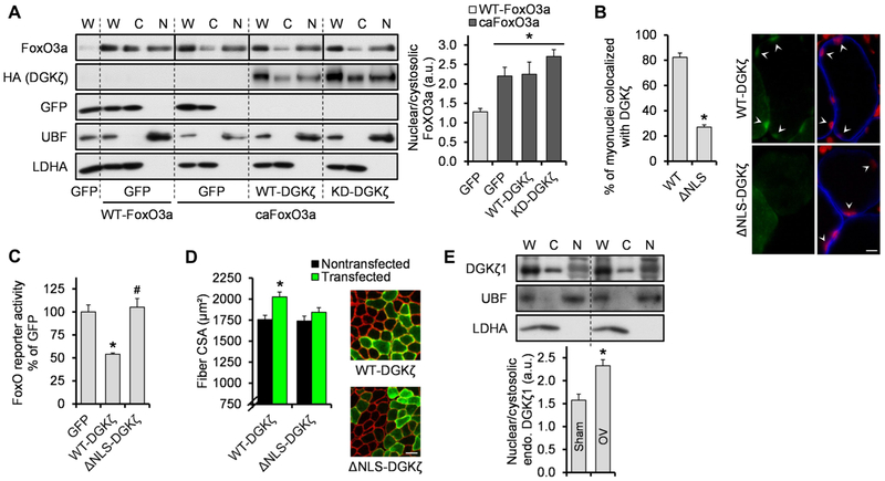
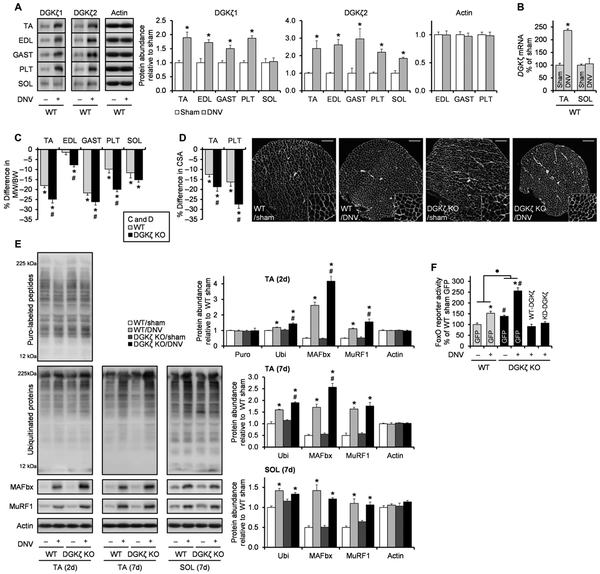
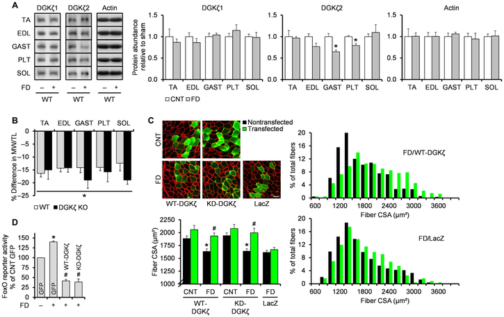
Skeletal muscle rapidly remodels in response to various stresses, and the resulting changes in muscle mass profoundly influence our health and quality of life. We identified a diacylglycerol kinase ζ (DGKζ)-mediated pathway that regulated muscle mass during remodeling. During mechanical overload, DGKζ abundance was increased and required for effective hypertrophy. DGKζ not only augmented anabolic responses but also suppressed ubiquitin-proteasome system (UPS)-dependent proteolysis. We found that DGKζ inhibited the transcription factor FoxO that promotes the induction of the UPS. This function was mediated through a mechanism that was independent of kinase activity but dependent on the nuclear localization of DGKζ. During denervation, DGKζ abundance was also increased and was required for mitigating the activation of FoxO-UPS and the induction of atrophy. Conversely, overexpression of DGKζ prevented fasting-induced atrophy. Therefore, DGKζ is an inhibitor of the FoxO-UPS pathway, and interventions that increase its abundance could prevent muscle wasting.


Figures

Fig. 1.
Fig. 1.. DGKζ is predominantly increased among DGK isoforms during mechanical overload.
WT and DGKζ KO mice were subjected to mechanical overload (OV+) or sham (OV−) surgery and the plantaris muscles (n) were collected at 7 days (7d) after surgery. (A to C) WT muscles were subjected to Western blotting (WB) to detect the indicated proteins (n = 4 from three mice per group) (A) or to DGK activity assay after immunoprecipitation (IP) of DGKζ (n = 3 to 4 from three mice per group) (B), in vitro treatment of R59949 (R5), or immunodepletion (Depl.) of DGKζ (n = 3 to 4 from three mice per group) (C). (D) WT and DGKζ KO muscles were subjected to Western blotting to detect DGKζ protein (right) or to DGK activity assay (left) (n = 4 to 6 from three mice per group). Values were expressed as means + SEM. *P < 0.05 compared to sham within the same immunoprecipitation (B), drug (C), immunodepletion (C), or genotype (D); #P < 0.05 compared to DGKζ (B), vehicle (C), IgG (C), or WT (D), within the same surgery, Student’s t test (A) or two-way analysis of variance (ANOVA) (B to D).
Fig. 2.
Fig. 2.. DGKζ is required for effective muscle growth during mechanical overload.
WT and DGKζ KO mice were subjected to mechanical overload (3d or 7d) or sham (0d) surgery, and the plantaris muscles (n) were collected at 3 or 7 days after surgery. (A) Muscle weight (MW)–to–body weight (BW) ratio (n = 6 to 8 from three to five mice per group). (B) Total protein content per muscle (M) normalized by BW (n = 5 to 6 from five to six mice per group). (C to F) Immunohistochemistry on cross sections with antibodies against laminin and type 2a myosin heavy chain (MHC), type 2× MHC, or type 2b MHC, to measure total fiber number per section (n = 6 to 9 from three to five mice per group) (C), and CSA in type 2a fibers (D), type 2× fibers (E), and type 2b fibers (F). For (D) to (F) (n = 5 to 8 from three to five mice per group), the right panels show representative images of the whole cross sections and their 2× magnified views (white, laminin; red, type 2a MHC; green, type 2× MHC; blue, type 2b MHC). Scale bars, 200 μm. (G) Immunohistochemistry on cross sections with propidium iodine (PI) and antibodies against dystrophin and type 2b MHC to measure the number of myonuclei (nuclei inside the dystrophin ring) per fiber cross section in non–type 2b fibers and type 2b fibers (n = 5 to 8 from three to four mice per group). The right panel shows representative images (red, dystrophin; blue, type 2b MHC; green, PI). Scale bar, 20 μm. (H) Immunohistochemistry on cross sections with PI and antibodies against laminin and F4/80 to measure the density of macrophages (as determined by nuclei colocalized with F4/80 outside the laminin ring) (n = 3 to 4 from three to four mice per group). The right panel shows representative images (red, PI; blue, laminin; green, F4/80). Scale bar, 20 μm. Values were expressed as means + SEM. *P < 0.05 compared to sham within the same genotype, #P < 0.05 compared to WT within the same surgery, two-way ANOVA (A to G) or Student’s t test (B, right, and H).
Fig. 3.
Fig. 3.. DGKζ contributes to the activation of mTOR signaling and protein synthesis and attenuates the activation of UPS-dependent protein degradation after the onset of mechanical overload.
WT and DGKζ KO mice were subjected to mechanical overload or sham surgery (A to D, F, and G), or a bout of maximal-intensity contractions (MIC+) or the control condition (MIC−) (E). The plantaris and tibialis anterior muscles (n) were collected at 2 days after surgery or immediately after MIC, respectively. (A) Western blotting to detect DGKζ protein with antibodies against the C terminus (C) or the N terminus (N; specific to splice variant 2) of DGKζ (n = 6 from three mice per group). (B) Quantitative reverse transcription polymerase chain reaction (qRT-PCR) to measure mRNA expression of DGKζ (n = 5 to 6 from three mice per group). (C) Measurement of protein synthesis rate as assessed by Western blotting of puromycin (puro)–labeled peptides (PLP) (n = 5 to 6 from three mice per group). (D and E) Western blotting to detect phosphorylated (P) and total (T) p70 [n = to 6 from three mice per group (D) and n = 3 to 4 from three to four mice per group (E)]. (F) Measurement of protein degradation rate as assessed by tyrosine release (n = 3 to 5 from three to five mice per group). (G) Western blotting to detect the indicated proteins (n = 5 to 8 from three to four mice per group). Values were expressed as means (+SEM in graphs). *P < 0.05 compared to sham within the same genotype, #P < 0.05 compared to WT within the same surgery (C, D, F, and G) or MIC+ (E), ·P < 0.05 compared to non-MG132 within the same genotype and surgery, Student’s t test (A to B) or two-way ANOVA (C to G). Ubi, ubiquitin.
Fig. 4.
Fig. 4.. DGKζ counteracts FoxO activity.
(A to C) WT and DGKζ KO mice were subjected to mechanical overload or sham surgery, and the plantaris muscles (n) were immediately cotransfected with pRL-SV40 Renilla luciferase (pRL) and FoxO response element firefly luciferase reporter (n = 4 to 5 from four to six mice per group) (A), NF-κB response element firefly luciferase reporter (n = 4 to 6 from four to six mice per group) (B), or left nontransfected (n = 5 to 8 from three to four mice per group) (C). Transfected muscles were collected at 2 days after surgery and subjected to dual-luciferase reporter assay. Nontransfected muscles were collected at 1 day after surgery and subjected to qRT-PCR to measure mRNA expression of the indicated genes. (D and E) Tibialis anterior muscles (n) from WT and DGKζ KO mice were cotransfected with GFP, hemagglutinin (HA)–tagged WT-DGKζ, or HA-tagged KD-DGKζ, and pRL and FoxO response element firefly luciferase reporter (n = 3 to 7 from three to six mice per group) (D) or MAFbx, MuRF1, or Gadd45a promoter firefly luciferase reporter (n = 3 to 8 from three to eight mice per group) (E). Muscles were collected at 2 days after transfection and subjected to dual-luciferase reporter assay. (F and G) Tibialis anterior muscles (n) from WT mice were cotransfected with GFP, HA-tagged WT-DGKζ, or HA-tagged KD-DGKζ, and pRL and FoxO response element firefly luciferase reporter ± constitutively active (ca) FoxO3a (n = 3 to 7 from three to six mice per group) (F) or the indicated promoter firefly luciferase reporter ± caFoxO3a (n = 4 to 8 from four to eight mice per group) (G). Muscles were collected at 2 days after transfection and subjected to dual-luciferase reporter assay. (H) Tibialis anterior muscles (n) from WT mice were transfected with LacZ, HA-tagged WT-DGKζ, or HA-tagged KD-DGKζ ± caFoxO3a, and collected at 7 days after transfection. Cross sections of the muscles were subjected to immunohistochemistry with antibodies against laminin (red) and LacZ (green) or HA (green) to measure CSA of the transfected and nontransfected fibers (n = 4 from three to four mice per group). The top panel shows representative images. Scale bar, 50 μm. Values were expressed as means + SEM. *P < 0.05 compared to sham within the same genotype (A to C), WT GFP (D and E), GFP (F and G), or LacZ (H); #P < 0.05 compared to WT mechanical overload (A to C), GFP + caFoxO3a (F and G), LacZ + caFoxO3a (H), two-way ANOVA (A to C), or one-way ANOVA (D to H).
Fig. 5.
Fig. 5.. The NLS of DGKζ is required for the inhibition of FoxO activity and the induction of hypertrophy.
(A) Tibialis anterior muscles (n) were cotransfected with WT-FoxO3a or caFoxO3a and GFP, HA-tagged WT-DGKζ, or HA-tagged KD-DGKζ. The muscles were collected at 2 days after transfection and subjected to Western blotting to detect FoxO3a, HA (DGKζ), GFP, upstream binding factor (UBF) (nuclear marker), and lactate dehydrogenase A (LDHA) (cytosolic marker), in whole homogenates (W), cytosolic fractions (C), and nuclear fractions (N) (n = 4 from four mice per group). Solid lines distinguish images from different gels. (B) Tibialis anterior muscles (n) were transfected with FLAG-tagged WT-DGKζ or a nuclear localization signal mutant (ΔNLS)–DGKζ and collected at 2 days after transfection. Cross sections of the muscles were subjected to immunohistochemistry with PI (red) and antibodies against dystrophin (blue) and FLAG (green) to measure myonuclei that colocalized with DGKζ (n = 4 from four mice per group). The right panel shows representative images. Scale bar, 10 μm. (C) Tibialis anterior muscles (n) were cotransfected with FoxO response element firefly luciferase reporter, pRL-SV40 Renilla luciferase, and GFP, FLAG-tagged WT-DGKζ, or FLAG-tagged ΔNLS-DGKζ. Muscles were collected at 2 days after transfection and subjected to dual-luciferase reporter assay (n = 4 to 7 from four to five mice per group. (D) Tibialis anterior muscles (n) were transfected with FLAG-tagged WT-DGKζ or FLAG-tagged ΔNLS-DGKζ and collected at 7 days after transfection. Cross sections of the muscles were subjected to immunohistochemistry with antibodies against laminin (red) and FLAG (green) to measure CSA of the transfected and nontransfected fibers (n = 6 from six mice per group). The right panel shows representative images. Scale bar, 50 μm. (E) Mice were subjected to mechanical overload or sham surgery, and the plantaris muscles (n) were collected at 2 days after surgery and subjected to Western blotting to detect the indicated proteins in different fractions as described in (A) (n = 5 from three mice per group). a.u., arbitrary units; endo., endogenous. Values were expressed as means + SEM. *P < 0.05 compared to GFP + WT-FoxO3a (A), WT-DGKζ (B), GFP (C), nontransfected (D), or sham (E); #P < 0.05 compared to WT-DGKζ, oneway ANOVA (A and C) or Student’s t test (B, D, and E).
Fig. 6.
Fig. 6.. DGKζ mitigates the activation of the FoxO-UPS pathway and muscle atrophy during denervation.
WT and DGKζ KO mice were subjected to denervation (DNV+) or sham (DNV−) surgery, and the tibialis anterior (TA), extensor digitorum longus (EDL), gastrocnemius (GAST), plantaris (PLT), and soleus (SOL) muscles (n) were collected at 7 days after surgery unless otherwise indicated. (A) Western blotting to detect DGKζ protein with antibodies against the C terminus of DGKζ1 (n = 4 to 6 from three to five mice per group) or the N terminus of DGKζ2 (n = 4 to 5 from three to five mice per group) in the indicated muscles. (B) qRT-PCR to measure mRNA expression of DGKζ in TA (n = 4 to 6 from three to four mice per group) and SOL (n = 5 from three to five mice per group). (C) Denervation-induced differences in the MW/BW ratio in the indicated muscles (n=9 to 13 from 9 to 13 mice per group).(D) Immunohistochemistry on cross sections with an antibody against laminin (white) to measure fiber CSA in TA (n = 5 to 6 from five to six mice per group) and PLT (n = 3 to 4 from three to four mice per group). The right panel shows representative images of the whole cross sections of the PLT muscles and their 2× magnified views. Scale bars, 200 μm. (E) Western blotting to detect the indicated proteins (n = 3 to 6 from three to six mice per group). (F) TA muscles were cotransfected with FoxO response element firefly luciferase reporter, pRL-SV40 Renilla luciferase, and GFP, HA-tagged WT-DGKζ, or HA-tagged KD-DGKζ at 5 days after denervation or sham surgery. Upon collection, the muscles were subjected to dual-luciferase reporter assay (n = 3 to 7 from three to six mice per group). Values were expressed as means + SEM. *P < 0.05 compared to sham [within the same genotype in (C) to (F)], #P < 0.05 compared to WT [within the same surgery in (E) and (F)], ·P < 0.05 in the magnitude of the denervation effect between genotypes, Student’s t test (A to D and F) or two-way ANOVA (E and F).
Fig. 7.
Fig. 7.. Overexpression of DGKζ inhibits food deprivation–induced muscle atrophy.
(A and B) WT and DGKζ KO mice were subjected to food deprivation (FD+) or the control (FD−) condition, and the TA, EDL, GAST, PLT, and SOL muscles (n) were collected at 2 days after FD. (A) Western blotting to detect DGKζ protein with antibodies against the C terminus of DGKζ1 or the N terminus of DGKζ2 in the indicated muscles (n = 5 to 6 from three mice per group). (B) FD-induced differences in the MW/TL (tibia length) ratio in the indicated muscles (n = 5 to 6 from three mice per group). (C) TA muscles (n) from WT mice were transfected with HA-tagged WT-DGKζ, HA-tagged KD-DGKζ, or LacZ immediately before being subjected to 2 days of FD or the control (CNT) condition. Cross sections of the muscles were subjected to immunohistochemistry with antibodies against laminin (red) and LacZ (green) or HA (green) to measure CSA of the transfected and nontransfected fibers (n = 4 to 5 from three to five mice per group). The top left panel shows representative images. Scale bar, 50 μm. The right panel shows the distribution of the CSA on a histogram. (D) TA muscles (n) were cotransfected with FoxO response element firefly luciferase reporter, pRL-SV40 Renilla luciferase, and GFP, HA-tagged WT-DGKζ, or HA-tagged KD-DGKζ, immediately before being subjected to 2 days of FD or the CNT condition. Upon collection, the muscles were subjected to dual-luciferase reporter assay (n = 3 from three mice per group). Values were expressed as means + SEM. *P < 0.05 compared to CNT (A and B) [within the same genotype in (B)] or CNT (C and D) [within the same transfection in (C)], #P < 0.05 compared to nontransfected within the same feeding and DNA (C) or GFP + FD (D), Student’s t test (A and B), two-way ANOVA (C), or one-way ANOVA (D).

References

    1. Evans WJ, Skeletal muscle loss: Cachexia, sarcopenia, and inactivity. Am. J. Clin. Nutr. 91, 1123S–1127S (2010). - PubMed
    1. Goldberg AL, Etlinger JD, Goldspink DF, Jablecki C, Mechanism of work-induced hypertrophy of skeletal muscle. Med. Sci. Sports 7, 185–198 (1975). - PubMed
    1. Izumiya Y, Hopkins T, Morris C, Sato K, Zeng L, Viereck J, Hamilton JA, Ouchi N, LeBrasseur NK, Walsh K, Fast/glycolytic muscle fiber growth reduces fat mass and improves metabolic parameters in obese mice. Cell Metab 7, 159–172 (2008). - PMC - PubMed
    1. Srikanthan P, Karlamangla AS, Relative muscle mass is inversely associated with insulin resistance and prediabetes. Findings from the third National Health and Nutrition Examination Survey. J. Clin. Endocrinol. Metab. 96, 2898–2903 (2011). - PubMed
    1. Srikanthan P, Karlamangla AS, Muscle mass index as a predictor of longevity in older adults. Am. J. Med. 127, 547–553 (2014). - PMC - PubMed

Publication types

MeSH terms