
Assessment of established techniques to determine developmental and malignant potential of human pluripotent stem cells

International Stem Cell Initiative. Nat Commun. .

Abstract

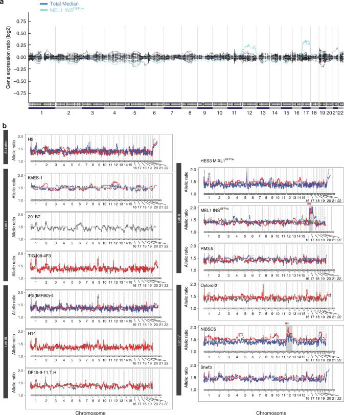
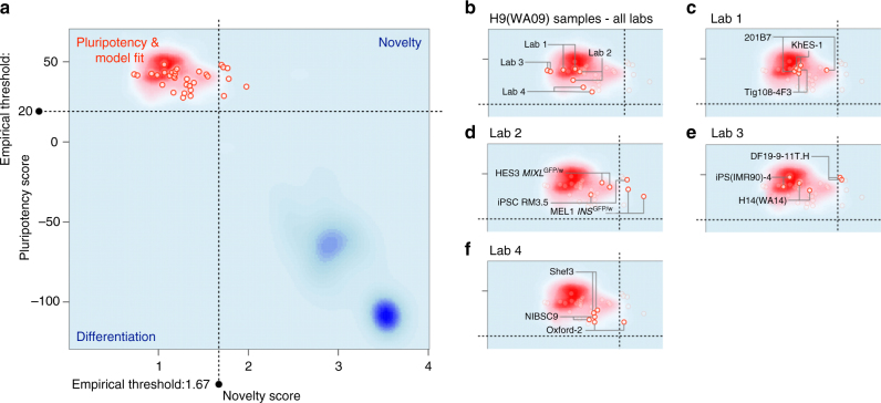
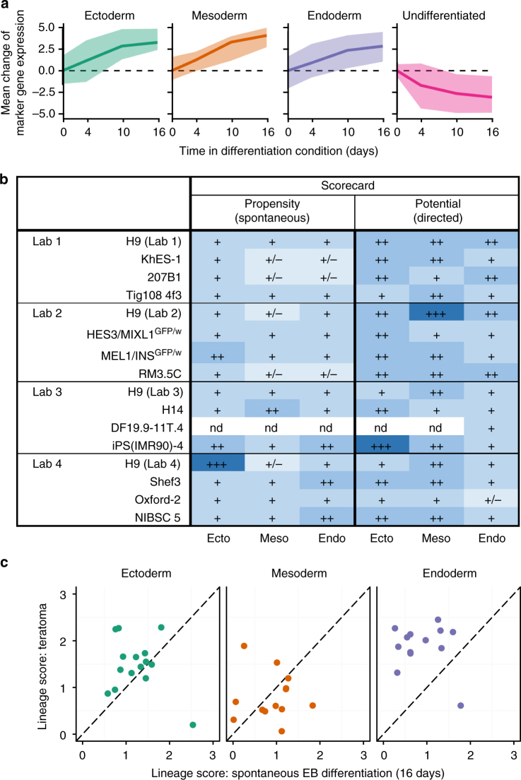
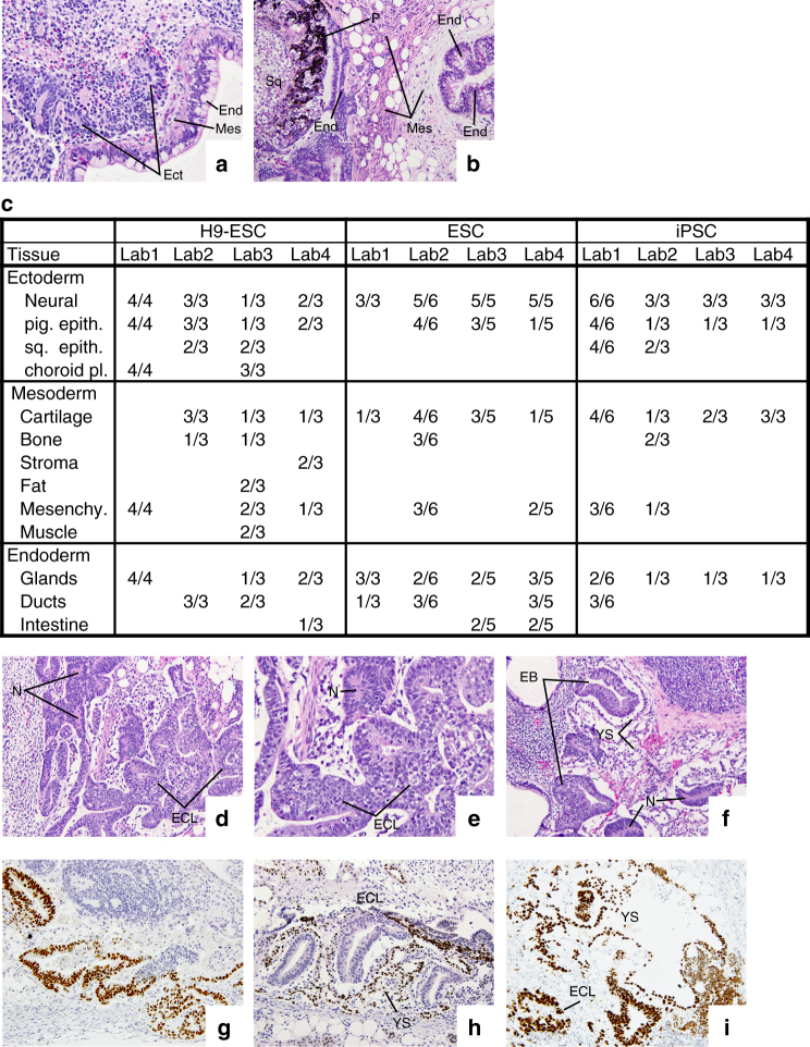
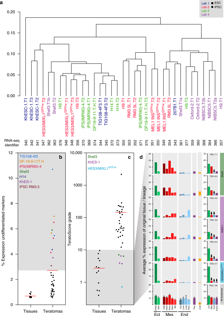
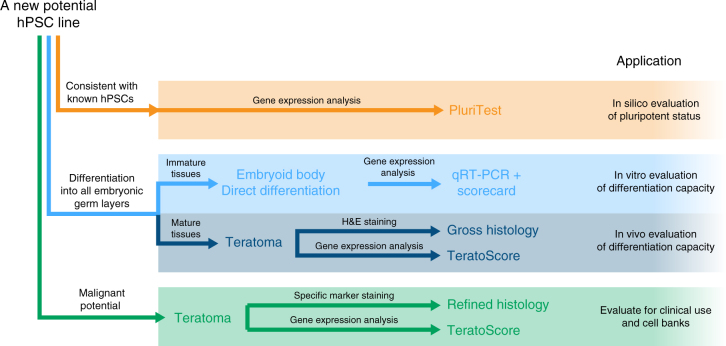
The International Stem Cell Initiative compared several commonly used approaches to assess human pluripotent stem cells (PSC). PluriTest predicts pluripotency through bioinformatic analysis of the transcriptomes of undifferentiated cells, whereas, embryoid body (EB) formation in vitro and teratoma formation in vivo provide direct tests of differentiation. Here we report that EB assays, analyzed after differentiation under neutral conditions and under conditions promoting differentiation to ectoderm, mesoderm, or endoderm lineages, are sufficient to assess the differentiation potential of PSCs. However, teratoma analysis by histologic examination and by TeratoScore, which estimates differential gene expression in each tumor, not only measures differentiation but also allows insight into a PSC's malignant potential. Each of the assays can be used to predict pluripotent differentiation potential but, at this stage of assay development, only the teratoma assay provides an assessment of pluripotency and malignant potential, which are both relevant to the pre-clinical safety assessment of PSCs.


Conflict of interest statement

C.L.M. is a co-founder of Pluriomics bv (from Sept 2017 Ncardia). N.N. is a director and shareholder of hPSC-related companies Stem Cell and Device Laboratory, Inc. and Kyoto Stem Cell Innovation, Inc. He is also a shareholder of ReproCELL, Inc. O.B. is a co-founder and owns equity of LIFE and BRAIN GmbH. S.Y. is a scientific advisor of iPS Academia Japan without salary. K.T. is a member of the scientific advisory board of I Peace Inc. without salary. The remaining authors declare no competing interests.

Figures

Fig. 1
Fig. 1
Detection of chromosomal aberrations in PSC and tumors using e-Karyotyping and eSNP-karyotyping. a e-Karyotyping: each line depicts the moving average plots of global gene expression in 13 different cell lines over 300-gene bins. The gene expression of 12 cell lines (black lines) was close to the total mean, suggesting a normal karyotype. In contrast, all replicates of the MEL1 INSGFP/w (cyan) cell line showed considerable upregulation of genes from both chromosomes 12 and chromosome 17, suggesting that it harbors an additional copy of these chromosomes. b eSNP-karyotyping: detection of chromosomal aberrations in tumors using eSNP-karyotyping. Each line depicts the moving average (over 151 SNPs) of gene expression generated from RNA-seq data of tumor derived from 13 different cell lines (one plot per source cell line). Colors represent tumor replicates. Only tumors derived from MEL1 INSGFP/w and NIBSC5 show an altered allele ratio in both replicates, suggesting an aberrant karyotype with additional copies of chromosomes 17 and 12, respectively
Fig. 2
Fig. 2
Pluritest. a All PluriTest results from this study (red circles) are based on normalization to the H9 samples and were plotted on the background of the empirical density distribution of all pluripotent (red cloud) and differentiated samples (blue clouds) in the PluriTest training dataset. bf highlight the subsets of samples included in this study: All results from the same hPSC line (H9) cultured at each laboratory (b). Samples from Lab 1 (c), Lab 2 (d), Lab 3 (e), Lab 4 (f) are highlighted specifically. All cell lines are above the Pluripotency Score threshold (θP >= 20). Both replicates of two cell lines MEL1 INSGFP/w in d and DF19-9-11T.H in e score above the Novelty threshold (θN >= 1.67) and thus would be highlighted for further investigation. Three cell lines show larger differences between the novelty scores of their respective replicate samples 201B7 in c, RM3.5 C in d, and Oxford-2 in f
Fig. 3
Fig. 3
Differentiation potential and propensity in EBs. a The line plots show the mean log2 expression change (relative to day 0) of marker genes (Supplementary table 3) as a function of time and averaged over all cell lines. The expression change is shown under ectoderm conditions for ectoderm markers, mesoderm conditions for mesoderm markers, endoderm conditions for endoderm markers, and across all conditions for markers of undifferentiated cells. Shaded contours indicate the minimum/maximum observed value. b A summary table of the lineage scorecard evaluation of the “propensity” (spontaneous differentiation, left) and “potential” (directed differentiation, right) for each cell line (rows) to differentiate into the respective lineage (columns). Colors and symbols indicate increased (blue) and limited (grading of lighter blues) preference for expression of lineage- specific marker genes. +++: score >3; ++: score 2–3; +: score 1–2; +/−: score <1. nd not analyzed due to RNA failing quality control criteria. c Scatterplots contrasting the lineage score after 16 days of EB differentiation (“propensity”; x-axis) with the lineage score for teratomas derived from the same cell lines (y-axis). The lineage scores for ectoderm (left), mesoderm (center), and endoderm (right) marker expression are shown separately
Fig. 4
Fig. 4
Histological evaluation of three embryonic germ layers and undifferentiated EC-Like and yolk sac elements in xenograft tumors. a Mucus secreting intestinal-like epithelium (End-endoderm), neural tube rosettes (Ect-ectoderm), and intervening stroma (Mes-mesoderm) (×240). b Intestinal-like epithelium (End-endoderm), surrounded by connective tissue, smooth muscle and fat cells (Mes-mesoderm). The left outer rim of mesoderm is lined by intestinal-like epithelium (End-endoderm). To the left there is pigmented epithelium (P), corresponding to retina (Ect-ectoderm), and a nest of glycogen rich squamous epidermal cells (Sq) (×120). c A summary of tissue types recorded per individual tumor piece surveyed from each laboratory; at least two pieces of each tumor were examined. d Lower magnification view of a teratoma containing undifferentiated stem cells (EC-Like, ECL), identified as embryonal carcinoma-like (ECL) cells, neural tube-like rosettes (N) and non-descript stromal cells (×120). e Higher magnification of the same xenograft. Undifferentiated ECL cells (ECL) are arranged into anastomosing cords. Dark dot-like cells are undergoing apoptosis. Compare the loosely structured chromatin of the ECL cells with the dark nuclei containing condensed chromatin in the neural rosettes (N) (×240). f Two embryoid bodies (EB) forming tubes lined by ECL cells, separated by a space from the surrounding yolk sac epithelium (YS). Both embryoid bodies contain prominent apoptotic bodies. Note the loosely textured yolk sac (YS) corresponding to the connective tissue that runs between the yolk sac and the blastocyst (magma reticulare) of early human embryos (×120). g Antibody to OCT3/4 staining ECL cell nuclei. h Antibody to the zinc-finger protein ZBTB16 reacts with the nuclei of yolk sac cells around three cylinders of ECL cells. i Antibody to SALL4 staining ECL cell nuclei and also the yolk sac (YS) cells in their vicinity
Fig. 5
Fig. 5
Teratoma RNA-seq expression data analysis. a Unsupervised hierarchical clustering analysis of RNA-seq expression of teratomas from four different laboratories (calculated using complete linkage and Spearman correlation distance). Tumors from the same laboratory appear in the same color. Label numbers (T1, T2, etc.) indicate teratoma replicates. Specific RNA-seq sample identifiers are indicated below the sample names. b Mean relative expression of human undifferentiated PSC/yolk sac markers within teratomas and normal tissues calculated with respect to their expression in PSCs. Eleven teratomas (highlighted by colored dots) showed an expression greater than teratoma overall average (2.5%). c TeratoScore grades, calculated from RNA-seq profiles of normal tissues and teratomas. Each grade represents expression of markers from the three embryonic germ layers and extra-embryonic membranes. Normal tissues provided a mean grade of 2.7 ± 0.2, while teratomas provided a mean grade of 145.0 ± 61.6. Six teratomas from three lines (Shef3, KhES-1 or HES3 MIXL1GFP/w) provided a grade lower than 10, the threshold reflecting sufficient representation of all lineages. Samples with a low TeratoScore grade are highlighted. d Distribution of aberrant tissue expression in teratomas. Shef3- derived teratomas show a low expression of endodermal and placental markers, whereas KhES-1 and HES3 MIXL1GFP/w teratomas show high expression of ectodermal markers and low expression of all other lineage markers. Arrows designate lineages with distinctly low expression (<4% of mean expression ratio). (TSG: TeratoScore Grade; Ect: Ectoderm; Mes: Mesoderm; End: Endoderm; CPNS: Central and Peripheral Nervous System; Adip: Adipose Tissue; Kdny: Kidney; Mscl: Skeletal Muscle; Bld: Blood; Panc: Pancreas; Plac: Placenta; Undf: Undifferentiated Markers; ExEm: Extraembryonic). Error bars represent SEM
Fig. 6
Fig. 6
Proposed strategy to analyze new human pluripotent stem cell lines depends on the information required. To first determine whether or not a cell line is pluripotent (orange lines), its signature can be compared to that of known pluripotent cells’ signatures using PluriTest. To confirm whether that cell line (blue lines), is capable of differentiating into derivatives of all three embryonic germ layers in vitro embryoid body (EB) formation in ‘neutral’ differentiation conditions, or by specific lineage-promoting differentiation conditions, combined with bioinformatic scorecard analysis, should be sufficient. If necessary differentiation to specific mature cells types may be also assessed in vivo by xenografting and teratoma formation followed by either histological analysis or RNA-seq analysis using Teratoscore. But to evaluate whether the cell line in question might have malignant potential (green lines) careful examination of histological sections of the teratoma using antibodies to specific markers or by focusing the RNA-seq and TeratoScore on specific markers associated with a malignant phenotype is suggested

References

    1. Friedman NB, Moore RA. Tumors of the testis; a report on 922 cases. Mil. Surg. 1946;99:573–593. - PubMed
    1. Stevens LC. Origin of testicular teratomas from primordial germ cells in mice. J. Natl Cancer Inst. 1967;38:549–552. - PubMed
    1. Stevens LC. The biology of teratomas. Adv. Morphog. 1967;6:1–31. doi: 10.1016/B978-1-4831-9953-5.50005-6. - DOI - PubMed
    1. Damjanov I, Solter D. Experimental teratoma. Curr. Top. Pathol. 1974;59:69–130. doi: 10.1007/978-3-642-65857-0_2. - DOI - PubMed
    1. Stevens LC, Little CC. Spontaneous testicular teratomas in an inbred strain of mice. Proc. Natl Acad. Sci. USA. 1954;40:1080–1087. doi: 10.1073/pnas.40.11.1080. - DOI - PMC - PubMed

Publication types