. 2018 May 15;8(1):7546.
doi: 10.1038/s41598-018-25774-1.

Loss of CENP-F Results in Dilated Cardiomyopathy with Severe Disruption of Cardiac Myocyte Architecture

Affiliations

Loss of CENP-F Results in Dilated Cardiomyopathy with Severe Disruption of Cardiac Myocyte Architecture

Annabelle Manalo et al. Sci Rep. .

Abstract

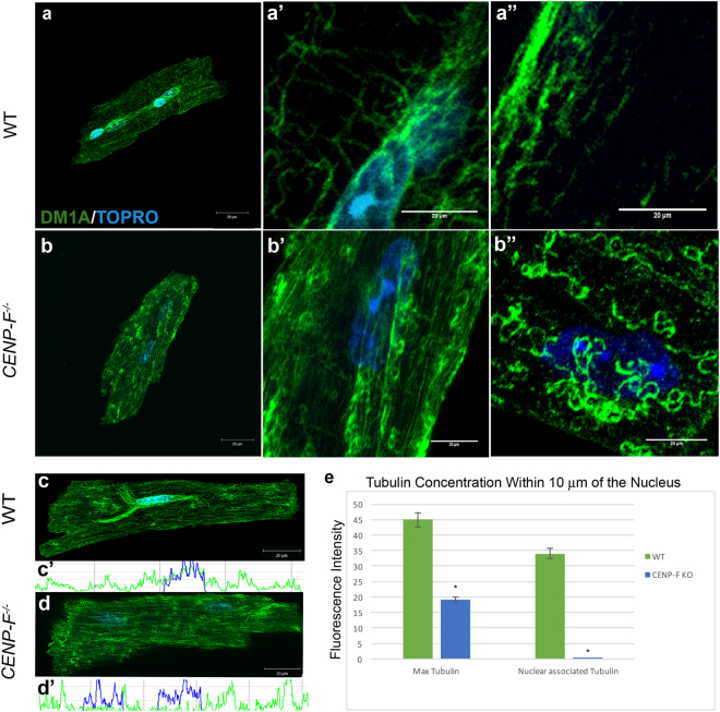
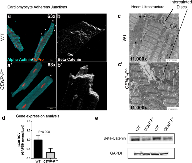
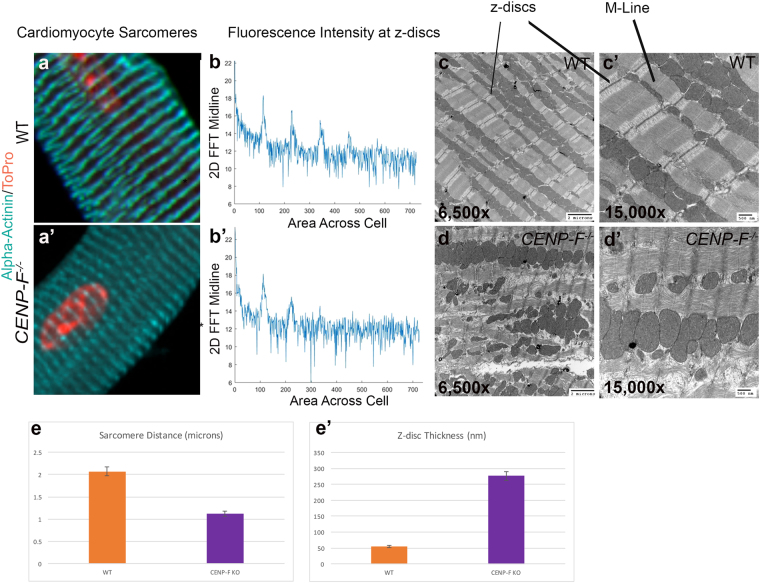
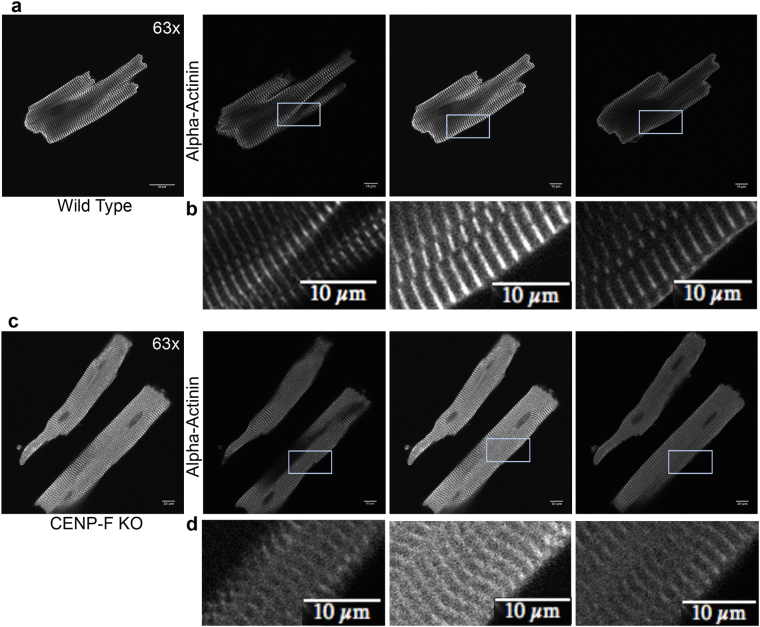
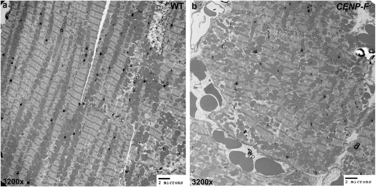
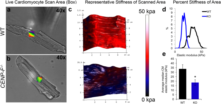
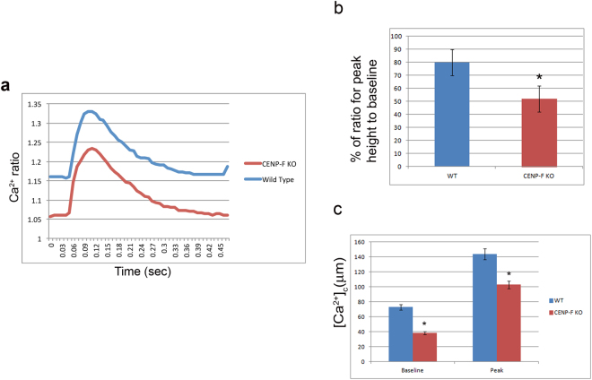
Centromere-binding protein F (CENP-F) is a very large and complex protein with many and varied binding partners including components of the microtubule network. Numerous CENP-F functions impacting diverse cellular behaviors have been identified. Importantly, emerging data have shown that CENP-F loss- or gain-of-function has critical effects on human development and disease. Still, it must be noted that data at the single cardiac myocyte level examining the impact of CENP-F loss-of-function on fundamental cellular behavior is missing. To address this gap in our knowledge, we analyzed basic cell structure and function in cardiac myocytes devoid of CENP-F. We found many diverse structural abnormalities including disruption of the microtubule network impacting critical characteristics of the cardiac myocyte. This is the first report linking microtubule network malfunction to cardiomyopathy. Importantly, we also present data demonstrating a direct link between a CENP-F single nucleotide polymorphism (snp) and human cardiac disease. In a proximate sense, these data examining CENP-F function explain the cellular basis underlying heart disease in this genetic model and, in a larger sense, they will hopefully provide a platform upon which the field can explore diverse cellular outcomes in wide-ranging areas of research on this critical protein.


Conflict of interest statement

The authors declare no competing interests.

Figures

Figure 1
Figure 1
Microtubules (MTs) form rings at plus ends and lose connection with nucleus in CENP-F−/− cardiomyocytes. Cardiomyocytes isolated from wild-type (a) and CENP-F−/− (b) mice were fixed and immunostained with DM1A antibody (green) and TOPRO (blue). A zoom in of the circumferentially-oriented MT network (a’,b’) and longitudinal array of MTs (a”) are shown in wild-type and CENP-F−/− mouse cardiomyocytes, respectively. Cardiomyocytes were treated with nocodazole to destabilize microtubules for phenotype comparison (b”). Immunostaining of MTs (green) and nucleus (blue) in wild-type cardiomyocytes show a microtubule-nuclei relationship (c), accompanied by a representative scan (c’). This relationship is completely gone in CENP-F−/− cardiomyocytes (d), accompanied by a representative scan (d’). In (e), quantification of the maximum tubulin and nuclear associated tubulin of wild-type vs. CENP-F−/− cardiomyocytes, show a tight relationship between microtubules and nuclei in wild-type cells opposed to a complete loss of association in CENP-F−/− cells. In wild-type and CENP-F−/− cells, n = 50 or more z-stack images analyzed for maximum tubulin. In wild-type and CENP-F−/−cells, n = 50 or more z-stack images analyzed for nuclear associated tubulin (e). Scale bars: 20 mm. *p < 0.001. Error bars indicate SEM.
Figure 2
Figure 2
Intercalated disc organization is disrupted with the loss of CENP-F in cardiomyocytes. Cardiomyocytes isolated from wild-type (a) and CENP-F−/− (a’) mice were fixed and immunostained with alpha-actinin 2 antibody (teal) and TOPRO (orange). In CENP-F+/+ cardiomyocytes, there are multiple junction ends, as indicated by * (a). However, in CENP-F−/− cardiomyocytes, the cell termini is blunted, indicated by * (a’). The intercalated discs were immunostained with beta-catenin antibody (white) and the images are zoomed in to depict the lining of the cardiomyocyte junctions. In wild-type mice, there is thin and distinct junction staining (b) opposed to disintegrated and thickened junction staining of the mutant cardiomyocytes (b’). An EM image of wild-type heart shows a clear depiction of an intercalated disc, see horizontal structure pointed to by line (c), In CENP-F−/− cardiomyocytes, the intercalated disc is highly disintegrated (c’). A gene expression analysis of beta-catenin in wild-type versus CENP-F−/− cardiomyocytes (d). A western blot analysis of beta-catenin in wild-type versus CENP-F−/− cardiomyocytes showed significant decrease in beta-catenin. 2 cropped samples are presented and n = 5, p < 0.05. (Full-length gels are presented in Supplementary Fig. 4). Scale bars: (a) 20 mm; (a’) 20 mm; (c) 2 microns; (c’) 2 microns. Error bars in (e) represent standard error of the mean (SEM).
Figure 3
Figure 3
Sarcomere architecture is disrupted with the loss of CENP-F in cardiomyocytes. Cardiomyocytes isolated from wild-type (a) and CENP-F−/− (a’) mice were fixed and immunostained with alpha-actinin antibody (teal) and TOPRO (orange). A zoom in view of the sarcomere structure displayed a distinct patterning in wild-type cardiomyocytes (a) versus a diffuse and thickened z-disc patterning in CENP-F−/− cardiomyocytes (a’). A 2-D spectrum analysis of the fluorescence regularity in alpha-actinin stained cardiomyocytes displayed high intensity points at regular positioning in wild type cardiomyocytes (b). There is much more variability in the fluorescence staining of the CENP-F−/− cardiomyocytes (b’). A TEM of a wild-type adult mouse heart shows the overall precise patterning of sarcomeric structure (c). The z-discs and M-lines are well defined, as depicted in a higher magnification (c’). In a CENP-F deleted mouse heart, the z-discs are disintegrated and there are breaks within the actomyosin network (d,d’). The distance between the z-discs is much shorter in CENP-F−/− cardiomyocytes in comparison to the wild-type population, *p < 0.001 (e). Additionally, the width of the z-discs is much greater in the mutant cardiomyocytes opposed to the wild-type, *p < 0.001 (f).Scale Bars: (c) 2 microns; (c’) 500 nm; (d) 2 microns; (d’) 500 nm. Error bars in (c,d) represent standard error of the mean (SEM).
Figure 4
Figure 4
CENP-F−/− cardiomyocytes have a disorganized z-disc structure compared to wild type cardiomyocytes. Individual slices of z-stack images in wild type cardiomyoctes show distinct z-disc staining with anti-alpha-actinin antibody (a,b). In CENP-F−/− cardiomyocytes, alpha-actinin staining is not sharp/distinct. Additionally, z-discs are widened and severely disintegrated (c,d). This is in agreement with quantification of z-disc width conducted on TEM images (see Supplementary Fig. 4) All images are adjusted at the same brightness and contrast levels. Images are in greyscale for proper comparison of image quality. Scale bars: (a) 20 um; (b) 10 um; (c) 20 um; (d) 10 um.
Figure 5
Figure 5
Mitochondria are misaligned and varied in size with the loss of CENP-F. Mitochondria are ordered, aligned, and relatively the same size in wild-type hearts (a). In CENP-F−/− hearts, mitochondria have no alignment and are varied in size (b). Scale bars: (a) 2 microns; (b) 2 microns.
Figure 6
Figure 6
Cardiomyocytes are softened with the loss of CENP-F. Live cardiomyocytes isolated from wild-type (a) and CENP-F−/− (b) mice were plated on laminin and analyzed for the elastic modulus of the cell surface. Representative topography plots (color bar) show a stiff surface across the 3D rendering of the wild-type cardiomyocyte (red), while, the CENP-F−/− cardiomyocyte (blue) is significantly softened (c). Distribution of cell stiffness from representative scans (d). The average median calculation of the elastic modulus of wild-type vs. CENP-F−/− cardiomyocytes is measured less (kpa) by an approximate 2-fold difference, p = 0.0124 (e). Error bars in (e) represent the standard error of the mean (SEM).
Figure 7
Figure 7
Calcium transients are decreased in CENP-F−/− live cardiomyocytes. Physiological analysis of live cardiomyocytes was conducted using Ionoptix Experimentation. Fluorescence ratio of FURA-2 binding to calcium was recorded from myocytes isolated from wild-type and CENP-F−/− hearts. Calcium ratio was measured during contraction/relaxation cycle resulting in a decreased ratio in CENP-F−/− cardiomyocytes (a). Calcium released during the contraction/relaxation cycle was decreased in CENP-F−/− cardiomyocytes, *p < 0.001 (b). In phases of the transient, both the pre-stimulation baseline value and maximal deflection from baseline (peak) was less, *p < 0.05 (c). Each bar represents mean SEM, n = 5–10 cardiomyocytes.

References

    1. McNally EM, Golbus JR, Puckelwartz MJ. Genetic mutations and mechanisms in dilated cardiomyopathy. J Clin Invest. 2013;123:19–26. doi: 10.1172/JCI62862. - DOI - PMC - PubMed
    1. Herman DS, et al. Truncations of titin causing dilated cardiomyopathy. N Engl J Med. 2012;366:619–628. doi: 10.1056/NEJMoa1110186. - DOI - PMC - PubMed
    1. Mohapatra B, et al. Mutations in the muscle LIM protein and alpha-actinin-2 genes in dilated cardiomyopathy and endocardial fibroelastosis. Mol Genet Metab. 2003;80:207–215. doi: 10.1016/S1096-7192(03)00142-2. - DOI - PubMed
    1. Du CK, et al. Knock-in mouse model of dilated cardiomyopathy caused by troponin mutation. Circ Res. 2007;101:185–194. doi: 10.1161/CIRCRESAHA.106.146670. - DOI - PubMed
    1. Dees E, et al. Cardiac-specific deletion of the microtubule-binding protein CENP-F causes dilated cardiomyopathy. Dis Model Mech. 2012;5:468–480. doi: 10.1242/dmm.008680. - DOI - PMC - PubMed

MeSH terms

Substances