. 2018 May 15;8(1):7551.
doi: 10.1038/s41598-018-25767-0.

TDP-43 regulation of stress granule dynamics in neurodegenerative disease-relevant cell types

Affiliations

TDP-43 regulation of stress granule dynamics in neurodegenerative disease-relevant cell types

Yousra Khalfallah et al. Sci Rep. .

Abstract

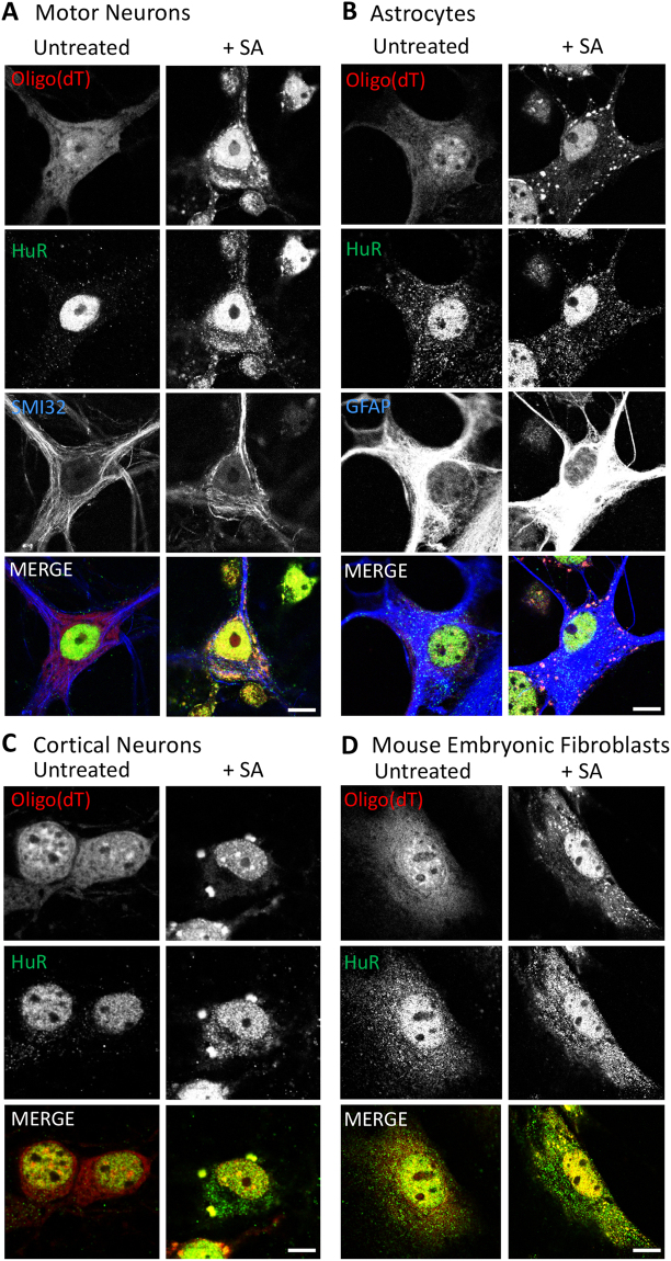
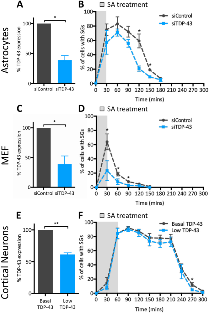
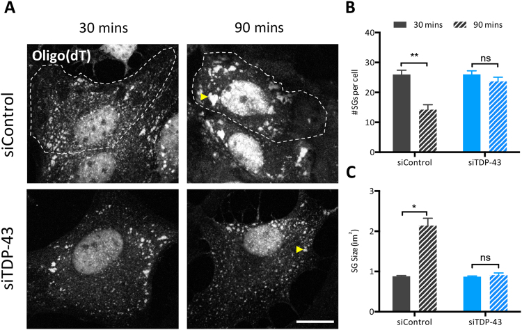
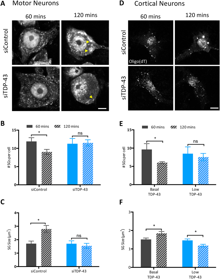
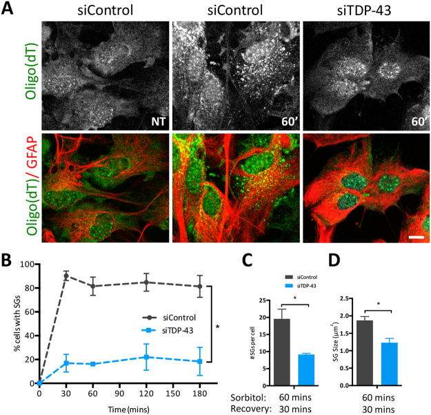
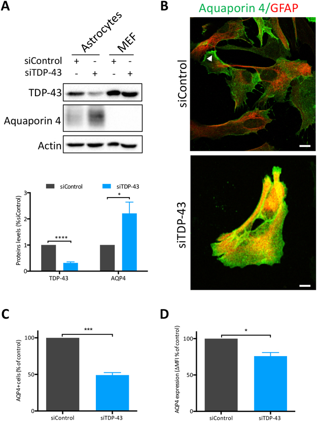
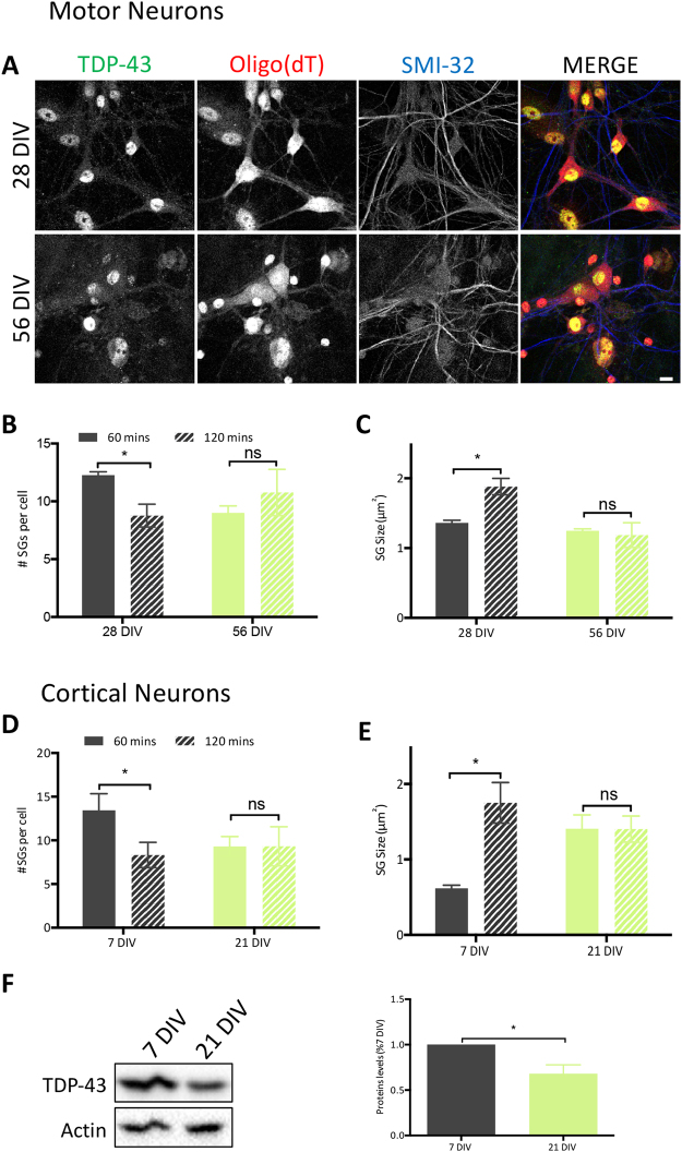
Stress granules (SGs) are cytoplasmic foci that form in response to various external stimuli and are essential to cell survival following stress. SGs are studied in several diseases, including ALS and FTD, which involve the degeneration of motor and cortical neurons, respectively, and are now realized to be linked pathogenically by TDP-43, originally discovered as a component of ubiquitin-positive aggregates within patients' neurons and some glial cells. So far, studies to undercover the role of TDP-43 in SGs have used primarily transformed cell lines, and thus rely on the extrapolation of the mechanisms to cell types affected in ALS/FTD, potentially masking cell specific effects. Here, we investigate SG dynamics in primary motor and cortical neurons as well as astrocytes. Our data suggest a cell and stress specificity and demonstrate a requirement for TDP-43 for efficient SG dynamics. In addition, based on our in vitro approach, our data suggest that aging may be an important modifier of SG dynamics which could have relevance to the initiation and/or progression of age-related neurodegenerative diseases.


Conflict of interest statement

The authors declare no competing interests.

Figures

Figure 1
Figure 1
Differences in SG morphology amongst cell types. Primary cultures of (A) motor neurons, (B) astrocytes, (C) cortical neurons and (D) mouse embryonic fibroblasts treated (or not) with 0.5 mM of sodium arsenite (+SA). Cytoplasmic SGs were co-labelled with an oligo(dT) probe to track polyadenylated mRNA, and an antibody against HuR (a known SG marker, that can also serve as a nuclear marker by its presence in the nucleus). Note, motor and cortical neurons were treated for 60 min while astrocytes and fibroblasts were treated for 30 min. Scale bar, 10 µm.
Figure 2
Figure 2
SG kinetics vary according to cell type and are modulated by TDP-43. (A,B) Primary astrocytes (n = 3, average N = 100 per time point), (C,D) mouse embryonic fibroblasts (MEF) (n = 3, average N = 100 per time point), and (E,F) cortical neurons (n = 4, average N = 70 per time point) were transfected with siRNA (siTDP-43 #1) and subjected to 0.5 mM SA. (A, C, E) TDP-43 expression levels expressed relative to control cultures, as determined by measurement of TDP-43 signal intensity. (B,D,F) Percentage of cells displaying SGs at different time points following SA exposure. Data of 3–4 independent experiments are expressed as the mean ± SEM; Student t test *p < 0.05.
Figure 3
Figure 3
TDP-43 is required for SG assembly in astrocytes. (A) Representative images of primary astrocytes transfected with TDP-43 siRNA (siTDP-43 #2) and exposed to SA with cytoplasmic SGs marked with an oligo(dT) probe. Scale bar, 10 µm. (B) Number of SGs per cell and (C) size of individual SGs measured at the indicated time points post-SA exposure (n = 3, N = 10). Data is expressed as the mean ± SEM; Student t test *p < 0.05, **p < 0.01, ns: not significant.
Figure 4
Figure 4
SG coalescence is mediated by TDP-43 in motor and cortical neurons. (A,D) Representative images of siRNA-microinjected motor neurons and siRNA transfected cortical neurons (both siTDP-43 #1) treated with SA and labelled for polyadenylated mRNA with oligo(dT). Arrowheads show SGs of interest. Scale bar, 10 µm. Number of SGs per cell and individual SG size in (B,C) motor neurons (n = 3, N = 10) and (E,F) cortical neurons (n = 4, N = 10) measured at the indicated time points post-SA treatment. Data is expressed as the mean ± SEM is shown; Student t test *p < 0.05, **p < 0.005, ns: not significant.
Figure 5
Figure 5
Astrocytes depleted of TDP-43 have abnormal SG kinetics and properties in response to hyperosmotic stress. (A) Representative images of siRNA transfected astrocytes (siTDP-43 #1) treated with 0.8 M D-Sorbitol. NT: non treated cells; 60′: 60 min of stress. Scale bar, 10 µm. (B) Number of cells with SGs at different time points during continual exposure to 0.8 M D-sorbitol (n = 3). (C) Number of SGs per cell and (D) size of individual SGs, induced with 0.8 M D-sorbitol for 60 min followed by 30 min of recovery (n = 3). Data is expressed as the mean ± SEM; Student t test *p < 0.05, **p < 0.005.
Figure 6
Figure 6
AQP4 is modulated by TDP-43. (A) Whole cell lysates of astrocytes and MEFs, treated with indicated siRNAs (siTDP-43 #1) were examined by immunoblotting for AQP4. Bands of interest were cropped from unmodified images, quantified via densitometry and normalized to Actin. (Uncropped blots are in Supplemental Material.) A representative experiment and quantification of 6 independent experiments are shown. (B) Immunofluorescence of siRNA-treated astrocytes showing localisation of AQP4 (green) on GFAP (red) positive cells. Arrowhead show astrocytic end feet. Scale bar, 10 µm. (C) AQP4 surface labelling of siRNA-treated astrocytes as assessed by flow cytometry (n = 4). (D) AQP4 median fluorescence intensity (ΔMFI over secondary control only) in astrocytes treated by siControl or siTDP-43 #1. Cells were analysed by flow cytometry (n = 4). Student t test *p < 0.05, ***p < 0.001, ****p < 0.0001.
Figure 7
Figure 7
Aging negatively impacts SG assembly in neurons in vitro. (A) Representative micrographs of primary motor neurons aged in culture for 28 and 56 days and then subjected to 0.5 mM SA. SGs are marked with oligo(dT) (red) and neuronal processes marked with phosphorylated neurofilaments (SMI32, blue). Scale bar, 10 µm. Quantification of the number of SGs per cell and individual SG size in (B,C) motor neurons cultured for 28DIV and 56DIV (n = 3) and (D,E) cortical neurons cultured for 7DIV and 21DIV (n = 3). (B,D) Number of SGs per cell. (C,E) Size of individual SGs. Number and size were measured at two time points with ImageJ. (F) Whole cell lysates of cortical neurons at 7DIV and 21DIV immunoblotted for TDP-43. Bands of interest were cropped from unmodified images, quantified via densitometry and normalized to Actin. (Uncropped blots are in Supplemental Material.) A representative blot and the mean ± SEM of densitometric quantification of three independent experiments are shown. Student t test *p < 0.05.

References

    1. Ou SH, Wu F, Harrich D, García-Martínez LF, Gaynor RB. Cloning and characterization of a novel cellular protein, TDP-43, that binds to human immunodeficiency virus type 1 TAR DNA sequence motifs. J. Virol. 1995;69:3584–96. - PMC - PubMed
    1. Buratti E, Baralle FE. Multiple roles of TDP-43 in gene expression, splicing regulation, and human disease. Front. Biosci. 2008;13:867–78. doi: 10.2741/2727. - DOI - PubMed
    1. Bose JK, Wang I-F, Hung L, Tarn W-Y, Shen C-KJ. TDP-43 Overexpression Enhances Exon 7 Inclusion during the Survival of Motor Neuron Pre-mRNA Splicing. J. Biol. Chem. 2008;283:28852–28859. doi: 10.1074/jbc.M805376200. - DOI - PMC - PubMed
    1. Ayala YM, et al. Human, Drosophila, and C.elegans TDP43: nucleic acid binding properties and splicing regulatory function. J. Mol. Biol. 2005;348:575–88. doi: 10.1016/j.jmb.2005.02.038. - DOI - PubMed
    1. Alami NH, et al. Axonal transport of TDP-43 mRNA granules is impaired by ALS-causing mutations. Neuron. 2014;81:536–43. doi: 10.1016/j.neuron.2013.12.018. - DOI - PMC - PubMed

Publication types

MeSH terms