. 2018 May 15;8(1):7604.
doi: 10.1038/s41598-018-25780-3.

Favipiravir (T-705) protects against Nipah virus infection in the hamster model

Affiliations

Favipiravir (T-705) protects against Nipah virus infection in the hamster model

Brian E Dawes et al. Sci Rep. .

Abstract

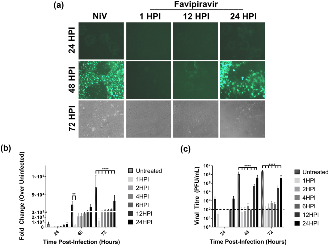
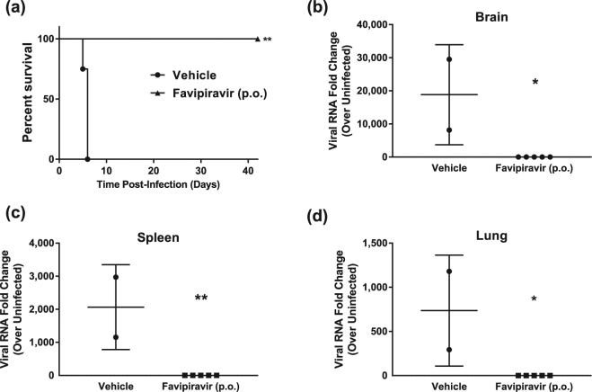
Nipah and Hendra viruses are recently emerged bat-borne paramyxoviruses (genus Henipavirus) causing severe encephalitis and respiratory disease in humans with fatality rates ranging from 40-75%. Despite the severe pathogenicity of these viruses and their pandemic potential, no therapeutics or vaccines are currently approved for use in humans. Favipiravir (T-705) is a purine analogue antiviral approved for use in Japan against emerging influenza strains; and several phase 2 and 3 clinical trials are ongoing in the United States and Europe. Favipiravir has demonstrated efficacy against a broad spectrum of RNA viruses, including members of the Paramyxoviridae, Filoviridae, Arenaviridae families, and the Bunyavirales order. We now demonstrate that favipiravir has potent antiviral activity against henipaviruses. In vitro, favipiravir inhibited Nipah and Hendra virus replication and transcription at micromolar concentrations. In the Syrian hamster model, either twice daily oral or once daily subcutaneous administration of favipiravir for 14 days fully protected animals challenged with a lethal dose of Nipah virus. This first successful treatment of henipavirus infection in vivo with a small molecule drug suggests that favipiravir should be further evaluated as an antiviral treatment option for henipavirus infections.


Conflict of interest statement

T.K. and Y.F. are employees of the Toyama Chemical Co., Ltd., the manufacturer of favipiravir. All other authors declare that no competing interests exist.

Figures

Figure 1
Figure 1
In vitro dose response of favipiravir against henipaviruses. Vero cells were infected with (a) NiV-M, (b) HeV, (c) NiV-B, (d) or rNiV-Gluc-eGFP at an MOI of 0.01. Cell culture media supplemented with serial 2-fold dilutions of favipiravir was added 1 hour post infection (HPI). Reduction in virus yield was determined at 48 HPI via plaque assay. (e) Vero cells were infected with NiV-M (MOI 0.01) and treated with 250 μM favipiravir alone or in combination with 400 μM adenosine or cytidine. Viral titres at 48HPI were then determined via plaque assay. Error bars are representing the S.D. from three individual experiments. Statistics are compared to untreated controls. *P < 0.05 and ****P < 0.0001.
Figure 2
Figure 2
Delayed treatment in vitro efficacy of favipiravir against NiV infection. (a) Microscopic analysis of Vero cells infected with rNiV-Gluc-eGFP (MOI 0.01). Favipiravir (250 μM) was added at 1, 12, and 24 HPI; eGFP expression, development of syncytia, and cytopathic effect were monitored by microscopy at 24, 48 and 72 HPI. (b) and (c) Vero cells were infected with rNiV-Gluc-eGFP at an MOI of 0.01 and treated with 250 μM favipiravir at the indicated time points post infection. Cell culture supernatant samples were assayed for (b) Gaussia luciferase activity relative to uninfected cells and (c) for viral titre. Data shown in (b) and (c) represent results from two separate experiments and thus luciferase activity is normalized to uninfected cells. **P < 0.01 and ****P < 0.0001.
Figure 3
Figure 3
In vivo efficacy of orally administered favipiravir against NiV infection in Syrian hamsters. Hamsters were infected with 104 PFU NiV Malaysia strain via the intraperitoneal route. Treatment with favipiravir was initiated immediately after infection. Favipiravir (n = 5) or vehicle (n = 4) was administered twice daily via oral gavage (about 12 hours apart) for 14 days. A 600 mg/kg loading dose was given on day 0, followed by 300 mg/kg/day on days 1–13 post-infection. (a) Survival graph of animals receiving favipiravir (black triangles) or vehicle (black circles). Graphs (b) to (d) show results from qRT-PCR analysis of tissue samples. NiV-M phosphoprotein gene copy numbers were determined in (b) brains, (c) spleens, and (d) lungs from euthanized control hamsters (vehicle) and survivors after favipiravir treatment. Copy numbers were quantified via comparison to a standard curve of purified NiV-M genome and normalized relative to uninfected tissues due to background detected in uninfected tissue. *P < 0.05 and **P < 0.01.
Figure 4
Figure 4
In vivo efficacy of subcutaneously administered favipiravir against NiV infection in Syrian hamsters. Hamsters were infected with 104 PFU NiV Malaysia strain via the intraperitoneal route. Treatment with favipiravir was initiated immediately after infection. Favipiravir (n = 5) or vehicle (n = 4) was administered once daily via the subcutaneous route for 14 days. A 600 mg/kg loading dose was given on day 0, followed by 300 mg/kg/day on days 1–13 post-infection. (a) Survival graph of animals receiving favipiravir (black squares) or vehicle (black circles). Graphs (b) to (d) show results from qRT-PCR analysis of tissue samples. NiV-M phosphoprotein gene copy numbers were determined in (b) brains, (c) spleens, and (d) lungs from euthanized control hamsters (vehicle) and survivors after favipiravir treatment. Copy numbers were quantified via comparison to a standard curve of purified NiV-M genome and normalized relative to uninfected tissues due to background detected in uninfected tissue. **P < 0.01.
Figure 5
Figure 5
Histopathology and immunohistochemistry. (a) Formalin fixed tissues were embedded in paraffin and H&E stained. Images represent brains, lungs, and spleens of vehicle control animals, favipiravir p.o. and favipiravir s.c. (b) Formalin fixed tissues were embedded in paraffin and stained with anti-NiV nucleoprotein antibodies. Images represent brains, lungs, and spleens of vehicle control animals, favipiravir p.o. and favipiravir s.c. V, blood vessels; Br, bronchioles; Fo, follicles; Arrows, NiV nucleoprotein antigens.

References

    1. Selvey LA, et al. Infection of humans and horses by a newly described morbillivirus. Med J Aust. 1995;162:642–645. - PubMed
    1. Chua KB, et al. Nipah virus: a recently emergent deadly paramyxovirus. Science. 2000;288:1432–1435. doi: 10.1126/science.288.5470.1432. - DOI - PubMed
    1. Ong KC, Wong KT. Henipavirus Encephalitis: Recent Developments and Advances. Brain Pathol. 2015;25:605–613. doi: 10.1111/bpa.12278. - DOI - PMC - PubMed
    1. Rockx B, Winegar R, Freiberg AN. Recent progress in henipavirus research: molecular biology, genetic diversity, animal models. Antiviral Res. 2012;95:135–149. doi: 10.1016/j.antiviral.2012.05.008. - DOI - PubMed
    1. Chua KB, et al. Fatal encephalitis due to Nipah virus among pig-farmers in Malaysia. Lancet. 1999;354:1257–1259. doi: 10.1016/S0140-6736(99)04299-3. - DOI - PubMed

Publication types

LinkOut - more resources