

Diverse Long-Range Axonal Projections of Excitatory Layer 2/3 Neurons in Mouse Barrel Cortex
- PMID: 29765308
- PMCID: PMC5938399
- DOI: 10.3389/fnana.2018.00033
Diverse Long-Range Axonal Projections of Excitatory Layer 2/3 Neurons in Mouse Barrel Cortex
Abstract
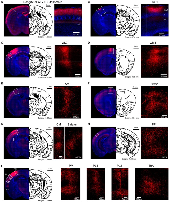
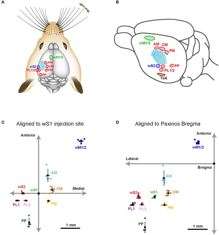
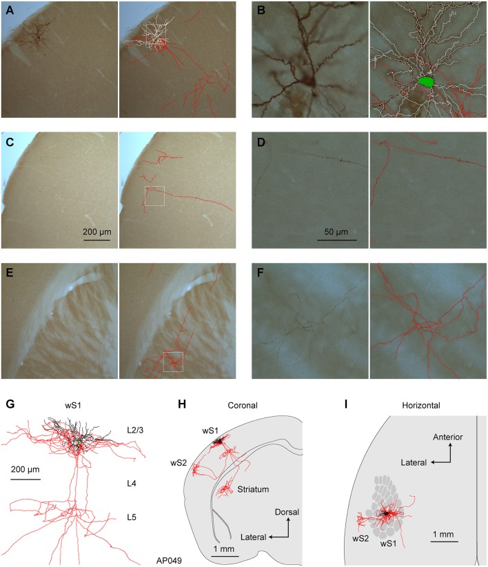
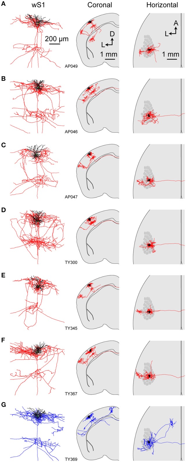
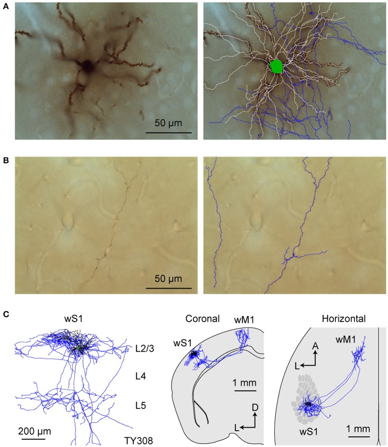
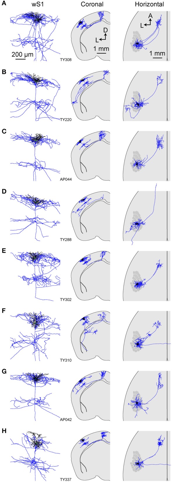
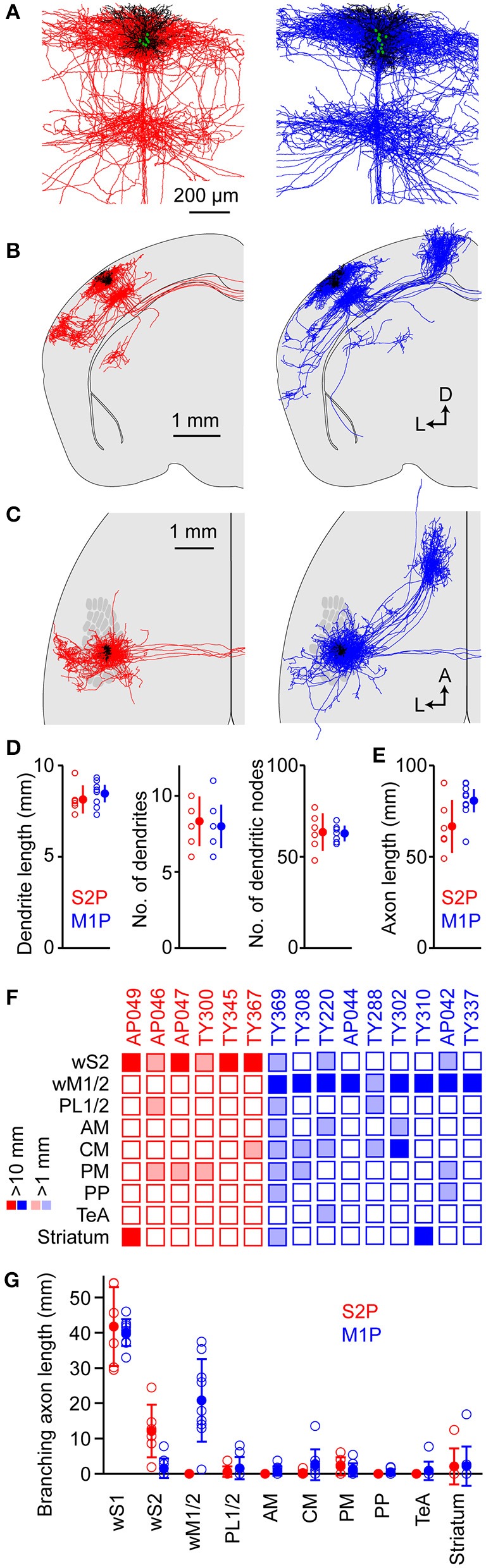
Excitatory projection neurons of the neocortex are thought to play important roles in perceptual and cognitive functions of the brain by directly connecting diverse cortical and subcortical areas. However, many aspects of the anatomical organization of these inter-areal connections are unknown. Here, we studied long-range axonal projections of excitatory layer 2/3 neurons with cell bodies located in mouse primary somatosensory barrel cortex (wS1). As a population, these neurons densely projected to secondary whisker somatosensory cortex (wS2) and primary/secondary whisker motor cortex (wM1/2), with additional axon in the dysgranular zone surrounding the barrel field, perirhinal temporal association cortex and striatum. In three-dimensional reconstructions of 6 individual wS2-projecting neurons and 9 individual wM1/2-projecting neurons, we found that both classes of neurons had extensive local axon in layers 2/3 and 5 of wS1. Neurons projecting to wS2 did not send axon to wM1/2, whereas a small subset of wM1/2-projecting neurons had relatively weak projections to wS2. A small fraction of projection neurons solely targeted wS2 or wM1/2. However, axon collaterals from wS2-projecting and wM1/2-projecting neurons were typically also found in subsets of various additional areas, including the dysgranular zone, perirhinal temporal association cortex and striatum. Our data suggest extensive diversity in the axonal targets selected by individual nearby cortical long-range projection neurons with somata located in layer 2/3 of wS1.
Keywords: axonal structure; barrel cortex; layer 2/3 pyramidal neuron; neocortex; projection neurons.
Figures
Similar articles
-
Interareal Synaptic Inputs Underlying Whisking-Related Activity in the Primary Somatosensory Barrel Cortex.J Neurosci. 2024 Jan 24;44(4):e1148232023. doi: 10.1523/JNEUROSCI.1148-23.2023. J Neurosci. 2024. PMID: 38050130 Free PMC article.
-
Cell class-specific long-range axonal projections of neurons in mouse whisker-related somatosensory cortices.Elife. 2024 Oct 11;13:RP97602. doi: 10.7554/eLife.97602. Elife. 2024. PMID: 39392390 Free PMC article.
-
Areal distributions of cortical neurons projecting to different levels of the caudal brain stem and spinal cord in rats.Somatosens Mot Res. 1990;7(3):315-35. doi: 10.3109/08990229009144711. Somatosens Mot Res. 1990. PMID: 2248004
-
Long-range connectivity of mouse primary somatosensory barrel cortex.Eur J Neurosci. 2010 Jun;31(12):2221-33. doi: 10.1111/j.1460-9568.2010.07264.x. Epub 2010 Jun 9. Eur J Neurosci. 2010. PMID: 20550566 Review.
-
Re: Woolsey TA, van der Loos H. 1970. The structural organization of layer IV in the somatosensory region (S I) of mouse cerebral cortex. Brain Res. 17: 205-242.Brain Res. 2016 Aug 15;1645:22-4. doi: 10.1016/j.brainres.2016.04.029. Epub 2016 Apr 14. Brain Res. 2016. PMID: 27086973 Review.
Cited by
-
Preferential superficial cortical layer activation during seizure propagation.Epilepsia. 2025 Mar;66(3):929-941. doi: 10.1111/epi.18239. Epub 2024 Dec 24. Epilepsia. 2025. PMID: 39718688 Free PMC article.
-
Cell-type-specific recruitment of GABAergic interneurons in the primary somatosensory cortex by long-range inputs.Cell Rep. 2021 Feb 23;34(8):108774. doi: 10.1016/j.celrep.2021.108774. Cell Rep. 2021. PMID: 33626343 Free PMC article.
-
Anatomically revealed morphological patterns of pyramidal neurons in layer 5 of the motor cortex.Sci Rep. 2020 May 13;10(1):7916. doi: 10.1038/s41598-020-64665-2. Sci Rep. 2020. PMID: 32405029 Free PMC article.
-
Cortical layer-specific modulation of neuronal activity after sensory deprivation due to spinal cord injury.J Physiol. 2021 Oct;599(20):4643-4669. doi: 10.1113/JP281901. Epub 2021 Sep 28. J Physiol. 2021. PMID: 34418097 Free PMC article.
-
Functional and structural features of L2/3 pyramidal cells continuously covary with pial depth in mouse visual cortex.Cereb Cortex. 2023 Mar 21;33(7):3715-3733. doi: 10.1093/cercor/bhac303. Cereb Cortex. 2023. PMID: 36017976 Free PMC article.
References
-
- Broser P., Grinevich V., Osten P., Sakmann B., Wallace D. J. (2008). Critical period plasticity of axonal arbors of layer 2/3 pyramidal neurons in rat somatosensory cortex: layer-specific reduction of projections into deprived cortical columns. Cereb. Cortex. 18, 1588–1603. 10.1093/cercor/bhm189 - DOI - PMC - PubMed
LinkOut - more resources
Full Text Sources
Other Literature Sources
Research Materials
Miscellaneous