. 2018 Feb 22;5(Pt 2):223-235.
doi: 10.1107/S2052252518001021. eCollection 2018 Mar 1.

Crystal structures of Lymphocytic choriomeningitis virus endonuclease domain complexed with diketo-acid ligands

Affiliations

Crystal structures of Lymphocytic choriomeningitis virus endonuclease domain complexed with diketo-acid ligands

Magali Saez-Ayala et al. IUCrJ. .

Abstract

The Arenaviridae family, together with the Bunyaviridae and Orthomyxoviridae families, is one of the three negative-stranded RNA viral families that encode an endonuclease in their genome. The endonuclease domain is at the N-terminus of the L protein, a multifunctional protein that includes the RNA-dependent RNA polymerase. The synthesis of mRNA in arenaviruses is a process that is primed by capped nucleotides that are 'stolen' from the cellular mRNA by the endonuclease domain in cooperation with other domains of the L protein. This molecular mechanism has been demonstrated previously for the endonuclease of the prototype Lymphocytic choriomeningitis virus (LCMV). However, the mode of action of this enzyme is not fully understood as the original structure did not contain catalytic metal ions. The pivotal role played by the cap-snatching process in the life cycle of the virus and the highly conserved nature of the endonuclease domain make it a target of choice for the development of novel antiviral therapies. Here, the binding affinities of two diketo-acid (DKA) compounds (DPBA and L-742,001) for the endonuclease domain of LCMV were evaluated using biophysical methods. X-ray structures of the LCMV endonuclease domain with catalytic ions in complex with these two compounds were determined, and their efficacies were assessed in an in vitro endonuclease-activity assay. Based on these data and computational simulation, two new DKAs were synthesized. The LCMV endonuclease domain exhibits a good affinity for these DKAs, making them a good starting point for the design of arenavirus endonuclease inhibitors. In addition to providing the first example of an X-ray structure of an arenavirus endonuclease incorporating a ligand, this study provides a proof of concept that the design of optimized inhibitors against the arenavirus endonuclease is possible.

Keywords: Arenaviridae; LCMV; Lymphocytic choriomeningitis virus; compound optimization; diketo acids; endonucleases; metal chelation.


Figures

Figure 1
Figure 1
Structures of 2,4-dioxo-4-phenylbutanoic acid [DPBA, (1)], (Z)-4-{1-benzyl-4-[(4-chlorophenyl)­methyl]­piperidin-4-yl}-2-hydroxy-4-oxobut-2-enoic acid [L-742,001, (2)], 2-hydroxy-4-(biphenyl-4-yl)-4-oxobut-2-enoic acid (3) and 2-hydroxy-4-oxo-4-(phenanthren-3-yl)but-2-enoic acid (4).
Figure 2
Figure 2
(a) Thermal stability of ENDO proteins determined by differential scanning fluorimetry (DSF). The melting temperatures (T m) of ENDO-WT, ENDO-D118A and ENDO-D88A (75 µM; P) with or without the indicated divalent cation (at 0.5 mM; P+Mg, P+Mn, P+Mg+Mn) and the compounds DPBA (1) (P+Mg+Mn+DPBA) and L-742,001 (2) (P+Mg+Mn+L-742,001) (at 450 µM, ligand:protein ratio = 6) were measured in a thermofluorescence experiment. (b) Polyacrylamide/8 M urea gels of inhibition of endonuclease activity by DPBA (1) and L-742,001 (2). Increasing concentrations of molecules (1) and (2) were incubated with 20 µM protein and 1 µM single-stranded RNA. The reaction products were analyzed in 20% polyacrylamide/8 M urea gels. The lane labelled NC lacked protein in the reaction mixture, while the lane labelled EDTA contained all reagents plus 5 mM EDTA.
Figure 3
Figure 3
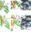
Crystal structures of the ENDO protein in complex with DPBA (1) and L-742,001 (2). LCMV endonuclease structures complexed with (1) (a, b, c) or (2) (d, e, f) are shown. Structures are represented as ribbons with helices in green and strands in gold, while the compound is represented as sticks. Mg2+ and Mn2+ ions are represented as light green and purple spheres, respectively. (b) and (e) show enlargements of the catalytic site showing the strong coordination of waters, ions and catalytic residues of the molecule. (c) and (f) show a 2F oF c OMIT map corresponding to the compound (1σ).
Figure 4
Figure 4
The five motifs (blue) that are conserved across all available sequences of the Arenaviridae endonuclease domain plotted on the LCMV structure (grey). Conserved motifs are shown in WebLogo representation. The size of the letter is representative of the frequency with which it observed in the alignment (with a probability of 1 being identity). The numbering of the residues refers to the sequence of LCMV. Motifs 1–4 represent the four positions that can be used to anchor a ligand over all Arenaviridae. Residues marked with a star correspond to key residues involved in the active site or in substrate binding, and their side chains are highlighted on the structure (orange).
Figure 5
Figure 5
(a) Structure of ENDO-WT complexed with Mg ions and the docking grid. The surface is coloured cyan and the conserved motif is coloured orange. Mg2+ ions are represented as light green spheres. The position of DPBA (1) is shown in the right panel. (b) Selection of the best docking poses of compounds (3) and (4). (c) Enlargement of the best pose on the cavity surface. (d) Enlargement of the best pose with the residues involved in binding. These are coloured according to hydrophobicity from cyan (least) to green (most). (e) Polyacrylamide/8 M urea gels of compounds (3) and (4) (50 µM) incubated with 20 µM protein and 1 µM single-stranded RNA. (f) Quantification of endonuclease activity in the presence of DMSO and compounds (1), (3) and (4).

Similar articles

Cited by

References

    1. Afonine, P. V., Grosse-Kunstleve, R. W., Echols, N., Headd, J. J., Moriarty, N. W., Mustyakimov, M., Terwilliger, T. C., Urzhumtsev, A., Zwart, P. H. & Adams, P. D. (2012). Acta Cryst. D68, 352–367. - PMC - PubMed
    1. Barton, L. L., Mets, M. B. & Beauchamp, C. L. (2002). Am. J. Obstet. Gynecol. 187, 1715–1716. - PubMed
    1. Bhatt, A., Gurukumar, K. R., Basu, A., Patel, M. R., Kaushik-Basu, N. & Talele, T. T. (2011). Eur. J. Med. Chem. 46, 5138–5145. - PMC - PubMed
    1. Blanc, E., Roversi, P., Vonrhein, C., Flensburg, C., Lea, S. M. & Bricogne, G. (2004). Acta Cryst. D60, 2210–2221. - PubMed
    1. Bonthius, D. J. (2012). Semin. Pediatr. Neurol. 19, 89–95. - PMC - PubMed