. 2018 May;8(5):256.
doi: 10.1007/s13205-018-1278-z. Epub 2018 May 14.

Designing of phenol-based β-carbonic anhydrase1 inhibitors through QSAR, molecular docking, and MD simulation approach

Affiliations

Designing of phenol-based β-carbonic anhydrase1 inhibitors through QSAR, molecular docking, and MD simulation approach

Shahzaib Ahamad et al. 3 Biotech. 2018 May.

Abstract

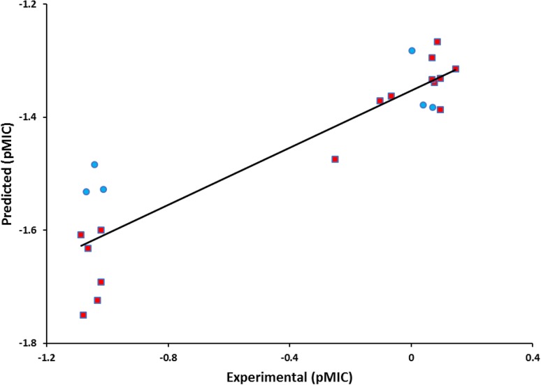
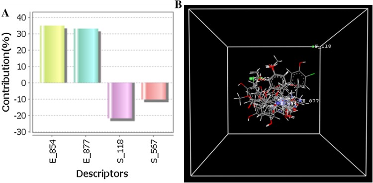
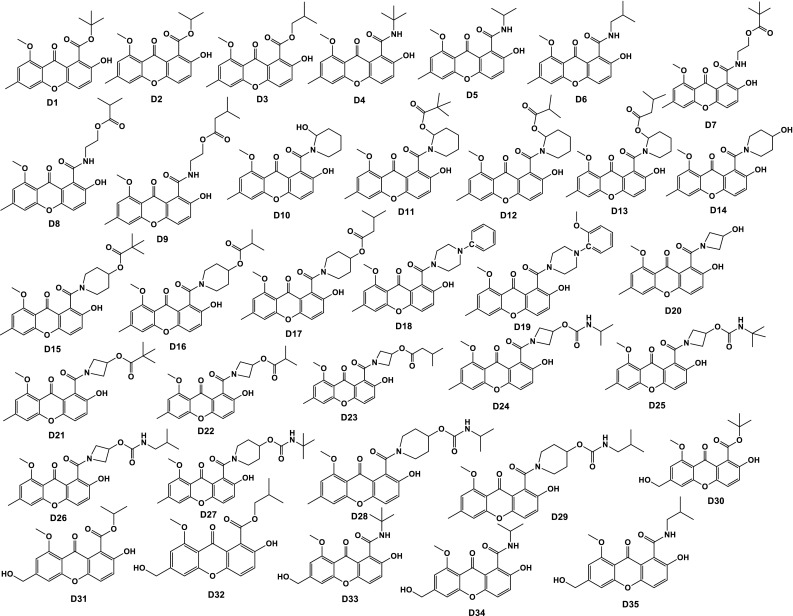
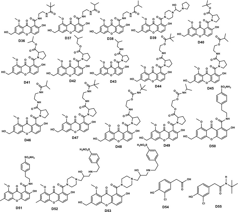
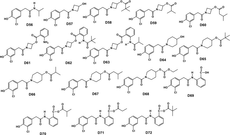
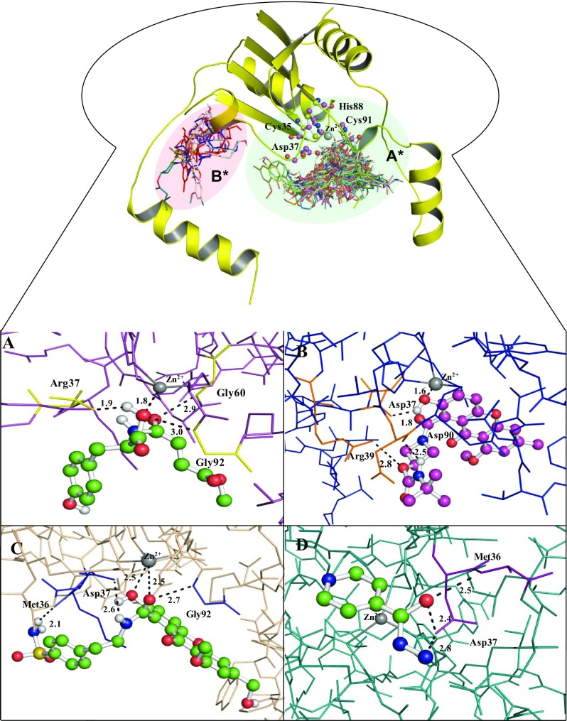
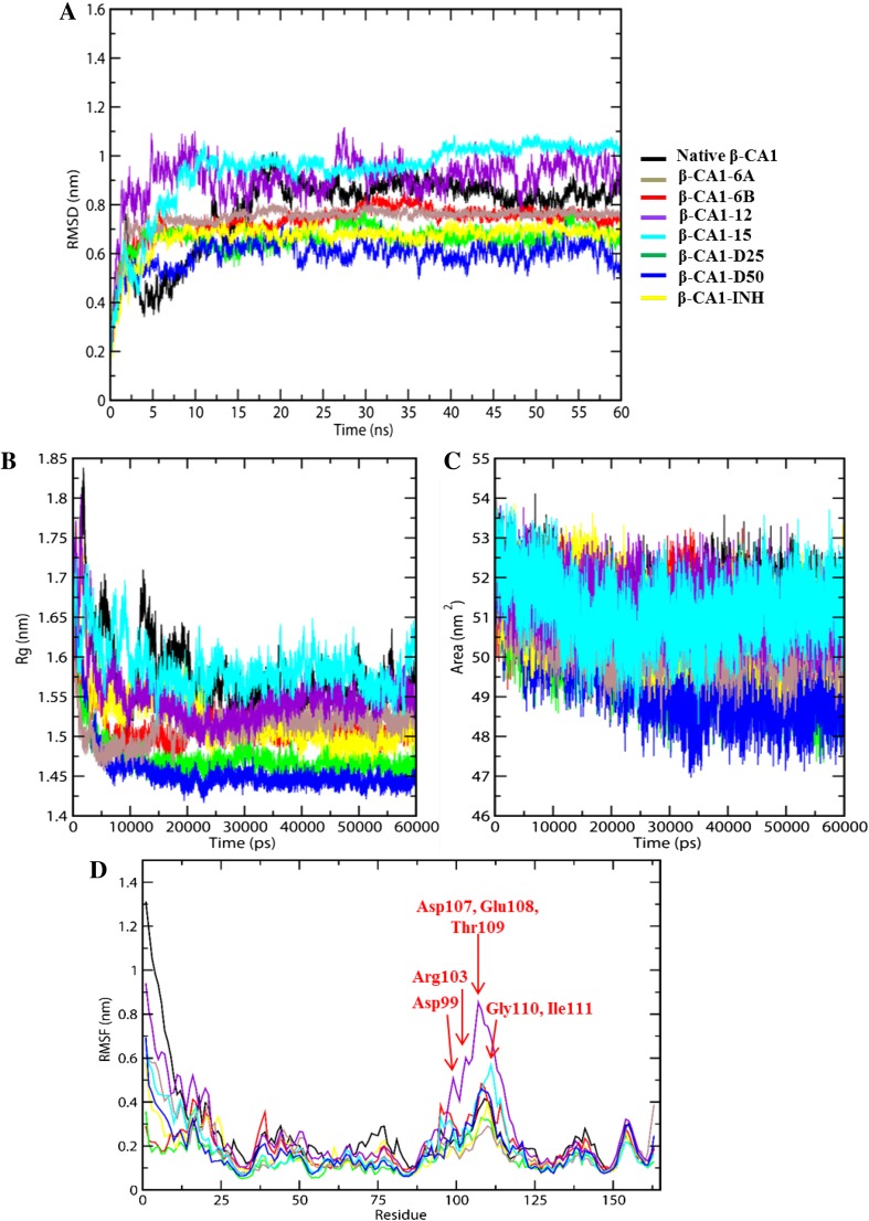
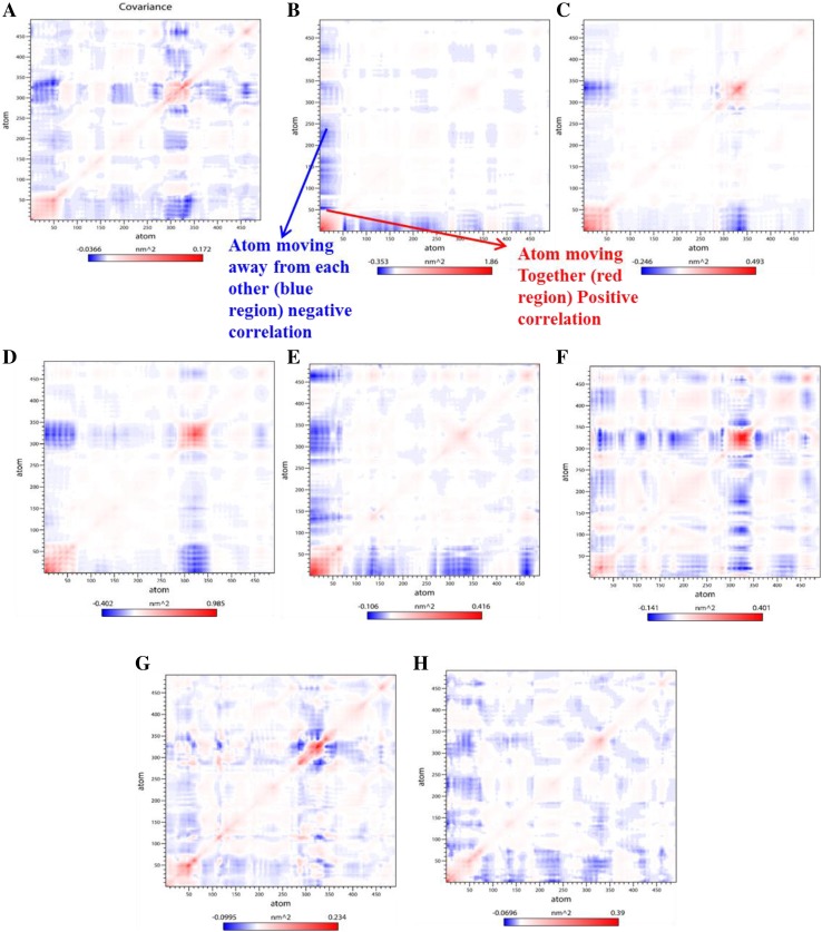
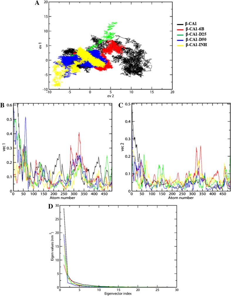
Tuberculosis (Tb) is an airborne infectious disease caused by Mycobacterium tuberculosis. Beta-carbonic anhydrase 1 (β-CA1) has emerged as one of the potential targets for new antitubercular drug development. In this work, three-dimensional quantitative structure-activity relationships (3D-QSAR), molecular docking, and molecular dynamics (MD) simulation approaches were performed on a series of natural and synthetic phenol-based β-CA1 inhibitors. The developed 3D-QSAR model (r2 = 0.94, q2 = 0.86, and pred_r2 = 0.74) indicated that the steric and electrostatic factors are important parameters to modulate the bioactivity of phenolic compounds. Based on this indication, we designed 72 new phenolic inhibitors, out of which two compounds (D25 and D50) effectively stabilized β-CA1 receptor and, thus, are potential candidates for new generation antitubercular drug discovery program.

Keywords: Docking; M. Tuberculosis; MD simulation; QSAR; β-Carbonic anhydrase 1.


Conflict of interest statement

Compliance with ethical standardsThere is no conflict of interests regarding the publication of this paper.

Figures

Fig. 1
Fig. 1
3D-QSAR Models, Fitness plot comparison between experimental vs predicted activity for training (red balls) and test set (blue balls) compounds
Fig. 2
Fig. 2
Contribution plot of the E_854, E_877, S_118, and S_567 descriptor participated in the model (a), stereo view of molecular grid around the superposed molecular units of phenols series of compounds using 3D-MLR method (b)
Chart 1
Chart 1
Chemical structures of 72 newly designed phenolic compounds with the help of QSAR study
Chart 1
Chart 1
Chemical structures of 72 newly designed phenolic compounds with the help of QSAR study
Chart 1
Chart 1
Chemical structures of 72 newly designed phenolic compounds with the help of QSAR study
Fig. 3
Fig. 3
Interaction of phenolic inhibitors with β-CA1. The binding mode of β-CA1 (line model) with compound 6B (green ball and stick model) (a), compound D25 (purple ball and stick model) (b), compound D50 (green ball and stick model) (c), and INH drug (green ball and stick model) interact with the active site residues of β-CA1 (d)
Fig. 4
Fig. 4
2D plot of the docked complexes of β-CA1 with compound 6B, D25, D50, and drug INH as shown in a, b, c, and d respectively. The 2D plot constructed by discovery studio 2.5
Fig. 5
Fig. 5
MD simulation of native β-CA1 and β-CA1-inhibitor complexes. Root-mean-square deviation plot (a), radius of gyration plot (b), solvent-accessible surface area plot (c), and root-mean-square fluctuations plot (d). The colors of all curves are indicated in the figure
Fig. 6
Fig. 6
Diagonalized covariance matrix depicted the correlated and anti-correlation motions of the β-CA1 and β-CA1-complexes within the residues. The matrix representing red-colored means residues moving together along with blue colored showing residues moving in opposite order. Matrix arranged native β-CA1 (a), β-CA1-6A complex (b), β-CA1-6B complex (c), β-CA1-12 complex (d), β-CA1-15 complex (e), β-CA1-D25 complex (f), β-CA1-D50 complex (g), and β-CA1-INH complex (h)
Fig. 7
Fig. 7
2D projection graph plotted between eigenvector 1 vs eigenvector 2 for the conformational space through the covariance matrix (a), graph plotted for the comparison between vec 1 and vec 2 vs atomic fluctuations (c and b), comparison of eigenvalues (nm2) plotted against the corresponding eigenvector index of the backbone by covariance matrix for the β-CA1 and its complexes (d). Same color scheme is applicable to all figures

References

    1. Ahamad S, Rahman S, Khan FI, Dwivedi N, Ali S, Kim J, Imtaiyaz Hassan M. QSAR based therapeutic management of M. tuberculosis. Arch Pharm Res. 2017 - PubMed
    1. Ali I, Haque A, Saleem K, Hsieh MF. Curcumin-I Knoevenagel’s condensates and their Schiff’s bases as anticancer agents: synthesis, pharmacological and simulation studies. Bioorg Med Chem. 2013;21(13):3808–3820. doi: 10.1016/j.bmc.2013.04.018. - DOI - PubMed
    1. Ali I, Haque A, Al Ajmi F, Hussain M, Marsin Sanagi A, Hussain M, Aboul-Enein IY. Supramolecular chiro-biomedical aspect of β-blockers in drug development. Curr Drug Targets. 2014;15(7):729–741. doi: 10.2174/1389450115666140429104516. - DOI - PubMed
    1. Ambriz-Pérez DL, Leyva-López N, Gutierrez-Grijalva EP, Heredia JB. Phenolic compounds: natural alternative in inflammation treatment. Rev Cogent Food Agric. 2016;2(1):1131412.
    1. Amiri-Kordestani L, Fojo T. Why do phase III clinical trials in oncology fail so often? J Natl Cancer Inst. 2012;104(8):568–569. doi: 10.1093/jnci/djs180. - DOI - PubMed

LinkOut - more resources