. 2018 Apr 18;5(2):ENEURO.0395-17.2018.
doi: 10.1523/ENEURO.0395-17.2018. eCollection 2018 Mar-Apr.

Post-stroke Intranasal (+)-Naloxone Delivery Reduces Microglial Activation and Improves Behavioral Recovery from Ischemic Injury

Affiliations

Post-stroke Intranasal (+)-Naloxone Delivery Reduces Microglial Activation and Improves Behavioral Recovery from Ischemic Injury

Jenni E Anttila et al. eNeuro. .

Abstract

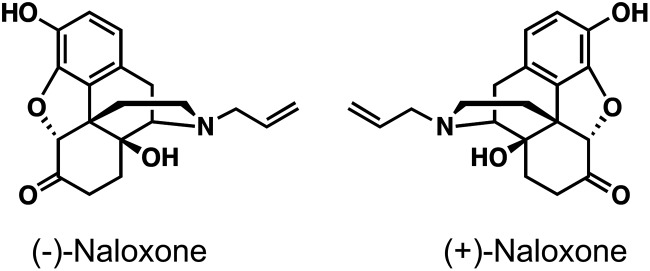
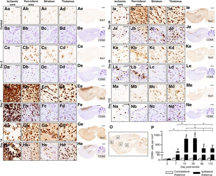
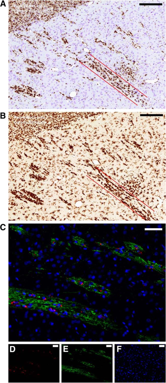
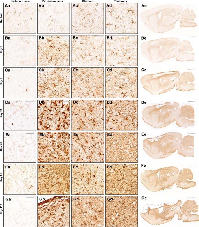
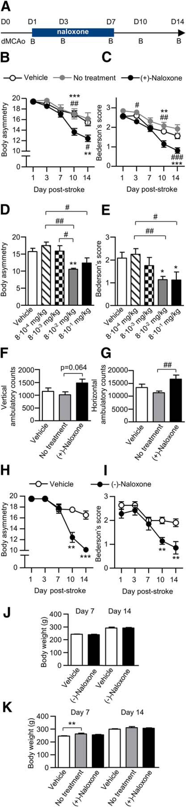
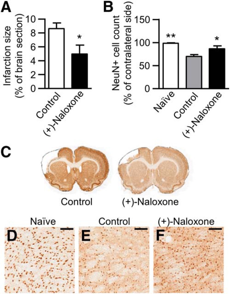
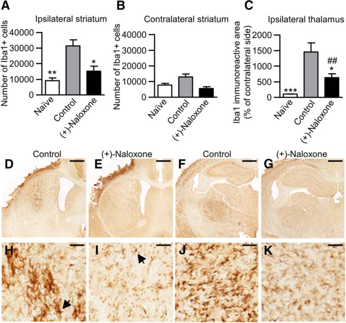
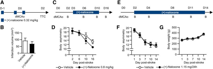
Ischemic stroke is the leading cause of disability, and effective therapeutic strategies are needed to promote complete recovery. Neuroinflammation plays a significant role in stroke pathophysiology, and there is limited understanding of how it affects recovery. The aim of this study was to characterize the spatiotemporal expression profile of microglial activation and whether dampening microglial/macrophage activation post-stroke facilitates the recovery. For dampening microglial/macrophage activation, we chose intranasal administration of naloxone, a drug that is already in clinical use for opioid overdose and is known to decrease microglia/macrophage activation. We characterized the temporal progression of microglia/macrophage activation following cortical ischemic injury in rat and found the peak activation in cortex 7 d post-stroke. Unexpectedly, there was a chronic expression of phagocytic cells in the thalamus associated with neuronal loss. (+)-Naloxone, an enantiomer that reduces microglial activation without antagonizing opioid receptors, was administered intranasally starting 1 d post-stroke and continuing for 7 d. (+)-Naloxone treatment decreased microglia/macrophage activation in the striatum and thalamus, promoted behavioral recovery during the 14-d monitoring period, and reduced neuronal death in the lesioned cortex and ipsilateral thalamus. Our results are the first to show that post-stroke intranasal (+)-naloxone administration promotes short-term functional recovery and reduces microglia/macrophage activation. Therefore, (+)-naloxone is a promising drug for the treatment of ischemic stroke, and further studies should be conducted.

Keywords: Microglia; NF-κB; middle cerebral artery occlusion; naloxone; secondary injury; stroke.


Figures

Figure 1.
Figure 1.
Chemical structure of (–)-naloxone and (+)-naloxone enantiomers. Ki of (–)-naloxone for antagonizing opioid receptors is in the 1 nM range, whereas (+)-naloxone has a very low affinity for opioid receptors, with a Ki of 10,000 nM (Iijima et al., 1978).
Figure 2.
Figure 2.
Time course of microglia/macrophage activation after cortical stroke. Representative images of immunostaining for all microglia/macrophages (Iba1) and phagocytic microglia/macrophages (CD68) from ischemic core (a), peri-infarct area (b), striatum (c), thalamus (d), and whole brain (e) sagittal sections at 2 (C, D), 7 (E, F), 14 (G, H), 28 (I, J), 56 (K, L), and 112 (M, N) days after 90-min dMCAO in rat. Control images (A, B) are from the contralateral hemisphere of the stroke brain. Scale bar is 50 µm (high magnification) and 2000 µm (low magnification). O, Example image of anti-NeuN immunostaining at post-stroke day 2 showing in more detail the regions a–d. P, Quantitation of CD68+ cells in the thalamus (d) at different time points showing the accumulation and clearance of phagocytic microglia/macrophages in the ipsilateral thalamus. *, p < 0.05 indicates statistical difference between the ipsilateral thalamus at different time points; #, p < 0.05 indicates statistical difference between the ipsilateral and contralateral thalamus in each time point, Mann–Whitney U test after Kruskal–Wallis test; n = 4 in each group. The data represent mean ± SEM.
Figure 3.
Figure 3.
Activated microglia in the striatum are lined up along axonal bundles after cortical stroke. Immunostaining of rat striatum at post-stroke day 14 for phagocytic microglia/macrophages (CD68; A) and all microglia/macrophages (Iba1; B). C–F, Double immunofluorescence staining of rat striatum at post-stroke day 14 for phagocytic microglia/macrophages (CD68; red; D) and myelin (MBP; green; E) with DAPI (blue; F). In A, B, scale bar is 200 µm; in C–F, scale bar is 50 µm.
Figure 4.
Figure 4.
Time course of astrocyte activation after cortical stroke. Representative images of immunostaining for astrocytes (GFAP) from the ischemic core (a), peri-infarct area (b), striatum (c), and thalamus (d) at 2 (B), 7 (C), 14 (D), 28 (E), 56 (F), and 112 (G) days after 90-min dMCAo in sagittal rat brain paraffin sections. Control images (A) are from the contralateral hemisphere of the stroke brain. Scale bar is 50 µm in high-magnification images and 2000 µm in low-magnification images. The regions analyzed (a–d) are shown in more detail in Fig. 2O.
Figure 5.
Figure 5.
Post-stroke intranasal administration of naloxone enantiomers promotes functional recovery. A, Experimental timeline. Intranasal naloxone (or vehicle) was administered twice daily for 7 d post-stroke. D1–D14, post-stroke days 1–14; B, behavioral assay. B, C, Effects of (+)-naloxone (0.32 mg/kg; n = 27), vehicle (n = 25), and no treatment (n = 13) on body asymmetry (B) and Bederson’s neurologic score test (C). **, p < 0.01 and ***, p < 0.001 indicate post hoc comparison between (+)-naloxone and vehicle groups, and #, p < 0.05, ##, p < 0.01, and ###, p < 0.001 indicate post hoc analysis between (+)-naloxone and no-treatment groups with Mann–Whitney U test after Kruskal–Wallis test. D, E, Effects of different doses of (+)-naloxone, 0.0008 mg/kg (n = 8), 0.008 mg/kg (n = 8), 0.08 mg/kg (n = 7), and 0.8 mg/kg (n = 8), compared to vehicle (n = 11) on day 14 post-stroke on body asymmetry (D) and Bederson’s neurologic score test (E). *, p < 0.05 and **, p < 0.01 indicate pairwise comparison with vehicle; #, p < 0.05 and ##, p < 0.01 indicate pairwise comparison with other (+)-naloxone doses with Mann–Whitney U test after Kruskal–Wallis test. F, G, Effects of (+)-naloxone (0.32 mg/kg; n = 16), vehicle (n = 16), and no treatment (n = 13) on vertical (F) and horizontal (G) activity measured for 24 h on day 14. ##, p < 0.01, Mann–Whitney U test after Kruskal–Wallis test. H–J, Effects of (–)-naloxone (0.32 mg/kg; n = 7) and vehicle (n = 11) on body asymmetry test (H), Bederson’s neurologic score test (I), and body weight (J). **, p < 0.01 and ***, p < 0.001 indicate comparison with vehicle group with Mann–Whitney U test. K, Effects of (+)-naloxone (0.32 mg/kg, n = 27), vehicle (n = 25), and no treatment (n = 13) on body weight on days 7 and 14 post-stroke. **, p < 0.01, one-way ANOVA, Bonferroni’s post hoc test. The data represent mean ± SEM.
Figure 6.
Figure 6.
Post-stroke intranasal (+)-naloxone decreases infarction area and neuronal loss in the thalamus. A, Average infarction size calculated from NeuN-negative area at day 14 post-stroke. *, p < 0.05, Student’s t test. B, Average number of neurons (NeuN+ cells) in the ipsilateral thalamus at day 14 post-stroke expressed as a percentage of the contralateral thalamus. *, p < 0.05 and **, p < 0.01 indicate pairwise comparison with the control group with Mann–Whitney U test following Kruskal–Wallis test. C, Representative photomicrographs of anti-NeuN immunostained brain sections, with infarction area delineated. D–F, Representative photomicrographs of anti-NeuN immunostaining of ipsilateral thalamus in naive (D), control (E), and (+)-naloxone–treated (F) rats. Scale bar is 150 µm. Naive, no-stroke rats (n = 6); control, stroke rats with vehicle or no treatment (n = 18); (+)-naloxone, 0.32–0.8 mg/kg (n = 10). The data represent mean ± SEM.
Figure 7.
Figure 7.
Post-stroke intranasal (+)-naloxone decreases microglia/macrophage activation in the striatum and thalamus. A, B, Microglia/macrophages (Iba1+ cells) were counted with unbiased stereology in the ipsilateral (A) and contralateral (B) striatum. *, p < 0.05 and **, p < 0.01 indicate pairwise comparison with the control group with Bonferroni’s post hoc test following one-way ANOVA. C, The area of Iba1+ cells in the ipsilateral thalamus expressed as a percentage of the contralateral thalamus. *, p < 0.05 and ***, p < 0.001 indicate pairwise comparison with the control group; ##, p < 0.01 indicates comparison with the naive group with Mann–Whitney U test following Kruskal–Wallis test. D–K, Representative photomicrographs of anti-Iba1 immunostaining of ipsilateral striatum (D, E) and thalamus (F, G) in control (D, F) and (+)-naloxone–treated (E, G) rats; H–K show high magnification. Black arrow shows a typical Iba1+ cell. Scale bars are 1000 µm (D–G) and 50 µm (H–K). Naive, no-stroke rats (n = 6); control, stroke rats with vehicle or no treatment (n = 18); (+)-naloxone, 0.32–0.8 mg/kg (n = 10). The data represent mean ± SEM.
Figure 8.
Figure 8.
Effect of different pre- and post-stroke treatment with (+)-naloxone on infarct volume and functional recovery. A, Experimental timeline. Intranasal (+)-naloxone or vehicle was administered three times: 12 and 1 h before dMCAo and immediately after reperfusion. Infarction volume was determined by TTC staining 2 d after stroke. B, Average infarction volume (mm3) on day 2 post-stroke in vehicle (n = 7) and (+)-naloxone (0.32 mg/kg; n = 8) pretreated rats. Prestroke intranasal administration of (+)-naloxone was not neuroprotective in 60-min dMCAo. C, Experimental timeline. (+)-Naloxone was delivered intranasally twice daily for 7 d post-stroke starting from post-stroke day 3. D, The effect of (+)-naloxone (0.8 mg/kg; n = 6) and vehicle (n = 7) treatment from post-stroke day 3 to post-stroke day 10 on body asymmetry test. E, Experimental timeline. (+)-Naloxone was delivered into the ventricle via mini-osmotic pumps for 12 d post-stroke starting from post-stroke day 2. F, G, The effects of 12-d continuous delivery of (+)-naloxone (1.15 mg/24 h; n = 7) and vehicle (n = 8) on body asymmetry test (F) and body weight (G). In A, C, and E: D, indicated post-stroke day; B, behavioral assay. The data represent mean ± SEM.
Figure 9.
Figure 9.
Naloxone reduced TNF-α secretion from CD11b+ cells. A, LPS induced TNF-α secretion from CD11b-expressing cells isolated from the infarct area of rat brain 7 d after dMCAo. **, p < 0.01, Mann–Whitney U test; control n = 6, LPS n = 5 in 2 independent experiments. B, CD11b+ cells were isolated from the infarct area and treated with different concentrations of naloxone as indicated for 20 h. *, p < 0.05 and **, p < 0.01 indicate pairwise comparison with the control group by Dunnett’s post hoc test following one-way ANOVA; control n = 9, naloxone n = 8–9 in 3 independent experiments. The culture medium was analyzed using TNF-α ELISA. The data represent mean ± SEM.

References

    1. Airavaara M, Chiocco MJ, Howard DB, Zuchowski KL, Peränen J, Liu C, Fang S, Hoffer BJ, Wang Y, Harvey BK (2010) Widespread cortical expression of MANF by AAV serotype 7: localization and protection against ischemic brain injury. Exp Neurol 225:104–113. 10.1016/j.expneurol.2010.05.020 - DOI - PMC - PubMed
    1. Airavaara M, Shen H, Kuo CC, Peränen J, Saarma M, Hoffer B, Wang Y (2009) Mesencephalic astrocyte-derived neurotrophic factor reduces ischemic brain injury and promotes behavioral recovery in rats. J Comp Neur 515:116–124. 10.1002/cne.22039 - DOI - PMC - PubMed
    1. Atkinson RL (1984) Endocrine and metabolic effects of opiate antagonists. J Clin Psychiatry 45:20–24. - PubMed
    1. Baskin DS, Hosobuchi Y (1981) Naloxone reversal of ischaemic neurological deficits in man. Lancet 2:272–275. - PubMed
    1. Bederson JB, Pitts LH, Tsuji M, Nishimura MC, Davis RL, Bartkowski H (1986) Rat middle cerebral artery occlusion: evaluation of the model and development of a neurologic examination. Stroke 17:472–476. - PubMed

Publication types

MeSH terms