Review
. 2018 Jun;9(3):313-324.
doi: 10.1007/s13244-018-0628-z. Epub 2018 May 15.

MR imaging findings in some rare neurological complications of paediatric cancer

Affiliations
Review

MR imaging findings in some rare neurological complications of paediatric cancer

Tetsuhiko Okabe et al. Insights Imaging. 2018 Jun.

Abstract

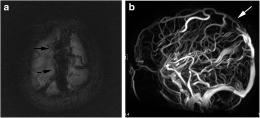
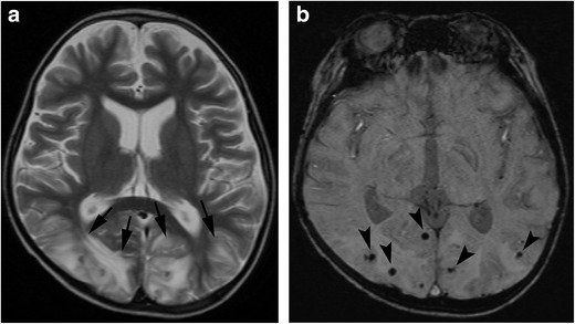
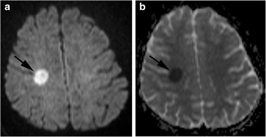
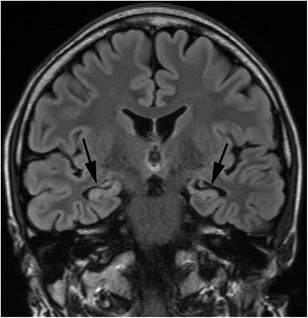
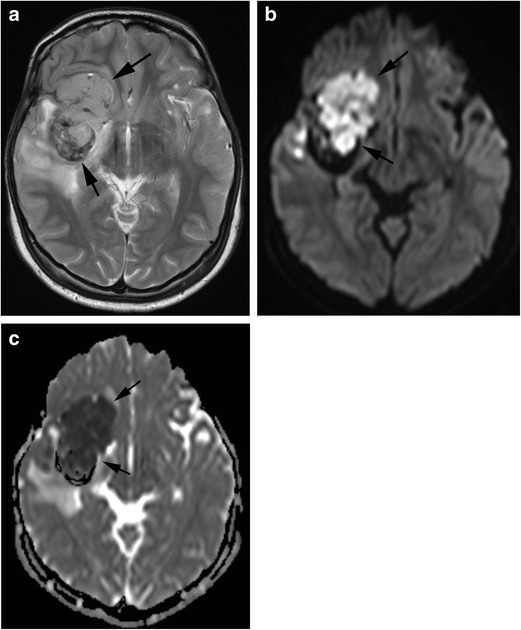
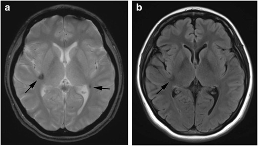
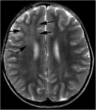
Neurological complications of paediatric cancers are a substantial problem. Complications can be primary from central nervous system (CNS) spread or secondary from indirect or remote effects of cancer, as well as cancer treatments such as chemotherapy and radiation therapy. In this review, we present the clinical and imaging findings of rare but important neurological complications in paediatric patients with cancer. Neurological complications are classified into three phases: pre-treatment, treatment and post-remission. Paraneoplastic neurological syndromes, hyperviscosity syndrome, haemophagocytic lymphohistiocytosis and infection are found in the pre-treatment phase, while Trousseau's syndrome, posterior reversible encephalopathy syndrome and methotrexate neurotoxicity are found in the treatment phase; though some complications overlap between the pre-treatment and treatment phases. Hippocampal sclerosis, radiation induced tumour, radiation induced focal haemosiderin deposition and radiation-induced white matter injury are found in the post-remission phase. With increasingly long survival after treatment, CNS complications have become more common. It is critical for radiologists to recognise neurological complications related to paediatric cancer or treatment. Magnetic resonance imaging (MRI) plays a significant role in the recognition and proper management of the neurological complications of paediatric cancer. TEACHING POINTS: • Neurological complications of paediatric cancer include various entities. • Neurological complications are classified into three phases: pre-treatment, treatment and post-remission. • Radiologists should be familiar with clinical and imaging findings of neurological complications. • MRI features may be characteristic and lead to early diagnosis and proper treatments.

Keywords: MRI, imaging; Neurological complication; Paediatric cancer; Treatment.


Conflict of interest statement

We declare that we have no conflicts of interest.

Figures

Fig. 1
Fig. 1
Paraneoplastic limbic encephalitis. A 17-year-old boy with recurrent medulloblastoma presented with seizure and altered mental status. a FLAIR image shows hyperintensities in the mesial temporal regions (arrows). b Contrast enhanced fat-suppressed T1-weighted image shows nodular meningeal enhancements in the left Sylvian fissure (arrowheads), suggestive of recurrence
Fig. 2
Fig. 2
Hyperviscosity syndrome. A 10-year-old girl with acute lymphocytic leukaemia, whose white blood cell count reached 400,000, presented with headache and vomiting. a A T1-weighted image shows hyperintensity in the pons and bilateral cerebellar hemispheres (arrows). b These lesions show hyperintensity with a hypointensity rim on T2-weighted image (arrows), indicating haemorrhage in the subacute phase. c T2*-weighted image demonstrates a number of microhaemorrhages which are not seen on T1- and T2-weighted images
Fig. 3
Fig. 3
Haemophagocytic lymphohistiocytosis. A 14-year-old girl with myelodysplastic syndrome presented with seizure. a A T2-weighted image shows patchy hyperintensities with swelling in the frontal and parietal lobes (arrows). b FLAIR image shows hyperintensity of these lesions (arrows). c T2*-weighted image shows a number of microhaemorrhages in these lesions (arrowheads). d Post-contrast image shows nodular enhancement along the leptmeninx (white arrows)
Fig. 4
Fig. 4
Pyogenic meningitis. An 8-year-old boy with acute myeloid leukaemia, who had received chemotherapy including intensification treatment, presented with esotropia. a FLAIR image shows hyperintensities along the surface of the brain stem (arrows). Communicating hydrocephalus is also seen (white arrows). b These lesions show enhancement (arrows)
Fig. 5
Fig. 5
Trousseau’s syndrome. (dural sinus thrombosis). A 5-year-old boy with acute lymphocytic leukaemia presented with seizure and altered mental status. a A T2*-weighted image shows hypointensity in the superior sagittal sinus (arrows). b MR venography shows signal loss in the superior sagittal sinus (white arrow)
Fig. 6
Fig. 6
Posterior reversible encephalopathy syndrome. (PRES). A 5-year-old boy with acute lymphocytic leukaemia, who received bone marrow transplantation, developed lethargy during administration of tacrolimus. a A T2-weighted image shows hyperintensities in the occiptal lobes (arrows). b A susceptibility-weighted image (SWI) shows punctate signal loss in these lesions, suggestive of microhaemorrhage (arrowheads)
Fig. 7
Fig. 7
Methotrexate neurotoxicity. (acute phase). A 14-year-old girl with acute lymphocytic leukaemia, who received intrathecal methotrexate, presented with left hemiplegia. a Diffusion-weighted image shows a small area of hyperintensity within the right centrum semiovale (arrow). b Apparent diffusion coefficient map shows restricted diffusion in this lesion (arrow)
Fig. 8
Fig. 8
Hippocampal sclerosis. A 24-year-old-man, who received bone marrow transplantation for malignant lymphoma at the age of six, presented with temporal lobe epilepsy. FLAIR image shows atrophy with high signal intensity in the bilateral amygdalae and hippocampi (arrows)
Fig. 9
Fig. 9
Radiation induced atypical teratoid/rhabdoid tumour. A 21-year-old man, who received radiation therapy for optic glioma during infancy, presented with headache, appetite loss and somnolence. a A T2-weighted image shows a lobulated, solid mass with internal low signal intensity, indicating a small haemorrhage in the middle cranial fossa to basal ganglia (arrows). b Diffusion weighted image shows hyperintensity in the tumour (arrows). c Apparent diffusion coefficient map shows restricted diffusion in the tumour (arrows)
Fig. 10
Fig. 10
Radiation induced focal haemosiderin deposition. A 29-year-old woman, who received radiation therapy for acute lymphocytic leukaemia, had no symptom. a A T2*-weighted image shows focal haemosiderin deposition in the right insular and left temporo-parietal lobe (arrows). b FLAIR image shows slight low intensity area in the right insular (arrowheads). Left temporo-parietal small lesion is not apparent (arrow)
Fig. 11
Fig. 11
Radiation induced white matter injury. A 5-year-old boy, who received radiation therapy for anaplastic ependymoma, was asymptomatic. A T2-weighted image shows hyperintensity in the right frontal lobe which was irradiated (arrows)

References

    1. Chemaitilly W, Sklar CA. Endocrine complications in long-term survivors of childhood cancers. Endocr Relat Cancer. 2010;17:R141–R159. doi: 10.1677/ERC-10-0002. - DOI - PubMed
    1. Kaderali Z, Lamberti-Pasculli M, Rutka JT. The changing epidemiology of paediatric brain tumours: a review from the Hospital for Sick Children. Childs Nerv Syst. 2009;25:787–793. doi: 10.1007/s00381-008-0771-9. - DOI - PubMed
    1. Soussain C, Ricard D, Fike JR, Mazeron JJ, Psimaras D, Delattre JY. CNS complications of radiotherapy and chemotherapy. Lancet. 2009;374:1639–1651. doi: 10.1016/S0140-6736(09)61299-X. - DOI - PubMed
    1. Wells EM, Dalmau J. Paraneoplastic neurologic disorders in children. Curr Neurol Neurosci Rep. 2011;11:187–194. doi: 10.1007/s11910-010-0169-4. - DOI - PubMed
    1. Alavi S. Paraneoplastic neurologic syndromes in children: a review article. Iran J Child Neurol. 2013;7:6–14. - PMC - PubMed

LinkOut - more resources