Review
. 2018 Mar;27(3):364-378.
doi: 10.1177/0963689717753378. Epub 2018 May 16.

The Role of Gene Editing in Neurodegenerative Diseases

Affiliations
Review

The Role of Gene Editing in Neurodegenerative Diseases

Hueng-Chuen Fan et al. Cell Transplant. 2018 Mar.

Abstract

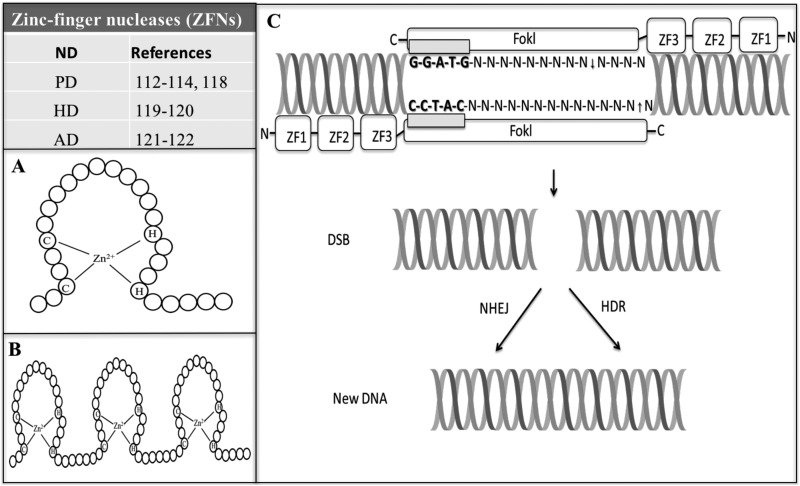
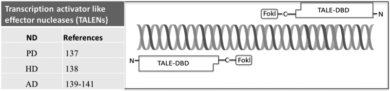
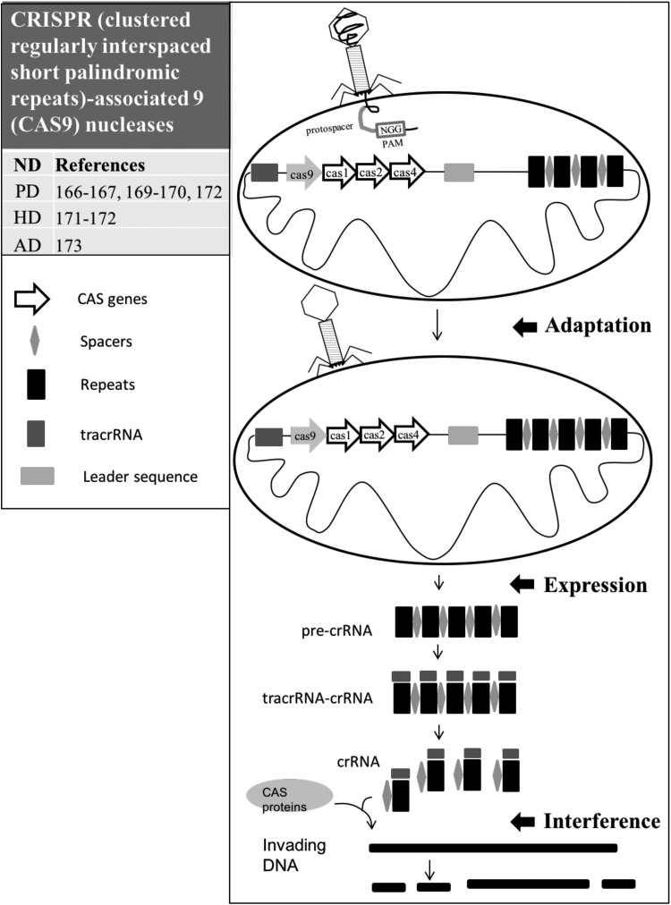
Neurodegenerative diseases (NDs), at least including Alzheimer's, Huntington's, and Parkinson's diseases, have become the most dreaded maladies because there are no precise diagnostic tools or definite treatments for these debilitating diseases. The increased prevalence and a substantial impact on the social-economic and medical care of NDs propel governments to develop policies to counteract the impact. Although the etiologies of NDs are still unknown, growing evidence suggests that genetic, cellular, and circuit alternations may cause the generation of abnormal misfolded proteins, which uncontrolledly accumulate to damage and eventually overwhelm the protein-disposal mechanisms of these neurons, leading to a common pathological feature of NDs. If the functions and the connectivity can be restored, alterations and accumulated damages may improve. The gene-editing tools including zinc-finger nucleases (ZFNs), transcription activator-like effector nucleases (TALENs), and clustered regularly interspaced short palindromic repeats-associated nucleases (CRISPR/CAS) have emerged as a novel tool not only for generating specific ND animal models for interrogating the mechanisms and screening potential drugs against NDs but also for the editing sequence-specific genes to help patients with NDs to regain function and connectivity. This review introduces the clinical manifestations of three distinct NDs and the applications of the gene-editing technology on these debilitating diseases.

Keywords: Alzheimer’s disease (AD); Huntington’s disease (HD); Parkinson’s diseases (PD); clustered regularly interspaced short palindromic repeats–associated nucleases (CRISPR/CAS); neurodegenerative diseases (NDs); transcription activator-like effector nucleases (TALENs); zinc-finger nucleases (ZFNs).


Conflict of interest statement

Declaration of Conflicting Interests: The author(s) declared the following potential conflicts of interest with respect to the research, authorship, and/or publication of this article: Paul R. Sanberg (PRS) is the coeditor in chief of Cell Transplantation. Neither PRS nor any of his colleagues were involved in the peer-review process or decision for this manuscript.

Figures

Fig. 1.
Fig. 1.
Schematic diagram of zinc-finger nucleases (ZFNs). Table indicates references regarding applications of ZFNs in neurodegenerative diseases (NDs) including Parkinson’s disease (PD), Huntington’s disease (HD), and Alzheimer’s disease (AD). (A) Structure of zinc finger (ZF). The paired cysteines (Cys) and histidines (His), (Cys2His2) tetrahedrally bind a zinc ion to form a compact structure. (B) Typical arrangement of C2H2 ZF motifs. The modular feature of the ZFs enables them to be assembled into a linear array to target DNA. (C) Structure of ZFNs. ZFNs consist of 2 functional domains, including a ZF DNA-binding domain includes a chain of 3 finger modules (ZF1 to ZF3). Each ZF can recognize a 3 bp of DNA; a DNA cleavage domain is comprised of the nuclease domain of the FokI. However, this technique requires 2 ZFNs to bind at or near the cleavage site because the FokI needs to form a dimer, and then it will function properly. Also, the target sequences must be separated by 5 to 7 base pairs to allow formation of the catalytically active FokI dimer, causing a double-strand break at a specific sequence to trigger the cell DNA repair machinery including homology-directed repair and nonhomologous end joining to repair the defects resulting in targeted gene disruptions or gene integration.
Fig. 2.
Fig. 2.
Schematic diagram of transcription activator-like effector nucleases (TALENs). Table indicates references regarding applications of TALENs in neurodegenerative diseases (NDs) including PD, HD, and AD. Structure of TALENs consists of 2 functional domains including transcription activator-like effector (TALE) and DNA cleavage domain (DCD). TALE is shown as long squares with a final carboxy-terminal truncated “half” repeat. TALE amino- and carboxy-terminal domains required for DNA-binding activity are shown as “N” and “C,” respectively. The DCD, including the FokI endonuclease, is shown as a small square.
Fig. 3.
Fig. 3.
Illustration of clustered regularly interspaced short palindromic repeats–associated nucleases (CRISPR/CAS). Table indicates references regarding applications of CRISPR/CAS in neurodegenerative diseases including PD, HD, and AD. CRISPR is an array of short repeated sequences (black rectangles) separated by spacers (gray diamonds) with unique sequences. When a segment of a bacteriophage’s genome invades and integrates into the cellular DNA, the processes of the CRISPR/CAS mediated immunity against the integration is initiated, including adaptation, expression, and interference. “Adaptation”—the invading bacteriophage’s DNA contains 2 to 5 bp protospacer adjacent motif (PAMs) acting as a recognition motif. The new single copy of spacer (green diamond) occurs at the leader side of the CRISPR array and is followed by its duplication. Any mutations in the protospacers or PAMs of the bacteriophage will interfere with the CRISPR/CAS-mediated reactions. “Expression”—the repeats, the invader DNA (green diamond), and spacer sequences are transcribed to the precursor of CRISPR RNAs (pre-crRNAs), which turn into the crRNA through the help of CAS proteins (CAS1, CAS2, CAS9, and CAS4) and the trans-activating crRNA (tracrRNA) molecule. The tracrRNA and crRNA form a tracrRNA-crRNA complex. “Interference”—crRNA guided CAS proteins to cleave the invader DNA into small DNA fragments.

References

    1. Fan HC, Chen SJ, Harn HJ, Lin SZ. Parkinson’s disease: from genetics to treatments. Cell Transplant. 2013;22(4):639–652. - PubMed
    1. Fan HC, Ho LI, Chi CS, Chen SJ, Peng GS, Chan TM, Lin SZ, Harn HJ. Polyglutamine (polyq) diseases: genetics to treatments. Cell Transplant. 2014;23(4–5):441–458. - PubMed
    1. Fan HC, Chi CS, Cheng SN, Lee HF, Tsai JD, Lin SZ, Harn HJ. Targeting new candidate genes by small molecules approaching neurodegenerative diseases. Int J Mol Sci. 2016;17(1): pii:E26. - PMC - PubMed
    1. Menken M, Munsat TL, Toole JF. The global burden of disease study: implications for neurology. Arch Neurol. 2000;57(3):418–420. - PubMed
    1. Scheper W, Hoozemans JJ. The unfolded protein response in neurodegenerative diseases: a neuropathological perspective. Acta Neuropathol. 2015;130(3):315–331. - PMC - PubMed

Publication types

MeSH terms

Substances