. 2018 Jul;40(1):165-178.
doi: 10.3892/or.2018.6438. Epub 2018 May 16.

Comparison of the synergistic anticancer activity of AlPcS4 photodynamic therapy in combination with different low‑dose chemotherapeutic agents on gastric cancer cells

Affiliations

Comparison of the synergistic anticancer activity of AlPcS4 photodynamic therapy in combination with different low‑dose chemotherapeutic agents on gastric cancer cells

Jing Xin et al. Oncol Rep. 2018 Jul.

Abstract

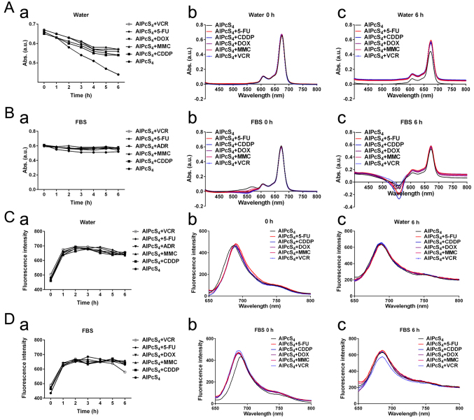
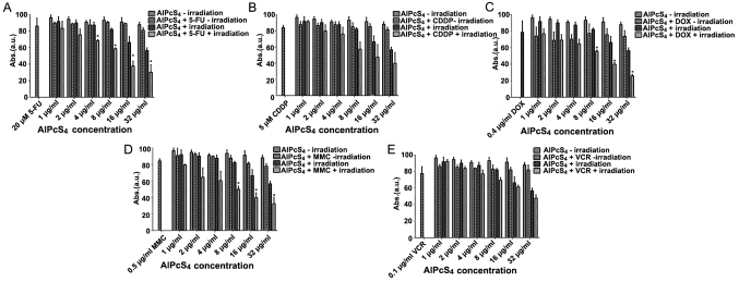
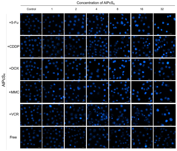
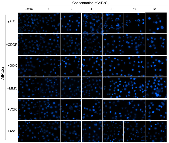
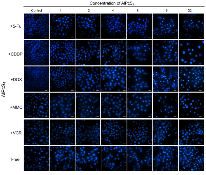
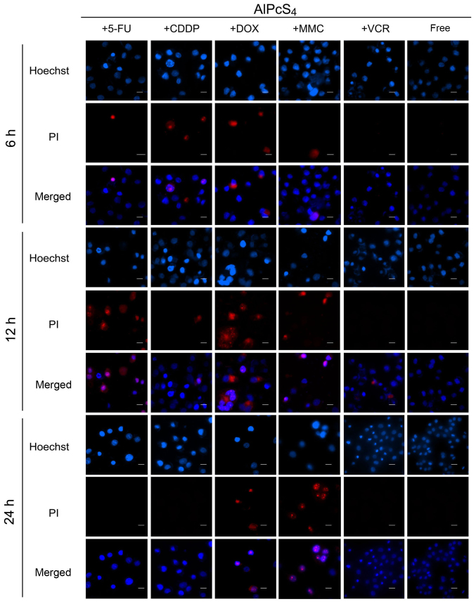
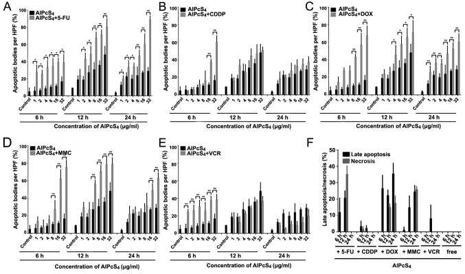
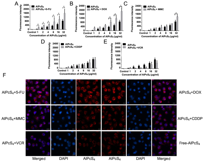
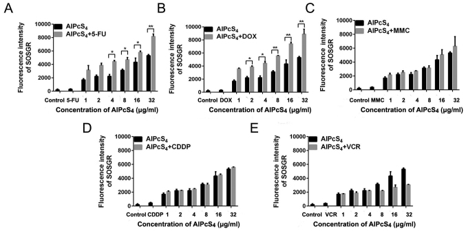
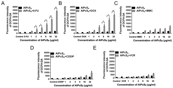
Limited cellular delivery and internalization efficiency of Al(III) phthalocyanine chloride tetrasulfonic acid (AlPcS4) induce poor penetration ability in cells and a slight photodynamic therapy (PDT) effect on gastric cancer. The combination treatment of AlPcS4/PDT with low‑dose chemotherapeutic agents may provide a promising treatment strategy to increase the weak delivery efficiency of AlPcS4, reducing the dose of chemical agents without reducing efficacy, and improving apoptosis‑inducing abilities, thereby increasing the antitumor effects and decreasing the noxious side effects on gastric cancer. We investigated and compared the synergistic antitumor growth effect on gastric cancer cells by combining AlPcS4/PDT treatment with different low‑dose chemotherapeutic agents, namely, 5‑fluorouracil (5‑FU), doxorubicin (DOX), cisplatin (CDDP), mitomycin C (MMC), and vincristine (VCR). The inhibitory effect was increased in treatments that combined AlPcS4/PDT with all the aforementioned low‑dose chemotherapeutic agents, to a different extent. An evident synergistic effect was obtained in the combination treatment of AlPcS4/PDT with low‑dose 5‑FU, DOX, and MMC by increasing AlPcS4 intracellular uptake ability, improving apoptosis‑inducing abilities, and prolonging apoptosis‑inducing time. The low‑dose chemotherapeutic agents prolonged the apoptosis‑inducing period of AlPcS4/PDT, and AlPcS4/PDT quickly improved apoptosis‑inducing abilities of chemotherapy even at low doses. Generally, the combination treatment of AlPcS4/PDT with low‑dose chemotherapeutic agents had significant antitumor growth effects in addition to a low dark‑cytotoxicity effect on gastric cancer, thereby representing an effective and feasible therapy method for gastric cancer.


Figures

Figure 1.
Figure 1.
UV-vis absorption spectra and fluorescence intensity of AlPcS4 mixture with 5-FU (20 µm), CDDP (5 µm), DOX (0.4 µm/ml), MMC (0.5 µm/ml), and VCR (0.1 µm/ml) or free-AlPcS4 at 8 µm/ml. (A-a and B-a) Maximum OD values near 675 nm absorption spectra of corresponding agents in deionized water and RPMI-1640 culture medium that contained FBS at 1–6 h. (A-b and B-b) UV-vis absorption spectra of corresponding agents in deionized water and RPMI-1640 culture medium that contained FBS at 0 h. (A-c and B-c) UV-Vis absorption spectra of corresponding agents in deionized water and RPMI-1640 culture medium that contained FBS at 6 h. (C-a and D-a) Maximum fluorescence intensity near 687 nm fluorescence spectra of corresponding agents in deionized water and RPMI-1640 culture medium that contained FBS at 1–6 h. (C-b and D-b) Fluorescence spectra of corresponding agents in deionized water and RPMI-1640 culture medium that contained FBS at 0 h. (C-c and D-c) Fluorescence spectra of corresponding agentsin deionized water and RPMI-1640 culture medium that contained FBS at 6 h. AlPcS4, Al(III) phthalocyanine chloride tetrasulfonic acid; 5-FU, 5-fluorouracil; DOX, doxorubicin; CDDP, cisplatin; MMC, mitomycin C; VCR, vincristine; ROS.
Figure 2.
Figure 2.
Antitumor growth effect on SGC-7901 cells in single and combination treatment therapy as determined by CCK-8 assay. Dark cytotoxicity and antitumor growth effect on SGC-7901 cells by (A) AlPcS4 + 5-FU, (B) AlPcS4 + CDDP, (C) AlPcS4 + DOX, (D) AlPcS4 + MMC or (E) AlPcS4 + VCR. The cells were treated with 1–32 µm/ml free-AlPcS4 or AlPcS4 + 5-FU (20 µm), AlPcS4 + CDDP (5 µm), AlPcS4 + DOX (0.4 µm/ml), AlPcS4 + MMC (0.5 µm/ml) or AlPcS4 + VCR (0.1 µm/ml) for 6 h. The cells were then incubated again for 24 h with or without 635-nm laser irradiation at 100 mW/cm2 illumination dosage for 5 min. *P<0.05, represents a statistical difference in antitumor effect between the combination of AlPcS4 with a chemical agent and free-AlPcS4. AlPcS4, Al(III) phthalocyanine chloride tetrasulfonic acid; 5-FU, 5-fluorouracil; DOX, doxorubicin; CDDP, cisplatin; MMC, mitomycin C; VCR, vincristine.
Figure 3.
Figure 3.
Apoptosis induced by AlPcS4 + 5-FU, AlPcS4 + CDDP, AlPcS4 + DOX, AlPcS4 + MMC, AlPcS4 + VCR and free-AlPcS4 in SGC-7901 cells after being irradiated for 6 h. The cells were then treated with 1–32 µm/ml free-AlPcS4 or AlPcS4 + 5-FU (20 µm), AlPcS4 + CDDP (5 µm), AlPcS4 + DOX (0.4 µm/ml), AlPcS4 + MMC (0.5 µm/ml), or AlPcS4 + VCR (0.1 µm/ml) for 6 h. These samples were then irradiated with 635-nm laser irradiation at 100 mW/cm2 illumination dosage for 5 min, incubated for 6 h, stained with Hoechst 33342 probe, and then imaged using afluorescence microscope. All the Hoechst staining images were acquired at an ×400 magnification. The scale bar represented 20 µm. AlPcS4, Al(III) phthalocyanine chloride tetrasulfonic acid; 5-FU, 5-fluorouracil; DOX, doxorubicin; CDDP, cisplatin; MMC, mitomycin C; VCR, vincristine.
Figure 4.
Figure 4.
Apoptosis induced by AlPcS4 + 5-FU, AlPcS4 + CDDP, AlPcS4 + DOX, AlPcS4 + MMC, AlPcS4 + VCR, and free-AlPcS4 in SGC-7901 cells after being irradiated for 12 h. The cells were treated with 1–32 µm/ml free-AlPcS4 or AlPcS4 + 5-FU (20 µm), AlPcS4 + CDDP (5 µm), AlPcS4 + DOX (0.4 µm/ml), AlPcS4 + MMC (0.5 µm/ml) or AlPcS4 + VCR (0.1 µm/ml) for 6 h. The cells were then irradiated with 635-nm laser irradiation at 100 mw/cm2 illumination dosage for 5 min, incubated for 12 h, stained with Hoechst 33342 probe, and then imaged using afluorescence microscope. All the Hoechst staining images were acquired at an ×400 magnification. The scale bar represented 20 µm. AlPcS4, Al(III) phthalocyanine chloride tetrasulfonic acid; 5-FU, 5-fluorouracil; DOX, doxorubicin; CDDP, cisplatin; MMC, mitomycin C; VCR, vincristine.
Figure 5.
Figure 5.
Apoptosis induced by AlPcS4 + 5-FU, AlPcS4 + CDDP, AlPcS4 + DOX, AlPcS4 + MMC, AlPcS4 + VCR and free-AlPcS4 in SGC-7901 cells after being irradiated for 24 h. The cells were then treated with 1–32 µm/ml free-AlPcS4 or AlPcS4 + 5-FU (20 µm), AlPcS4 + CDDP (5 µm), AlPcS4 + DOX (0.4 µm/ml), AlPcS4 + MMC (0.5 µm/ml) or AlPcS4 + VCR (0.1 µm/ml) for 6 h. The cells were then irradiated with 635-nm laser irradiation at 100 mW/cm2 illumination dosage for 5 min, incubated for 24 h, stained with Hoechst 33342 probe, and then imaged using afluorescence microscope. All the Hoechst staining images were acquired at an ×400 magnification. The scale bar represented 20 µm. AlPcS4, Al(III) phthalocyanine chloride tetrasulfonic acid; 5-FU, 5-fluorouracil; DOX, doxorubicin; CDDP, cisplatin; MMC, mitomycin C; VCR, vincristine.
Figure 6.
Figure 6.
Apoptosis and necrosis induced by AlPcS4 + 5-FU, AlPcS4 + CDDP, AlPcS4 + DOX, AlPcS4 + MMC, AlPcS4 + VCR and free-AlPcS4 in SGC-7901 cells after being irradiated for 6, 12 and 24 h. The cells were treated with 1–32 µm/ml free-AlPcS4 or AlPcS4 + 5-FU (20 µm), AlPcS4 + CDDP (5 µm), AlPcS4 + DOX (0.4 µm/ml), AlPcS4 + MMC (0.5 µm/ml) or AlPcS4 + VCR (0.1 µm/ml) for 6 h. The cells were then irradiated with 635-nm laser irradiation at 100 mW/cm2 illumination dosage for 5 min. The cells were incubated for 6, 12 and 24 h, stained with Hoechst 33342 and PI probes, and imaged usingfluorescence microscopy. Below 32 µm/ml, no necrosis was obtained. Therefore, the Hoechst staining and PI images are shown at 32 µm/ml. All the Hoechst staining and PI images were acquired at an ×400 magnification. The scale bar represented 20 µm. AlPcS4, Al(III) phthalocyanine chloride tetrasulfonic acid; 5-FU, 5-fluorouracil; DOX, doxorubicin; CDDP, cisplatin; MMC, mitomycin C; VCR, vincristine.
Figure 7.
Figure 7.
Statistical analysis of apoptosis and necrosis induced by AlPcS4 + 5-FU, AlPcS4 + CDDP, AlPcS4 + DOX, AlPcS4 + MMC, AlPcS4 + VCR and free-AlPcS4 in SGC-7901 cells after being irradiated for 6, 12 and 24 h. (A-E) The histograms represent the percentage of cells with apoptotic and necrotic characteristics among 200 cells at a high-power field. The data represent the average of three experiments. The bar is the SD. *P<0.05 and **P<0.01 represented a statistically significant difference in the number of apoptotic bodies between the combination of AlPcS4 with a chemical agent and free-AlPcS4. AlPcS4, Al(III) phthalocyanine chloride tetrasulfonic acid; 5-FU, 5-fluorouracil; DOX, doxorubicin; CDDP, cisplatin; MMC, mitomycin C; VCR, vincristine.
Figure 8.
Figure 8.
Fluorescence intensity analysis and fluorescence imaging of AlPcS4 in SGC-7901 cells after treatment with AlPcS4 + 5-FU, AlPcS4 + DOX, AlPcS4 + MMC, AlPcS4 + CDDP, AlPcS4 + VCR and free-AlPcS4. (A-E) Fluorescent intensity analysis of AlPcS4 in SGC-7901 cells after treatment with 1–32 µm/ml free-AlPcS4, AlPcS4 + 5-FU (20 µm), AlPcS4 + DOX (0.4 µm), AlPcS4 + MMC (0.5 µm/ml), AlPcS4 + CDDP (5 µm) or AlPcS4 + VCR (0.1 µm/ml) for 6 h and measured by using a fluorescence spectrophotometer. The data represents the average of three experiments and the bar is the SD. *P<0.05 and **P<0.01 represented a statistically significant difference in the fluorescence intensity of AlPcS4 in cells between the combination therapy of AlPcS4 with a chemical agent and single therapy of free-AlPcS4. (F) The fluorescent images of AlPcS4 in SGC-7901 cells after treatment with 32 µm/ml free-AlPcS4, AlPcS4 + 5-FU (20 µm), AlPcS4 + DOX (0.4 µm/ml), AlPcS4 + MMC (0.5 µm/ml), AlPcS4 + CDDP (5 µm) or AlPcS4 + VCR (0.1 µm/ml) for 6 h and measured using a fluorescence microscope. All the images were acquired at an ×400 magnification. The scale bar represented 20 µm. AlPcS4, Al(III) phthalocyanine chloride tetrasulfonic acid; 5-FU, 5-fluorouracil; DOX, doxorubicin; CDDP, cisplatin; MMC, mitomycin C; VCR, vincristine.
Figure 9.
Figure 9.
SOG production in SGC-7901 cells treated with AlPcS4 + 5-FU, AlPcS4 + CDDP, AlPcS4 + DOX, AlPcS4 + MMC, AlPcS4 + VCR and free-AlPcS4. (A-E) Fluorescence intensities of SOSGR probes were measured to analyze SOG in SGC-7901 cells after treatment with 1–32 µm free-AlPcS4. (A) AlPcS4 + 5-FU (20 µm), (B) AlPcS4 + DOX (0.4 µm/ml), (C) AlPcS4 + MMC (0.5 µm/ml), (D) AlPcS4 + CDDP (5 µm) or (E) AlPcS4 + VCR (0.1 µm/ml) and irradiation with 635-nm laser light for 5 min. Data represent the average of three experiments and the bar is the SD. *P<0.05 and **P<0.01 represent a statistically significant difference in the fluorescence intensity of SOSGR in cells between the combination therapy of AlPcS4 with a chemical agent and single therapy of free-AlPcS4. AlPcS4, Al(III) phthalocyanine chloride tetrasulfonic acid; 5-FU, 5-fluorouracil; DOX, doxorubicin; CDDP, cisplatin; MMC, mitomycin C; VCR, vincristine; SOG, singlet oxygen.
Figure 10.
Figure 10.
ROS production in SGC-7901 cells treated with AlPcS4 + 5-FU, AlPcS4 + CDDP, AlPcS4 + DOX, AlPcS4 + MMC, AlPcS4 + VCR and free-AlPcS4. (A-E) Fluorescence intensities of DCFH-DA probes were measured to analyze ROS in SGC-7901 cells after treatment with 1–32 µm/ml free-AlPcS4. (A) AlPcS4 + 5-FU (20 µm/ml), (B) AlPcS4 + DOX (0.4 µm/ml), (C) AlPcS4 + MMC (0.5 µm/ml), (D) AlPcS4 + CDDP (5 µm) or (E) AlPcS4 + VCR (0.1 µm/ml) and irradiation with 635-nm laser light for 5 min. Data represent the average of three experiments and the bar is the SD. *P<0.05 and **P<0.01 represent a statistically significant difference in the fluorescence intensity of DCFH-DA in cells between the combination therapy of AlPcS4 with a chemical agent and single therapy of free-AlPcS4. AlPcS4, Al(III) phthalocyanine chloride tetrasulfonic acid; 5-FU, 5-fluorouracil; DOX, doxorubicin; CDDP, cisplatin; MMC, mitomycin C; VCR, vincristine; ROS, reactive oxygen species.

Similar articles

Cited by

References

    1. Torre LA, Bray F, Siegel RL, Ferlay J, Lortet-Tieulent J, Jemal A. Global cancer statistics, 2012. CA Cancer J Clin. 2015;65:87–108. doi: 10.3322/caac.21262. - DOI - PubMed
    1. Dougherty TJ, Gomer CJ, Henderson BW, Jori G, Kessel D, Korbelik M, Moan J, Peng Q. Photodynamic therapy. J Natl Cancer Inst. 1998;90:889–905. doi: 10.1093/jnci/90.12.889. - DOI - PMC - PubMed
    1. Vrouenraets MB, Visser GW, Snow GB, van Dongen GA. Basic principles, applications in oncology and improved selectivity of photodynamic therapy. Anticancer Res. 2003;23:505–522. - PubMed
    1. Shafirstein G, Battoo A, Harris K, Baumann H, Gollnick SO, Lindenmann J, Nwogu CE. Photodynamic therapy of non-small cell lung cancer. Narrative review and future directions. Ann Am Thorac Soc. 2016;13:265–275. - PMC - PubMed
    1. Moor AC. Signaling pathways in cell death and survival after photodynamic therapy. J Photochem Photobiol B. 2000;57:1–13. doi: 10.1016/S1011-1344(00)00065-8. - DOI - PubMed