. 2018 Sep 1;315(3):C310-C318.
doi: 10.1152/ajpcell.00183.2017. Epub 2018 May 16.

Does partial titin degradation affect sarcomere length nonuniformities and force in active and passive myofibrils?

Affiliations

Does partial titin degradation affect sarcomere length nonuniformities and force in active and passive myofibrils?

V Joumaa et al. Am J Physiol Cell Physiol. .

Abstract

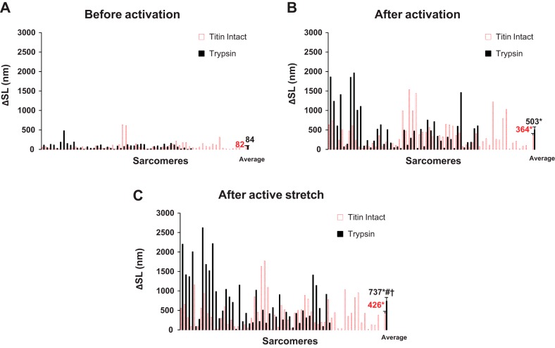
The aim of this study was to determine the role of titin in preventing the development of sarcomere length nonuniformities following activation and after active and passive stretch by determining the effect of partial titin degradation on sarcomere length nonuniformities and force in passive and active myofibrils. Selective partial titin degradation was performed using a low dose of trypsin. Myofibrils were set at a sarcomere length of 2.4 µm and then passively stretched to sarcomere lengths of 3.4 and 4.4 µm. In the active condition, myofibrils were set at a sarcomere length of 2.8 µm, activated, and actively stretched by 1 µm/sarcomere. The extent of sarcomere length nonuniformities was calculated for each sarcomere as the absolute difference between sarcomere length and the mean sarcomere length of the myofibril. Our main finding is that partial titin degradation does not increase sarcomere length nonuniformities after passive stretch and activation compared with when titin is intact but increases the extent of sarcomere length nonuniformities after active stretch. Furthermore, when titin was partially degraded, active and passive stresses were substantially reduced. These results suggest that titin plays a crucial role in actively stretched myofibrils and is likely involved in active and passive force production.

Keywords: Z-disk; active stretch; force regulation; passive stretch; sarcomere stability.


Figures

Fig. 1.
Fig. 1.
Sarcomere length (SL) and stress as a function of time for titin-intact and trypsin-treated myofibrils passively stretched from a SL of 2.4 µm to SLs of 3.4 µm (A) and 4.4 µm (B). Sarcomere length nonuniformities increased after stretch in titin-intact and trypsin-treated myofibrils.
Fig. 2.
Fig. 2.
The extent of individual sarcomere length nonuniformities (ΔSL) at a SL of 2.4 µm before passive stretch (A) and at the steady-state after passive stretch to SLs of 3.4 µm (B) and 4.4 µm (C) in titin-intact and trypsin-treated myofibrils. The extent of individual sarcomere length nonuniformities increased after passive stretch but was not different between titin-intact and trypsin-treated myofibrils. *Significant difference from the same group at a SL of 2.4 µm.
Fig. 3.
Fig. 3.
Length difference between half-sarcomeres at a sarcomere length (SL) of 2.4 µm before passive stretch (A) and at the steady-state after passive stretch to SLs of 3.4 µm (B) and 4.4 µm (C) in titin-intact and trypsin-treated myofibrils. *Significant difference from the same group at a SL of 2.4 µm. †Significant difference from the titin-intact group at the same SL.
Fig. 4.
Fig. 4.
Loss in stress at the steady-state after activation at a sarcomere length (SL) of 2.8 µm (A) and after active stretch by 1 µm/sarcomere (B) as a function of the loss in passive stress in the trypsin-treated myofibrils. There is a correlation between the loss in active stress and the loss in passive stress.
Fig. 5.
Fig. 5.
Sarcomere length (SL) and stress as a function of time for titin-intact (A) and trypsin-treated (B) myofibrils activated at a SL of 2.8 µm and actively stretched by 1 µm/sarcomere. Sarcomere length nonuniformities increased after activation and active stretch in titin-intact and trypsin-treated myofibrils.
Fig. 6.
Fig. 6.
The extent of individual sarcomere length nonuniformities (ΔSL) at a SL of 2.8 µm before activation (A) and at the steady-state after activation (B) and active stretch by 1 µm/sarcomere (C) in titin-intact and trypsin-treated myofibrils. ΔSL increased to the same extent in titin-intact and trypsin-treated myofibrils after activation, but while active stretch did not further increase ΔSL in titin-intact myofibrils, it resulted in a significant increase in ΔSL in the trypsin-treated myofibrils compared with before active stretch and the titin-intact myofibrils. *Significant difference from the same group before activation; #significant difference from the same group after activation; †significant difference from the titin-intact group after active stretch.
Fig. 7.
Fig. 7.
A 2% SDS electrophoresis gel (A) showing titin degradation after treatment with trypsin for 5 min and a 12% SDS electrophoresis gel (B) showing no changes in the contractile proteins after trypsin treatment. MHC, myosin heavy chain; Skinned, skinned samples; T1, intact titin; T2, degradation molecule of titin; Tryp 5′, trypsin-treated samples for 5 min.
Fig. 8.
Fig. 8.
Mass spectrometry analysis shows no apparent degradation of contractile and regulatory proteins when rabbit psoas muscle samples were incubated in trypsin solution (0.25 µg/ml in relaxing solution) for 5 min, whereas degradation was clear after overnight incubation (inset). Strips of rabbit psoas muscle were incubated with 0.25 µg/ml trypsin in relaxing solution for 5 min and overnight. The samples were then removed and the incubation solutions were used for mass spectrometry analysis. MLC, myosin light chain.

Similar articles

Cited by

References

    1. Ait-Mou Y, Hsu K, Farman GP, Kumar M, Greaser ML, Irving TC, de Tombe PP. Titin strain contributes to the Frank-Starling law of the heart by structural rearrangements of both thin- and thick-filament proteins. Proc Natl Acad Sci USA 113: 2306–2311, 2016. doi:10.1073/pnas.1516732113. - DOI - PMC - PubMed
    1. Brechenmacher L, Nguyen TH, Zhang N, Jun TH, Xu D, Mian MA, Stacey G. Identification of soybean proteins and genes differentially regulated in near isogenic lines differing in resistance to aphid infestation. J Proteome Res 14: 4137–4146, 2015. doi:10.1021/acs.jproteome.5b00146. - DOI - PubMed
    1. Brooks SV, Faulkner JA. The magnitude of the initial injury induced by stretches of maximally activated muscle fibres of mice and rats increases in old age. J Physiol 497: 573–580, 1996. doi:10.1113/jphysiol.1996.sp021790. - DOI - PMC - PubMed
    1. Cazorla O, Wu Y, Irving TC, Granzier H. Titin-based modulation of calcium sensitivity of active tension in mouse skinned cardiac myocytes. Circ Res 88: 1028–1035, 2001. doi:10.1161/hh1001.090876. - DOI - PubMed
    1. DuVall MM, Gifford JL, Amrein M, Herzog W. Altered mechanical properties of titin immunoglobulin domain 27 in the presence of calcium. Eur Biophys J 42: 301–307, 2013. doi:10.1007/s00249-012-0875-8. - DOI - PubMed

Publication types

LinkOut - more resources