

Structural re-positioning, in silico molecular modelling, oxidative degradation, and biological screening of linagliptin as adenosine 3 receptor (ADORA3) modulators targeting hepatocellular carcinoma
- PMID: 29768061
- PMCID: PMC6010121
- DOI: 10.1080/14756366.2018.1462801
Structural re-positioning, in silico molecular modelling, oxidative degradation, and biological screening of linagliptin as adenosine 3 receptor (ADORA3) modulators targeting hepatocellular carcinoma
Abstract
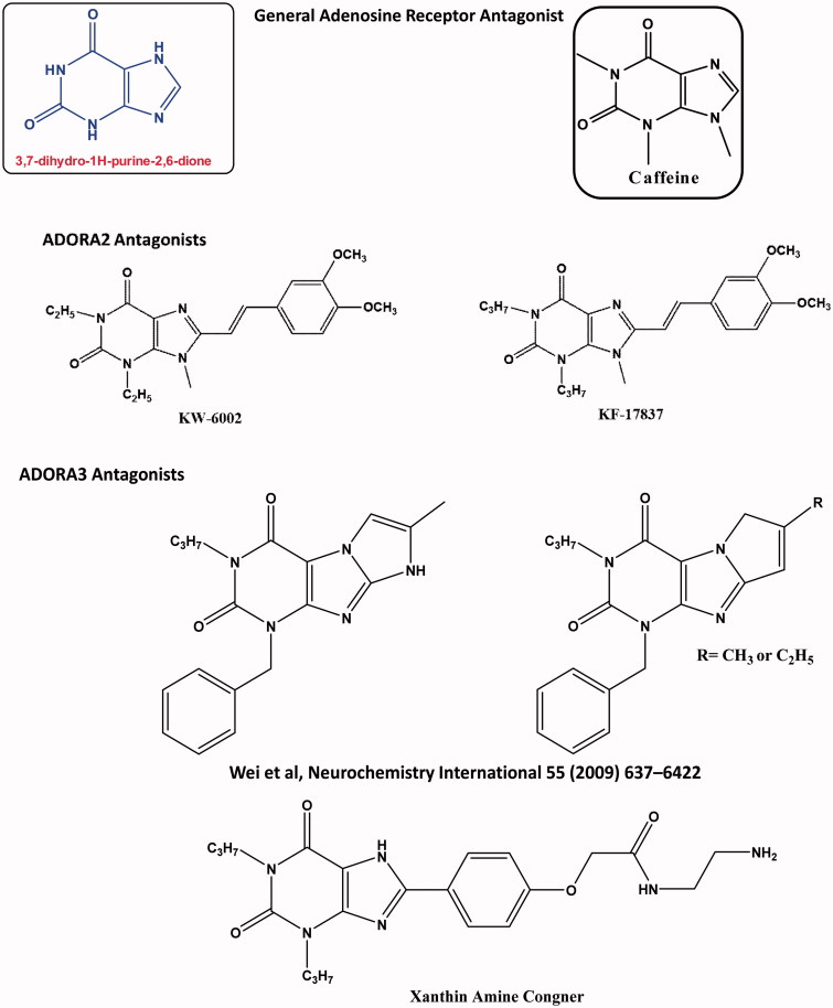
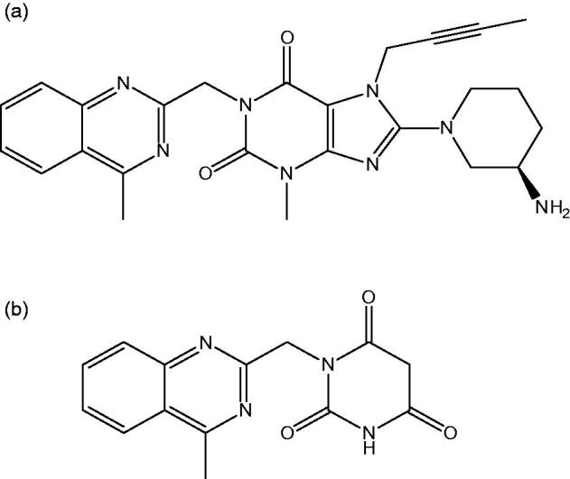
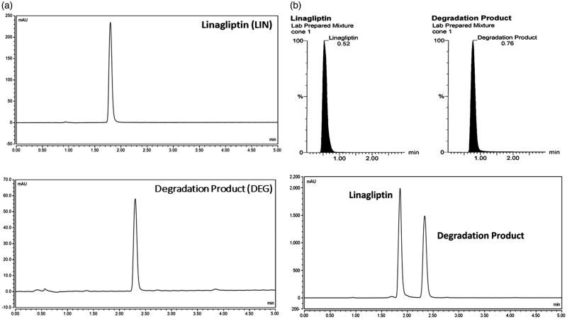
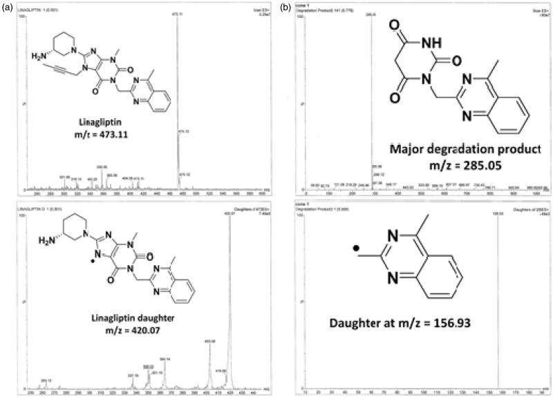
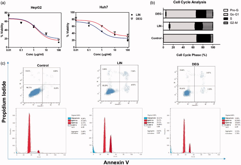
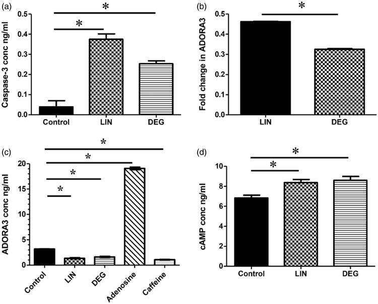
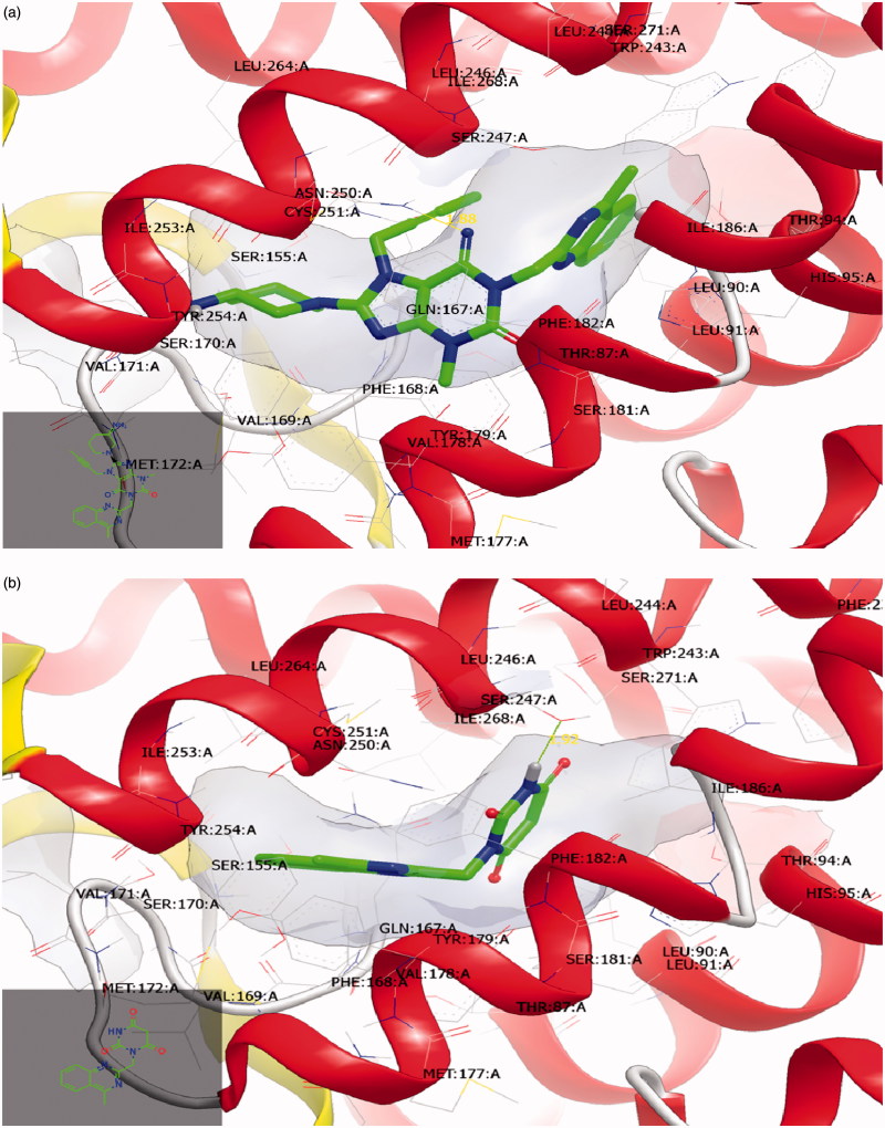
Chemical entities with structural diversity were introduced as candidates targeting adenosine receptor with different clinical activities, containing 3,7-dihydro-1H-purine-2,6-dione, especially adenosine 3 receptors (ADORA3). Our initial approach started with pharmacophore screening of ADORA3 modulators; to choose linagliptin (LIN), approved anti-diabetic drug as Dipeptidyl peptidase-4 inhibitors, to be studied for its modulating effect towards ADORA3. This was followed by generation, purification, analytical method development, and structural elucidation of oxidative degraded product (DEG). Both of LIN and DEG showed inhibitory profile against hepatocellular carcinoma cell lines with induction of apoptosis at G2/M phase with increase in caspase-3 levels, accompanied by a downregulation in gene and protein expression levels of ADORA3 with a subsequent increase in cAMP. Quantitative in vitro assessment of LIN binding affinity against ADORA3 was also performed to exhibit inhibitory profile at Ki of 37.7 nM. In silico molecular modelling showing binding affinity of LIN and DEG towards ADORA3 was conducted.
Keywords: Drug repositioning; adenosine 3 receptor; hepatocellular carcinoma; linagliptin.
Figures
Similar articles
-
Design, synthesis, biological evaluation, and modeling studies of novel conformationally-restricted analogues of sorafenib as selective kinase-inhibitory antiproliferative agents against hepatocellular carcinoma cells.Eur J Med Chem. 2021 Jan 15;210:113081. doi: 10.1016/j.ejmech.2020.113081. Epub 2020 Dec 4. Eur J Med Chem. 2021. PMID: 33310290
-
Design, synthesis and biological evaluation of novel hybrids targeting mTOR and HDACs for potential treatment of hepatocellular carcinoma.Eur J Med Chem. 2021 Dec 5;225:113824. doi: 10.1016/j.ejmech.2021.113824. Epub 2021 Sep 3. Eur J Med Chem. 2021. PMID: 34509167
-
Design, synthesis and biological evaluation of novel 1,3,4-trisubstituted pyrazole derivatives as potential chemotherapeutic agents for hepatocellular carcinoma.Bioorg Chem. 2018 Aug;78:149-157. doi: 10.1016/j.bioorg.2018.03.014. Epub 2018 Mar 16. Bioorg Chem. 2018. PMID: 29567429
-
Synthesis, biological activity and multiscale molecular modeling studies of bis-coumarins as selective carbonic anhydrase IX and XII inhibitors with effective cytotoxicity against hepatocellular carcinoma.Bioorg Chem. 2019 Jun;87:838-850. doi: 10.1016/j.bioorg.2019.03.003. Epub 2019 Mar 7. Bioorg Chem. 2019. PMID: 31003041
-
Discovery of 5-aryl-3-thiophen-2-yl-1H-pyrazoles as a new class of Hsp90 inhibitors in hepatocellular carcinoma.Bioorg Chem. 2020 Jan;94:103433. doi: 10.1016/j.bioorg.2019.103433. Epub 2019 Nov 19. Bioorg Chem. 2020. PMID: 31785857
Cited by
-
Therapeutic potential of adenosine receptor modulators in cancer treatment.RSC Adv. 2025 Jun 17;15(26):20418-20445. doi: 10.1039/d5ra02235e. eCollection 2025 Jun 16. RSC Adv. 2025. PMID: 40530308 Free PMC article. Review.
-
Anti-Diabetic Therapies and Cancer: From Bench to Bedside.Biomolecules. 2024 Nov 20;14(11):1479. doi: 10.3390/biom14111479. Biomolecules. 2024. PMID: 39595655 Free PMC article. Review.
-
Repositioning of dipeptidyl peptidase-4 inhibitors and glucagon like peptide-1 agonists as potential neuroprotective agents.Neural Regen Res. 2019 May;14(5):745-748. doi: 10.4103/1673-5374.249217. Neural Regen Res. 2019. PMID: 30688255 Free PMC article. Review.
-
Repositioning of Hypoglycemic Drug Linagliptin for Cancer Treatment.Front Pharmacol. 2020 Mar 3;11:187. doi: 10.3389/fphar.2020.00187. eCollection 2020. Front Pharmacol. 2020. PMID: 32194417 Free PMC article.
-
In silico approaches for drug repurposing in oncology: a scoping review.Front Pharmacol. 2024 Jun 11;15:1400029. doi: 10.3389/fphar.2024.1400029. eCollection 2024. Front Pharmacol. 2024. PMID: 38919258 Free PMC article.
References
MeSH terms
Substances
LinkOut - more resources
Full Text Sources
Other Literature Sources
Medical
Research Materials