. 2018 May 16;13(5):e0196186.
doi: 10.1371/journal.pone.0196186. eCollection 2018.

Remote ischemic preconditioning STAT3-dependently ameliorates pulmonary ischemia/reperfusion injury

Affiliations

Remote ischemic preconditioning STAT3-dependently ameliorates pulmonary ischemia/reperfusion injury

Nanfu Luo et al. PLoS One. .

Abstract

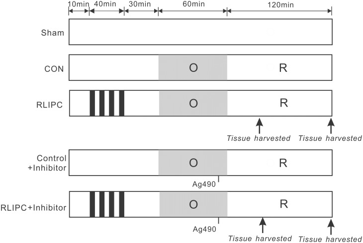
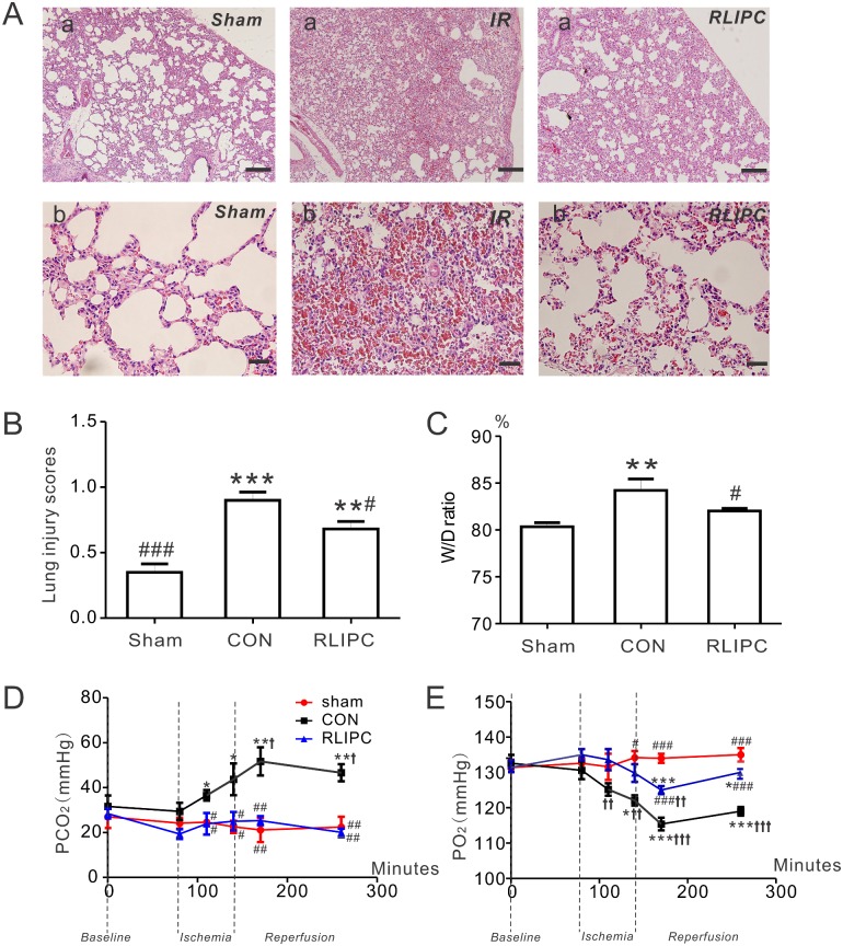
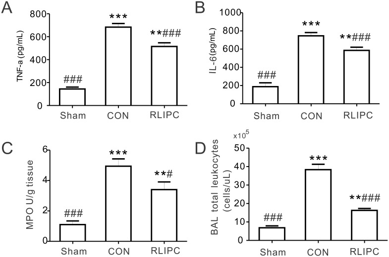
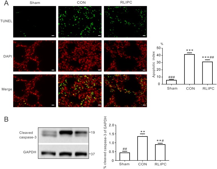
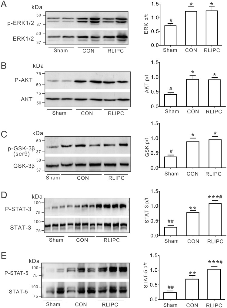
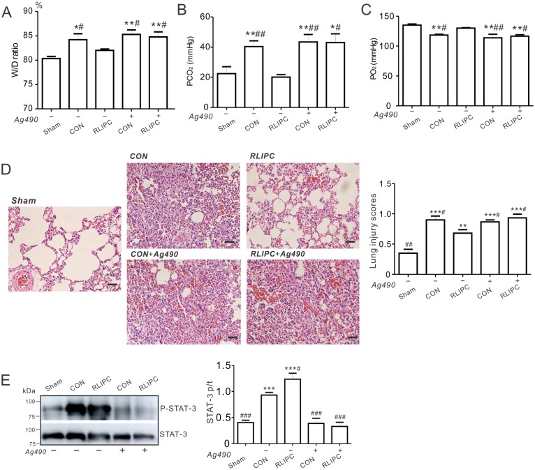
The lungs are highly susceptible to injury, including ischemia/reperfusion (I/R) injury. Pulmonary I/R injury can occur when correcting conditions such as primary pulmonary hypertension, and is also relatively common after lung transplantation or other cardiothoracic surgery. Methods to reduce pulmonary I/R injury are urgently needed to improve outcomes following procedures such as lung transplantation. Remote liver ischemic preconditioning (RLIPC) is an effective cardioprotective measure, reducing damage caused by subsequent cardiac I/R injury, but little is known about its potential role in pulmonary protection. Here, we analyzed the efficacy and mechanistic basis of RLIPC in a rat model of pulmonary I/R injury. RLIPC reduced lung I/R injury, lessening structural damage, inflammatory cytokine production and apoptosis. In addition, RLIPC preserved pulmonary function compared to controls following lung I/R injury. RLIPC stimulated phosphorylation of pulmonary STAT3, a component of the SAFE signaling pathway, but not phosphorylation of RISK pathway signaling proteins. Accordingly, STAT3 inhibition using AG490 eliminated the pulmonary protection afforded by RLIPC. Our data demonstrate for the first time that RLIPC protects against pulmonary I/R injury, via a signaling pathway requiring STAT3 phosphorylation.


Conflict of interest statement

Competing Interests: The authors have declared that no competing interests exist.

Figures

Fig 1
Fig 1. Experimental protocols.
All lungs were subjected to a 60-min ligation of the left pulmonary hilus (O), followed by 2 h of reperfusion (R), except for the sham-operated rats. For remote liver ischemic preconditioning (RLIPC), four cycles of 5 min of liver ischemia with 5min intermittent reperfusions were conducted before pulmonary ischemia. Thirty minutes of acute memory phase were allowed before being followed by a 60-min left pulmonary hilus ischemia. The STAT3 inhibitor Ag490 was applied 5 min before reperfusion. Arrows indicate the time points at which tissue samples were harvested.
Fig 2
Fig 2. RLIPC ameliorates pulmonary injury after experimentally imposed pulmonary IR.
A. Representative images of hematoxylin and eosin-stained left lung sections. Lung histopathology changes were examined under a light microscope; n = 4–5 rats per group. Panel (a) scale bars: 100 μm; panel (b) scale bars, 20 μm. B. Morphological evaluation of lung sections of sham-operated, CON and RLIPC rats. Sham animals did not undergo liver stimulus; CON, control, RLIPC, remote liver ischemic preconditioning; n = 4–5 rats per group. **P<0.01, ***P<0.001, compared with sham-operated lungs, #P<0.05 compared with CON (by One-way ANOVA). C. Water content (%) of left lung sections from sham-operated, CON and RLIPC rats post-pulmonary I/R injury; n = 6–7. All data were expressed as mean ± SEM. **P<0.05 compared with sham-operated lungs, #P<0.05 compared with CON (by One-way ANOVA). D. Changes in PCO2 values from sham-operated, CON and RLIPC rats; n = 5–7, *P<0.05, **P<0.01, compared with sham-operated lungs, #P<0.05,##P<0.01, compared with CON, P<0.05 compared with baseline. E. Changes in PO2 values from sham-operated, CON and RLIPC rats; n = 5–7, *P<0.05, ***P<0.001, compared with sham-operated lungs, #P<0.05,###P<0.001, compared with CON,††P<0.01, †††P<0.001 compared with baseline.
Fig 3
Fig 3. RLIPC decreases the production of inflammatory mediators after pulmonary I/R injury.
A. TNF-α content in BAL fluids at the end of 2 h of reperfusion from sham-operated, CON and RLIPC rats; n = 6 rats per group. Sham, sham-operated group; CON, control; RLIPC, remote liver ischemic preconditioning. **P<0.01, ***P<0.001 compared with sham-operated lungs, ###P<0.001, compared with CON (by One-way ANOVA). B. IL-6 content in BAL fluids at the end of 2h of reperfusion from sham-operated, CON and RLIPC rats; n = 6 rats per group. **P<0.01, ***P<0.001 compared with sham-operated lungs, ###P<0.001, compared with CON (by One-way ANOVA). C. Lung tissue harvested at the end of 2h reperfusion from sham-operated, CON and RLIPC rats were homogenized and assessed for myeloperoxidase (MPO) activity; n = 6 rats per group. **P<0.01, ***P<0.001 compared with sham-operated lungs, #P<0.05, ###P<0.001, compared with CON (by One-way ANOVA). D. Total leukocytes were quantitated in BAL fluids at the end of 2h of reperfusion from sham-operated, CON and RLIPC rats; n = 5 rats per group. **P<0.01, ***P<0.001, compared with sham-operated lungs, ###P<0.001 compared with CON (by One-way ANOVA).
Fig 4
Fig 4. RLIPC reduces pulmonary apoptosis post I/R.
A. Terminal transferase dUTP nick end labeling (TUNEL) technique was applied for the analysis of pulmonary apoptosis. Left, TUNEL-positive nuclei in the left lungs obtained from sham-operated (sham), liver conditioned (RLIPC) and control (CON) rats after pulmonary reperfusion injury were stained green (TUNEL) and nuclei were stained red (DAPI). Scale bars, 20μm. Right, Bar graph showing the TUNEL-positive nuclei expressed as a percentage of total nuclei in the left lung sections; n = 4–6. All data were expressed as mean ± SEM. ***P<0.001 compared with sham-operated lungs, ##P<0.01, ###P<0.001, compared with CON (by One-way ANOVA). B. Representative Western blots (left) and quantification (right) of cleaved caspase-3 protein band density (normalized to GAPDH) in sham, CON, and liver-conditioned rat left lungs are shown; n = 5. **P<0.05, compared with sham-operated rats; #P<0.05, ##P<0.01, compared with CON (by One-way ANOVA).
Fig 5
Fig 5. RLIPC affects pulmonary STAT3 pathway induction.
A. Western blots (left) and quantification (right) of ERK1/2 (p) and ERK1/2 (t) protein band density in sham, CON, and liver-conditioned rat left lungs are shown; n = 5–7. *P<0.05, compared with sham-operated rats; #P<0.05, compared with CON (by One-way ANOVA). B. Left, representative western blots of phospho- (p) AKT and total (t) AKT isolated from sham, CON, and liver-conditioned rat left lungs; one rat per lane. Right, mean ratio of pAKT/tAKT band density from blots as in left; n = 7. *P<0.05, compared with sham-operated rats; #P<0.05, compared with CON (by One-way ANOVA). C. Left, representative western blots of phospho- (p) GSK3β and total (t) GSK3β isolated from sham, CON, and liver-conditioned rat left lungs. Right, mean ratio of pGSK3β/tGSK3β band density from blots as in left; n = 6. *P<0.05, compared with sham-operated rats; #P<0.05, compared with CON (by One-way ANOVA). D. Left, representative western blots of phospho- (p) STAT3 and total (t) STAT3 isolated from sham, CON, and liver-preconditioned rat left lungs. Right, mean ratio of pSTAT3/tSTAT3 band density from blots as in left; n = 6. **P<0.01, ***P<0.001 compared with sham-operated rats; #P<0.05, ##P<0.01 compared with CON (by One-way ANOVA). E. Left, representative western blots of phospho- (p) STAT5 and total (t) STAT5 isolated from sham, CON, and liver-preconditioned rat left lungs. Right, mean ratio of pSTAT5/tSTAT5 band density from blots as in left; n = 5–6. **P<0.01, ***P<0.001 compared with sham-operated rats; #P<0.05, ##P<0.01 compared with CON (by One-way ANOVA).
Fig 6
Fig 6. Pharmacological inhibition of STAT3 abolishes the RLIPC-induced protection against pulmonary I/R injury.
A. Water content (%) of left lung sections from sham-operated, CON and RLIPC rats with (+) or without (-) Ag490 post-pulmonary I/R injury; n = 6–7. Values for sham-operated, CON and RLIPC rats are repeated from Fig 2C for comparison. Sham, sham-operated group; CON, control; RLIPC, remote liver ischemic preconditioning. W/D ratio was expressed as wet weight-dry weight) / wet weight x 100. All data were expressed as mean ± SEM. *P<0.05, **P<0.01, compared with sham-operated lungs, #P<0.05 compared with RLIPC lungs without Ag490 (by One-way ANOVA). B. Changes in PCO2 values from left lungs of rats with (+) or without (-) Ag490 after 180 min of pulmonary I/R injury; n = 6–7. Values for sham-operated, CON and RLIPC rats are repeated from Fig 1D for comparison. All data were expressed as mean ± SEM. *P<0.05, **P<0.01, compared with sham-operated lungs, #P<0.05, ##P<0.01, compared with RLIPC lungs without Ag490 (by One-way ANOVA). C. Changes in PO2 values from left lungs of rats with (+) or without (-) Ag490 after 180 min of pulmonary I/R injury; n = 6–7. Values for sham-operated, CON and RLIPC rats are repeated from Fig 1E for comparison. All data were expressed as mean ± SEM. **P<0.01, compared with sham-operated lungs, #P<0.05, ##P<0.01, compared with RLIPC lungs without Ag490 (by One-way ANOVA). D. Left, representative histological H&E-stained micrographs of lung sections from rat left lungs with or without Ag490 post-IR. Representative of n = 4–5 rats per group. Scale bars, 20 μm. Right, morphological evaluation of pulmonary damage after reperfusion in left lungs (n = 4–5 rats per group). The type and severity of pulmonary damage were graded as shown in the ‘Methods’ section. Values for CON and RLIPC rats are repeated from Fig 2B for comparison. All data were expressed as mean ± SEM. **P<0.01, ***P<0.001, compared with sham-operated lungs, #P<0.05 compared with RLIPC lungs without AG490 (by One-way ANOVA). E. Left, representative Western blots of phospho- (p) STAT3 and total (t) STAT3 isolated from left lungs of rats with (+) or without (-) Ag490. Right, mean ratio of pSTAT3/tSTAT3 band density from blots as in left; n = 5–6 each group. ***P<0.001 compared with sham-operated lungs; #P<0.05, ###P<0.001 compared with CON (by One-way ANOVA).

References

    1. Braunwald E, Kloner RA. Myocardial reperfusion: a double-edged sword? The Journal of clinical investigation. 1985;76(5):1713–9. doi: 10.1172/JCI112160 - DOI - PMC - PubMed
    1. Templeton AW, Garrotto LJ. Acquired extracardiac causes of pulmonary ischemia. Dis Chest. 1967;51(2):166–71. . - PubMed
    1. den Hengst WA, Gielis JF, Lin JY, Van Schil PE, De Windt LJ, Moens AL. Lung ischemia-reperfusion injury: a molecular and clinical view on a complex pathophysiological process. American journal of physiology Heart and circulatory physiology. 2010;299(5):H1283–99. doi: 10.1152/ajpheart.00251.2010 . - DOI - PubMed
    1. Weyker PD, Webb CA, Kiamanesh D, Flynn BC. Lung ischemia reperfusion injury: a bench-to-bedside review. Semin Cardiothorac Vasc Anesth. 2013;17(1):28–43. doi: 10.1177/1089253212458329 . - DOI - PubMed
    1. Garcia-de-la-Asuncion J, Garcia-del-Olmo E, Perez-Griera J, Marti F, Galan G, Morcillo A, et al. Oxidative lung injury correlates with one-lung ventilation time during pulmonary lobectomy: a study of exhaled breath condensate and blood. Eur J Cardiothorac Surg. 2015;48(3):e37–44. doi: 10.1093/ejcts/ezv207 . - DOI - PubMed

Publication types

Substances