

Finite element analysis of the tibial bone graft in cementless total knee arthroplasty
- PMID: 29769146
- PMCID: PMC5956944
- DOI: 10.1186/s13018-018-0830-1
Finite element analysis of the tibial bone graft in cementless total knee arthroplasty
Abstract
Background: Achieving stability of the tibial implant is essential following cementless total knee arthroplasty with bone grafting. We investigated the effects of bone grafting on the relative micromotion of the tibial implant and stress between the tibial implant and adjacent bone in the immediate postoperative period.
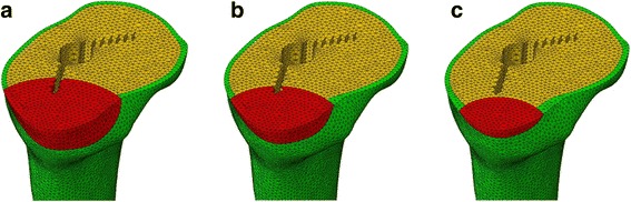
Methods: Tibial implant models were developed using a nonlinear, three-dimensional, finite element method. On the basis of a preprepared template, several bone graft models of varying sizes and material properties were prepared.
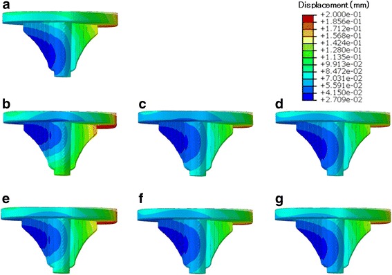
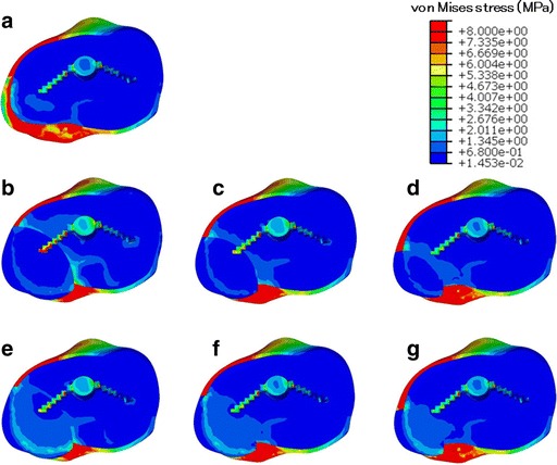
Results: Micromotion was larger in the bone graft models than in the intact model. Maximum micromotion and excessive stress in the area adjacent to the bone graft were observed for the soft and large graft models. With hard bone grafting, increased load transfer and decreased micromotion were observed.
Conclusions: Avoidance of large soft bone grafts and use of hard bone grafting effectively reduced micromotion and undue stress in the adjacent area.
Keywords: Bone graft; Finite element analysis; Total knee arthroplasty.
Conflict of interest statement
Ethics approval and consent to participate
This study was approved by the Ethics Committee of the University of Miyazaki.
Informed consent to participate in the study was obtained from the participant.
Competing interests
The authors declare that they have no competing interests.
Publisher’s Note
Springer Nature remains neutral with regard to jurisdictional claims in published maps and institutional affiliations.
Figures


Similar articles
-
Finite-element analysis of the proximal tibial sclerotic bone and different alignment in total knee arthroplasty.BMC Musculoskelet Disord. 2019 Dec 26;20(1):617. doi: 10.1186/s12891-019-3008-z. BMC Musculoskelet Disord. 2019. PMID: 31878972 Free PMC article.
-
An Anterior Spike Decreases Bone-Implant Micromotion in Cementless Tibial Baseplates for Total Knee Arthroplasty: A Biomechanical Study.J Arthroplasty. 2024 May;39(5):1323-1327. doi: 10.1016/j.arth.2023.11.020. Epub 2023 Nov 22. J Arthroplasty. 2024. PMID: 38000515
-
Computationally efficient prediction of bone-implant interface micromotion of a cementless tibial tray during gait.J Biomech. 2014 May 7;47(7):1718-26. doi: 10.1016/j.jbiomech.2014.02.018. Epub 2014 Feb 19. J Biomech. 2014. PMID: 24642351
-
Analysis of bone-prosthesis interface micromotion for cementless tibial prosthesis fixation and the influence of loading conditions.J Biomech. 2010 Apr 19;43(6):1074-80. doi: 10.1016/j.jbiomech.2009.12.006. Epub 2010 Mar 1. J Biomech. 2010. PMID: 20189576
-
Cementless fixation issues in revision total knee arthroplasty.Instr Course Lect. 1999;48:177-82. Instr Course Lect. 1999. PMID: 10098043 Review.
Cited by
-
[Effects of elastic modulus of the metal block on the condylar-constrained knee prosthesis tibial fixation stability].Sheng Wu Yi Xue Gong Cheng Xue Za Zhi. 2025 Aug 25;42(4):782-789. doi: 10.7507/1001-5515.202410039. Sheng Wu Yi Xue Gong Cheng Xue Za Zhi. 2025. PMID: 40887194 Free PMC article. Chinese.
-
Model Properties and Clinical Application in the Finite Element Analysis of Knee Joint: A Review.Orthop Surg. 2024 Feb;16(2):289-302. doi: 10.1111/os.13980. Epub 2024 Jan 4. Orthop Surg. 2024. PMID: 38174410 Free PMC article. Review.
-
Finite-element analysis of the proximal tibial sclerotic bone and different alignment in total knee arthroplasty.BMC Musculoskelet Disord. 2019 Dec 26;20(1):617. doi: 10.1186/s12891-019-3008-z. BMC Musculoskelet Disord. 2019. PMID: 31878972 Free PMC article.
References
-
- Dorr LD, Ranawat CS, Sculco TA, McKaskill B, Orisek BS. Bone graft for tibial defects in total knee arthroplasty. Clin Orthop Relat Res. 1986;205:153–65. - PubMed
-
- Naim S, Toms AD. Impaction bone grafting for tibial defects in knee replacement surgery results at two years. Acta Orthop Belg. 2013;79:205–210. - PubMed
MeSH terms
Grants and funding
LinkOut - more resources
Full Text Sources
Other Literature Sources
Medical
