. 2018 Jun 6;38(23):5429-5440.
doi: 10.1523/JNEUROSCI.2484-17.2018. Epub 2018 May 16.

Reciprocal Negative Regulation Between Lmx1a and Lmo4 Is Required for Inner Ear Formation

Affiliations

Reciprocal Negative Regulation Between Lmx1a and Lmo4 Is Required for Inner Ear Formation

Yanhan Huang et al. J Neurosci. .

Abstract

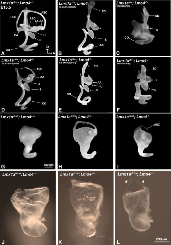
LIM-domain containing transcription factors (LIM-TFs) are conserved factors important for embryogenesis. The specificity of these factors in transcriptional regulation is conferred by the complexes that they form with other proteins such as LIM-domain-binding (Ldb) proteins and LIM-domain only (LMO) proteins. Unlike LIM-TFs, these proteins do not bind DNA directly. LMO proteins are negative regulators of LIM-TFs and function by competing with LIM-TFs for binding to Ldb's. Although the LIM-TF Lmx1a is expressed in the developing mouse hindbrain, which provides many of the extrinsic signals for inner ear formation, conditional knock-out embryos of both sexes show that the inner ear source of Lmx1a is the major contributor of ear patterning. In addition, we have found that the reciprocal interaction between Lmx1a and Lmo4 (a LMO protein within the inner ear) mediates the formation of both vestibular and auditory structures. Lmo4 negatively regulates Lmx1a to form the three sensory cristae, the anterior semicircular canal, and the shape of the utricle in the vestibule. Furthermore, this negative regulation blocks ectopic sensory formation in the cochlea. In contrast, Lmx1a negatively regulates Lmo4 in mediating epithelial resorption of the canal pouch, which gives rise to the anterior and posterior semicircular canals. We also found that Lmx1a is independently required for the formation of the endolymphatic duct and hair cells in the basal cochlear region.SIGNIFICANCE STATEMENT The mammalian inner ear is a structurally complex organ responsible for detecting sound and maintaining balance. Failure to form the intricate 3D structure of this organ properly during development most likely will result in sensory deficits on some level. Here, we provide genetic evidence that a transcription factor, Lmx1a, interacts with its negative regulator, Lmo4, to pattern various vestibular and auditory components of the mammalian inner ear. Identifying these key molecules that mediate formation of this important sensory organ will be helpful for designing strategies and therapeutics to alleviate hearing loss and balance disorders.

Keywords: cochlea; cristae; development; genetics; inner ear; semicircular canals.


Figures

Figure 1.
Figure 1.
Lmx1a conditional mouse knock-out scheme. A, Genomic organization of mouse Lmx1a gene and Lmx1alox alleles are shown. LoxP sites were positioned to flank exon 2 of the target gene using the ET recombination technique. Homologous recombination events in R1 ES cells were selected by genomic Southern analysis. B, Using 5′ and 3′ DNA probes against genomic DNA from wild-type (WT) and various ES clones (1–5) digested with NdeI and BglI, respectively.
Figure 2.
Figure 2.
Dysmorphic inner ears have normal Lmx1a immunoreactivities in the hindbrain of Foxg1cre/+;Lmx1adr/lox and Sox9cre/+;Lmx1adr/lox CKO mutants. A, B, Paint-filled inner ears of control Foxg1+/+;Lmx1adr/lox (A, n = 4) and Foxg1cre/+;Lmx1adr/lox CKO (B, n = 8) embryos at E15.5. The dysmorphic inner ear in the conditional mutant is indistinguishable from that of the dreher. CF, Lmx1a immunostaining (C, D) and Atoh1 expression (E, F) in the hindbrain of Foxg1+/+;Lmx1a+/lox control (C, E) and Foxg1cre/+;Lmx1adr/lox CKO (D, F) embryos at E10.5. C, D, Lmx1a immunostaining is markedly reduced in the otic epithelium (arrowheads) but maintained in the hindbrain (arrows) of Foxg1cre/+;Lmx1adr/lox CKO. No downregulation of Atoh1 expression is observed in the rhombic lip of Foxg1cre/+;Lmx1adr/lox CKO hindbrains (F, n = 3) compared with Foxg1cre/+;Lmx1a+/lox controls. GI, Inner ears and hindbrain phenotypes of Sox9cre/+;Lmx1adr/lox CKO mutants. G, H, Anti-Lmx1a staining in the Sox9cre/+;Lmx1adr/lox CKO mutants (H) is comparable to the Sox9cre/+;Lmx1a+/lox controls (G) in the dorsal hindbrain (arrows), but is much reduced in the inner ear (arrowheads). I, J, Msx1 expression in the rhombic lip of a control (I) and Sox9cre/+;Lmx1adr/lox CKO mutants (J). The diamond-shaped roof plate is slightly smaller in the CKO mutants. AA, Anterior ampulla; ASC, anterior semicircular canal; CO, cochlear duct; ED, endolymphatic duct; LA, lateral ampulla; LSC, lateral semicircular canal, PA, posterior ampulla; PSC, posterior semicircular canal; S, saccule; U, utricle. Orientations: A, anterior; D, dorsal; L, lateral. Anterior is toward the top of the panel for E, F, I, and J.
Figure 3.
Figure 3.
Sensory defects in Foxg1cre/+;Lmx1adr/lox CKO mutants. AE, Hair cells labeled with anti-myosin VIIa antibodies are in green, actin labeled with rhodamine-phalloidin are in red, and pillar cells labeled with anti-P75Ngfr are in blue. GI, Developing calyxes labeled with anti-neurofilament antibodies (2H3) are in green. A, B, Whole-mount (A) and dissected (B) inner ears of Foxg1+/+;Lmx1adr/lox controls. Vestibular and auditory sensory organs are compartmentalized in chambers (A; n = 9). CE, Whole mount of dreher (C; n = 2) and whole (D; n = 5) and flat (E; n = 5) mounts of Foxg1cre/+;Lmx1adr/lox CKO ears showing individual sensory organs are distinguishable but often fused with each other. The double arrowheads indicate the saccular macula and the organ of Corti are continuous with each other. In addition to the lack of a distinct lateral crista, the posterior crista is malformed (D, E). Sensory organs are identified based on their locations in the inner ear. D and E are composites of images taken at a higher magnification. F, Percentages of the length of P75Ngfr-positive cochlear region of Foxg1cre/+;Lmx1adr/lox CKO (n = 5) relative to that of Foxg1+/+;Lmx1adr/lox and Foxg1cre/+;Lmx1alox/+ controls (n = 8, p < 0.005 (p = 0.0012, Student's t test)). G, Higher magnification of the square region in E. Continuous P75Ngfr expression only starts at the midbase (G′′) and not at the base (G′) of the cochlea. At the base of the CKO cochlea, 2H3-positive staining surrounding the hair cells is observed (G′; n = 3), which resembles developing calyxes in the maculae of controls (H; n = 4). Multiple rows of hair cells are present in the midbase of mutants (G′′) compared with the one row of inner hair cells (IHC) and three rows of outer hair cells (OHC) in Foxg1+/+;Lmx1adr/lox controls taken from a comparable basal cochlear region (I). AC, Anterior crista; OC, organ of Corti; LC, lateral crista; PC, posterior crista; SM, saccular macula; UM, utricular macula. For orientations, please refer to Figure 2.
Figure 4.
Figure 4.
Paint-filled and dissected inner ears of Lmx1a and Lmo4 compound mutants. A, Lmx1adr/+;Lmo4+/− inner ears are normal (n = 4). B, Lmx1a+/+;Lmo4−/− inner ear in an embryo without exencephaly lacking semicircular canals, ampullae, and a well defined utricle (asterisk, n = 4/8). C, Lmx1a+/+;Lmo4−/− inner ear in an embryo with exencephaly showing the presence of a saccule, a distorted cochlear duct and the absence of canals (C, n = 8/9). DF, Most Lmx1adr/+;Lmo4−/− inner ears in embryos without exencephaly consist of an anterior ampulla (AA), an anterior canal (asc), and a well demarcated utricle (D, n = 8/11), but some only show an ampulla without a canal (E, n = 2/11). In contrast, a well defined utricle (asterisk), ampulla, and canal are not recovered in Lmx1adr/+;Lmo4−/− ears in embryos with exencephaly (F, n = 5/8). The other three specimens have no utricle. Some Lmx1adr/dr;Lmo4+/− (H, n = 7/11) and Lmx1adr/dr;Lmo4−/− (I, n = 1/2, L, n = 1) show the presence of a canal compared with the dreher mutants (G, n = 4). Other Lmx1adr/dr;Lmo4+/− (J, n = 2) and Lmx1adr/dr;Lmo4−/− (K, n = 1) dissected ears resemble the dreher. JL, Dissected membranous labyrinths of Lmx1adr/dr;Lmo4+/− (J, n = 2) and Lmx1adr/dr;Lmo4−/− specimens (K, L). Asterisks in L show the presence of a canal. ASC, Anterior semicircular canal; LSC, lateral semicircular canal; PSC, posterior semicircular canal. For other abbreviations, please refer to Figure 2.
Figure 5.
Figure 5.
Crista and ectopic organ of Corti formation are inversely related to the presence of Lmx1a. AD, Anti-myosin VIIa staining is shown in green, actin staining with rhodamine-phalloidin in red, and cristae are marked with red asterisks. Insets show the anterior crista of control and the anteriorly located crista in mutants. A, Lmx1adr/+;Lmo4+/− inner ears are normal showing six myosin VIIa-positive sensory organs (n = 4). Cruciatum divides the anterior and posterior crista into two equal halves (arrowhead in inset). B, Lmx1a+/+;Lmo4−/− inner ears lack all three sensory cristae (n = 2/3), whereas a crista-like sensory organ is present in Lmx1adr/+;Lmo4−/− ears (C, n = 4/4). D, Anterior and posterior located cristae are present in Lmx1adr/dr;Lmo4−/− ears (asterisk, n = 4/4). It is not clear whether the two patches of crista tissues in the anterior region represent a small anterior and lateral crista or two halves of the anterior crista. EJ, Cryosections of the crista probed for Sox2 (EG) and cochlea probed for Myosin15 (HJ) transcripts in Lmx1adr/+;Lmo4+/− (E, H), Lmx1a+/+;Lmo4−/− (F, I), and Lmx1adr/+;Lmo4−/− (G, J) sections. M, Medial. For other abbreviations, please refer to Figures 2 and 3.
Figure 6.
Figure 6.
Overlapping expression domains of Lmx1a and Lmo4 in the developing crista and cochlea. AA″ and BB″, are adjacent sections of the inner ear at E11.5 taken at the level of vertical canal pouch (AA″) and presumptive anterior crista (BB″, AC). In the Bmp4-negative, vertical canal pouch (A), Lmo4 is expressed strongly in the rim (A′, arrow) but weak in the center (A′, arrowheads) of the canal pouch. In contrast, Lmx1a is strongly expressed in the center (A″, arrowheads) but weaker at the rim (arrow) of the canal pouch. In the Bmp4-positive anterior crista (B, double arrows), Lmo4 (B′) expression is strong and Lmx1a (B″) expression is weaker. CC″, Expression pattern of Lmo4 and Lmx1a overlap in the lateral cochlear duct at E16.5 (C′, C″, arrows). Lunatic Fringe (Lfng) labels the organ of Corti (C, black bar). For orientations, please refer to Figure 2.
Figure 7.
Figure 7.
Ectopic Lmx1b expression in the presumptive anterior crista of the chicken inner ear downregulates crista markers Bmp4, Jag1, and Sox2. Sections of chicken inner ear electroporated with pMES-GFP (AD) or pMES-Lmx1b-GFP (EH) plasmids at E3.5 and were harvested 24 h later and processed for anti-Jag1 (B, F) and anti-Sox2 (C, G) immunostaining and Bmp4 gene expression (D, H). The levels and domains of Jag1 and Sox2 immunoreactivities and Bmp4 hybridization signals in the prospective anterior crista (marked by arrowheads) are reduced in specimens electroporated with pMES-Lmx1b-GFP (EH) compared with pMES-GFP controls (AD).
Figure 8.
Figure 8.
Summary diagram of the requirements of Lmx1a and Lmo4 in inner ear formation. The vertical canal pouch (right) gives rise to the anterior and posterior canals. The epithelial cells in the center region of each prospective canal (tan color) fuse and resorb resulting in the rim of the canal pouch (blue color) forming the anterior and posterior canals. Lmo4 negatively regulates Lmx1a to form the three cristae (red stripes), anterior canal, and proper shape of the utricle (yellow color) and to inhibit ectopic sensory tissue formation in the cochlear duct. In contrast, Lmx1a negatively regulates Lmo4 to mediate resorption in the vertical canal pouch. The endolymphatic duct formation requires Lmx1a independent of Lmo4, whereas the hair cells in the basal cochlear region may require Lmx1a independently or its negative regulation of Lmo4. Organ of Corti is shown in gray and the saccule in green. For orientations, please refer to Figure 2.

Similar articles

Cited by

References

    1. Abelló G, Khatri S, Radosevic M, Scotting PJ, Giráldez F, Alsina B (2010) Independent regulation of Sox3 and Lmx1b by FGF and BMP signaling influences the neurogenic and non-neurogenic domains in the chick otic placode. Dev Biol 339:166–178. 10.1016/j.ydbio.2009.12.027 - DOI - PubMed
    1. Abraira VE, Del Rio T, Tucker AF, Slonimsky J, Keirnes HL, Goodrich LV (2008) Cross-repressive interactions between Lrig3 and netrin 1 shape the architecture of the inner ear. Development 135:4091–4099. 10.1242/dev.029330 - DOI - PMC - PubMed
    1. Akiyama H, Kim JE, Nakashima K, Balmes G, Iwai N, Deng JM, Zhang Z, Martin JF, Behringer RR, Nakamura T, de Crombrugghe B (2005) Osteo-chondroprogenitor cells are derived from Sox9 expressing precursors. Proc Natl Acad Sci U S A 102:14665–14670. 10.1073/pnas.0504750102 - DOI - PMC - PubMed
    1. Bach I. (2000) The LIM domain: regulation by association. Mech Dev 91:5–17. 10.1016/S0925-4773(99)00314-7 - DOI - PubMed
    1. Chang W, Lin Z, Kulessa H, Hébert J, Hogan BL, Wu DK (2008) Bmp4 is essential for the formation of the vestibular apparatus that detects angular head movements. PLoS Genet 4: e1000050. 10.1371/journal.pgen.1000050 - DOI - PMC - PubMed

Publication types

MeSH terms

LinkOut - more resources