. 2018 Jul 6;293(27):10692-10706.
doi: 10.1074/jbc.RA117.000999. Epub 2018 May 16.

The chloroplast division protein ARC6 acts to inhibit disassembly of GDP-bound FtsZ2

Affiliations

The chloroplast division protein ARC6 acts to inhibit disassembly of GDP-bound FtsZ2

Min Woo Sung et al. J Biol Chem. .

Abstract

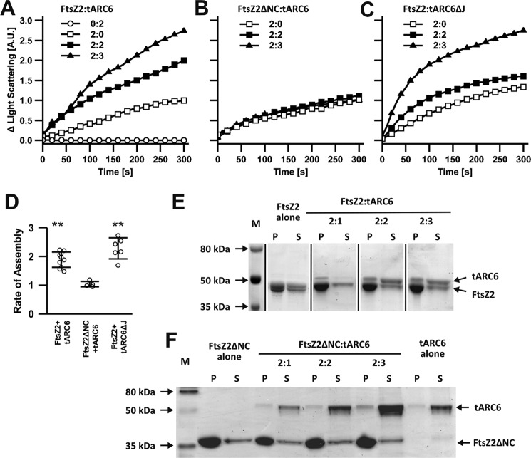
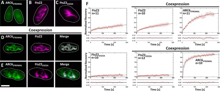
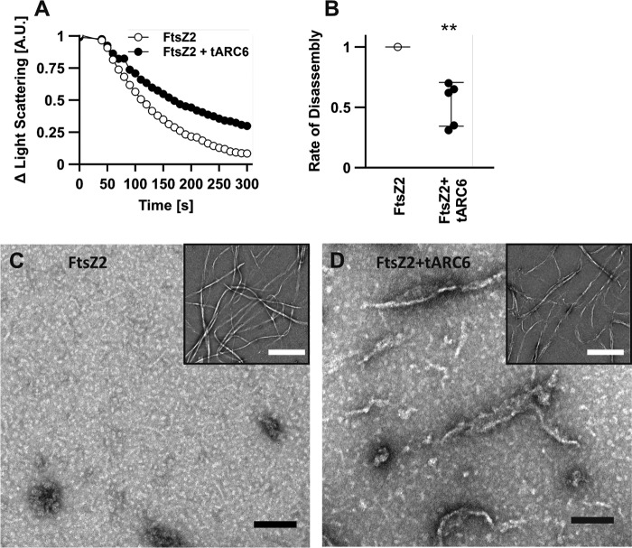
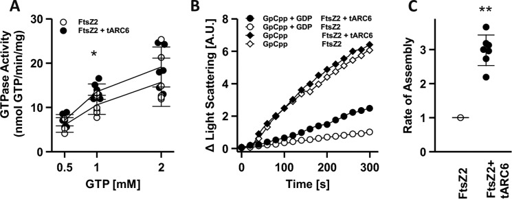
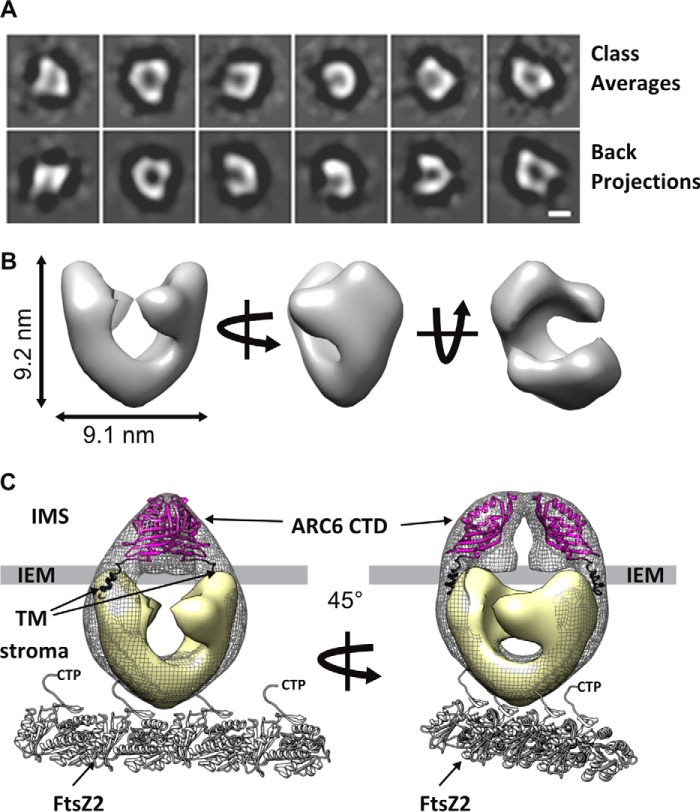
Chloroplasts host photosynthesis and fulfill other metabolic functions that are essential to plant life. They have to divide by binary fission to maintain their numbers throughout cycles of cell division. Chloroplast division is achieved by a complex ring-shaped division machinery located on both the inner (stromal) and the outer (cytosolic) side of the chloroplast envelope. The inner division ring (termed the Z ring) is formed by the assembly of tubulin-like FtsZ1 and FtsZ2 proteins. ARC6 is a key chloroplast division protein that interacts with the Z ring. ARC6 spans the inner envelope membrane, is known to stabilize or maintain the Z ring, and anchors the Z ring to the inner membrane through interaction with FtsZ2. The underlying mechanism of Z ring stabilization is not well-understood. Here, biochemical and structural characterization of ARC6 was conducted using light scattering, sedimentation, and light and transmission EM. The recombinant protein was purified as a dimer. The results indicated that a truncated form of ARC6 (tARC6), representing the stromal portion of ARC6, affects FtsZ2 assembly without forming higher-order structures and exerts its effect via FtsZ2 dynamics. tARC6 prevented GDP-induced FtsZ2 disassembly and caused a significant net increase in FtsZ2 assembly when GDP was present. Single particle analysis and 3D reconstruction were performed to elucidate the structural basis of ARC6 activity. Together, the data reveal that a dimeric form of tARC6 binds to FtsZ2 filaments and does not increase FtsZ polymerization rates but rather inhibits GDP-associated FtsZ2 disassembly.

Keywords: Arabidopsis; GTPase; chloroplast; chloroplast division; cytoskeleton; single particle analysis; structural model.


Conflict of interest statement

The authors declare that they have no conflicts of interest with the contents of this article.

Figures

Figure 1.
Figure 1.
Arc6 and FtsZ2 constructs, expression phenotypes, and protein purification. A, ARC6 structural elements. TP, transit peptide; J-like, J-like domain; TM, transmembrane domain; PDV2, PDV2-binding domain (aa 679–774). Constructs encode the full-length mature form of ARC6(89–801), stromal portion tARC6(89–519), tARC6ΔJ(157–519) lacking the J-like domain, tARC6ΔC(89–310), tARC6ΔN(310–519), GFP-tagged tARC6, and mVenus-tagged stromal portion of ARC6, ARC6STROMAL. B, FtsZ2 structural elements. CTP, conserved C-terminal peptide of FtsZ2(463–478). Constructs encode full-length mature form of FtsZ2(49–476) and FtsZ2ΔNC(118–424) lacking both the C- and N-terminal regions, and the eCFP-tagged version of FtsZ2 and of the GTPase-deficient mutant version of FtsZ2, FtsZ2D322A. C, phase contrast images of E. coli carrying the expression vectors before (left panels) and after (right panels) overnight growth at 18 °C in the presence of 0.5 mm IPTG. Scale bar, 20 μm. D, gel-permeation chromatography purification of tARC6, FtsZ2, and FtsZ2ΔNC on SEC 650 column (top row) and of ARC6 and tARC6ΔJ purification on Superdex-6 column (bottom row), showing a single peak with relative molecular mass MR corresponding to the dimeric form of the protein. The rightmost panels show elution volume calibration on each column. Standards and their MR are as follows: 1, thyroglobulin (670 kDa); 2, bovine γ-globulin (158 kDa); 3, chicken ovalbumin (44 kDa), and 4, equine myoglobin (17 kDa).
Figure 2.
Figure 2.
tARC6 facilitates FtsZ2 assembly through interaction. A and B, light-scattering assay of FtsZ2 (A) or FtsZ2ΔNC (B) in the absence/presence of tARC6. C, light-scattering assay of FtsZ2 assembly in the absence/presence of tARC6ΔJ. Results from representative experiments are shown. Number of replicates is given below. D, initial rate of assembly of FtsZ2 or FtsZ2ΔNC in the presence of equimolar amounts of tARC6 or tARC6ΔJ, normalized to control reactions without tARC6 or tARC6ΔJ. Error bars represent standard deviation; n = 10 (FtsZ2 + tARC6), n = 5 (FtsZ2ΔNC + tARC6), and n = 6 (FtsZ2+ tARC6ΔJ). ** indicates significant difference from the control reactions without the respective ARC6 protein construct (p < 0.01, Student's t test). E and F, sedimentation assay FtsZ2 (E) or FtsZ2ΔNC (F) co-pelleting with tARC6 after assembly at FtsZ/tARC6 molar ratios as indicated. The spliced images in E are all from the same gel. M, molecular mass markers; P, pellet; S, supernatant.
Figure 3.
Figure 3.
Effect of ARC6 on FtsZ2 filament morphology. A and B, FtsZ2 filament bundles in the absence (A) and presence (B) of equimolar amounts of tARC6. Insets show the same samples at higher magnification. Scale bar, 1 μm and 100 nm in the insets, respectively. C, filaments and higher order structures formed by bacterial FtsZ in the presence of SepF or FzlA. Micrographs adapted from Refs. , . Scale bars, 100 nm. D, width; E, length of FtsZ2 filament bundles in the absence or presence of tARC6.
Figure 4.
Figure 4.
Filament assembly and subunit exchange in S. pombe. A–E, epifluorescence micrographs of ARC6STROMAL–mVenus (A), FtsZ2-eCFP (B), FtsZ2D322A–eCFP (C), ARC6STROMAL–mVenus coexpressed with FtsZ2-eCFP (D), and ARC6STROMAL–mVenus coexpressed with FtsZ2D322A–eCFP (E). Fluorescent signals from ARC6STROMAL–mVenus are pseudo-colored green, and those of FtsZ2–eCFP and FtsZ2D322A–eCFP are pseudo-colored magenta. The white color in merged images represents regions where the two fluorescence signals overlap and colocalize. White lines denote cell boundaries. Scale bar, 5 μm. F, FRAP analysis of FtsZ2–eCFP and FtsZ2D322A–eCFP expressed alone and with ARC6STROMAL–mVenus. Graphs of the normalized fluorescence recovery versus time (seconds) are shown on top. The residuals of the fit shown below each recovery plot are well within 0.10 normalized recovery units (10%). Data are normalized to the pre-bleach fluorescence intensity (1 on the y axis) and the fluorescence intensity at the time of photobleaching (0 on the y axis). n represents the number of independent FRAP experiments performed. Error bars, S.E. at each time point.
Figure 5.
Figure 5.
Stabilization of FtsZ2 filaments by ARC6 under disassembly condition. A, light-scattering assay of FtsZ2 disassembly in the absence/presence of tARC6. After 5 min of assembly in the presence of GTP, disassembly was initiated by the addition of excess GDP. B, rate of disassembly in FtsZ2 + tARC6 reactions relative to FtsZ2-alone reactions, from five pairwise comparisons. Error bar, S.D. ** indicates significant difference from the FtsZ2-alone control reactions (p < 0.01, Student's t test). C and D, electron micrograph of the FtsZ2-only (C) and FtsZ2 + tARC6 (D) reactions after disassembly. Insets show lower magnification view of the assembled FtsZ filament bundles before disassembly. Scale bar, 100 nm and 1 μm in the insets, respectively.
Figure 6.
Figure 6.
GTPase activity of FtsZ2 and effect of ARC6 and nucleotide on FtsZ2 assembly. A, GTPase activity of FtsZ2 at increasing concentrations of GTP in the absence/presence of equimolar amounts (2 μm) of tARC6. Assay for each GTP concentration was repeated four to seven times. * indicates significant difference from the FtsZ2-alone control reactions (p < 0.05, Student's t test). B, light-scattering assay of FtsZ2 polymerization in the absence/presence of equimolar amount (2 μm) of tARC6 in reactions containing 0.1 mm GpCpp or 0.1 mm GpCpp + 0.2 mm GDP. Addition of GDP serves to induce slow disassembly of FtsZ. Representative plot is shown, and the assay was repeated six times with similar results. C, comparison of the rates of FtsZ2 assembly in reactions containing GpCpp + GDP; rates in FtsZ2 + tARC6 reactions are expressed relative to reactions with FtsZ2 alone. n = 7; ** indicates significant difference from the FtsZ2-alone control reactions (p < 0.01, Student's t test). Error bars, S.D.
Figure 7.
Figure 7.
Single particle analysis and 3D reconstruction of tARC6 and ARC6, model of ARC6 membrane topology, and FtsZ2 protofilament binding. A, gallery of representative reference-free class averages of tARC6 (top row) and corresponding back projections from the final 3D reconstructions under similar orientations (bottom row). B, 3D reconstruction of a tARC6 dimer. C, 3D reconstruction of tARC6 dimer (pale yellow) superimposed on the full-length mature form, ARC6, dimer density map (black mesh). Crystal structures of ARC6 CTD (magenta) (36) are fitted into the top part of ARC6 dimer density map. Two single helices corresponding to the transmembrane domain (TM) of ARC6 (black) are located between the CTDs of ARC6 and the tARC6 dimer reconstruction. Each molecule of the stroma-localized portion of ARC6 dimer is depicted interacting with the flexible C terminus of adjacent FtsZ2 molecules in a protofilament. The C terminus of FtsZ2 contains the ARC6-binding CTP. Scale bar, 5 nm in A.

Similar articles

Cited by

References

    1. Martin W., and Kowallik K. V. (1999) Annotated English translation of Mereschkowsky's 1905 paper “Über Natur und Ursprung der Chromatophoren im Pflanzenreiche”. Eur. J. Phycol. 34, 287–296
    1. Okazaki K., Kabeya Y., and Miyagishima S. Y. (2010) The evolution of the regulatory mechanism of chloroplast division. Plant Signal. Behav. 5, 164–167 10.4161/psb.5.2.10461 - DOI - PMC - PubMed
    1. Osteryoung K. W., and McAndrew R. S. (2001) The plastid division machine. Annu. Rev. Plant Physiol. Plant Mol. Biol. 52, 315–333 10.1146/annurev.arplant.52.1.315 - DOI - PubMed
    1. Osteryoung K. W., Stokes K. D., Rutherford S. M., Percival A. L., and Lee W. Y. (1998) Chloroplast division in higher plants requires members of two functionally divergent gene families with homology to bacterial ftsZ. Plant Cell 10, 1991–2004 10.1105/tpc.10.12.1991 - DOI - PMC - PubMed
    1. Osteryoung K. W., and Vierling E. (1995) Conserved cell and organelle division. Nature 376, 473–474 10.1038/376473b0 - DOI - PubMed

Publication types

MeSH terms