Case Reports
. 2018 Jul 6;293(27):10810-10824.
doi: 10.1074/jbc.RA118.002583. Epub 2018 May 16.

O-GlcNAc transferase missense mutations linked to X-linked intellectual disability deregulate genes involved in cell fate determination and signaling

Affiliations
Case Reports

O-GlcNAc transferase missense mutations linked to X-linked intellectual disability deregulate genes involved in cell fate determination and signaling

Nithya Selvan et al. J Biol Chem. .

Abstract

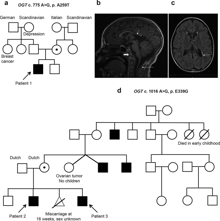
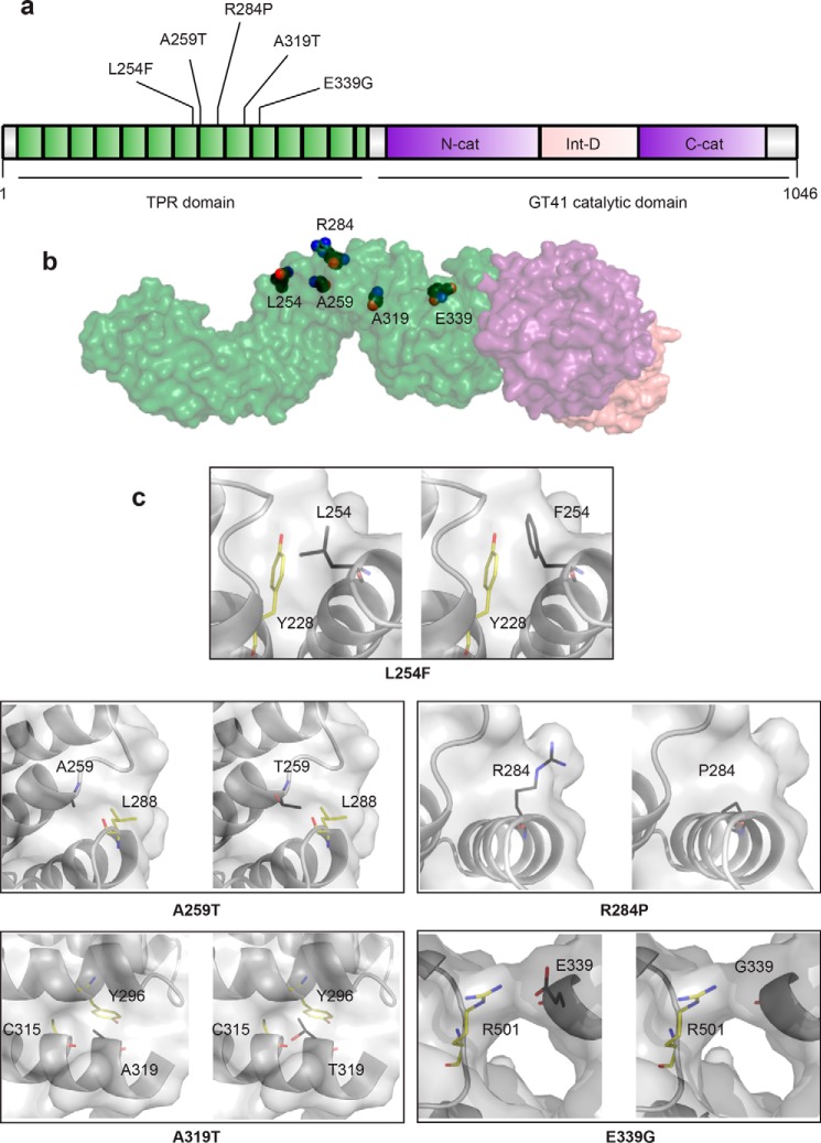
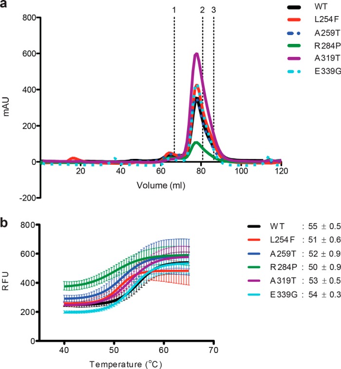
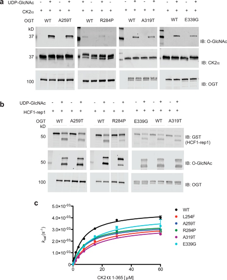
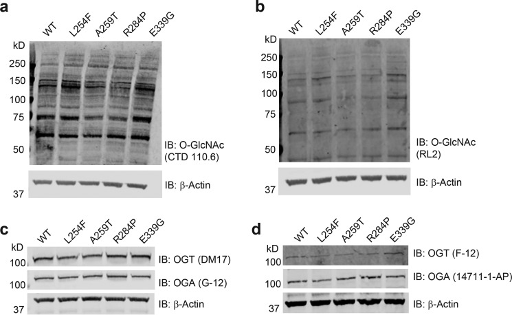
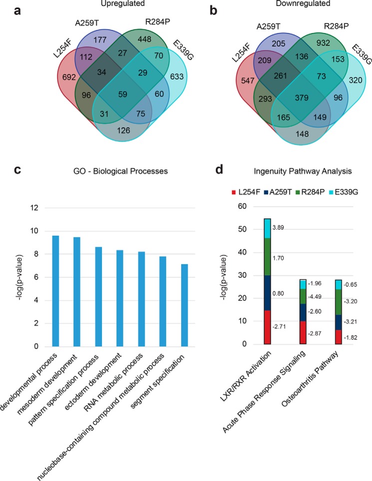
It is estimated that ∼1% of the world's population has intellectual disability, with males affected more often than females. OGT is an X-linked gene encoding for the enzyme O-GlcNAc transferase (OGT), which carries out the reversible addition of N-acetylglucosamine (GlcNAc) to Ser/Thr residues of its intracellular substrates. Three missense mutations in the tetratricopeptide (TPR) repeats of OGT have recently been reported to cause X-linked intellectual disability (XLID). Here, we report the discovery of two additional novel missense mutations (c.775 G>A, p.A259T, and c.1016 A>G, p.E339G) in the TPR domain of OGT that segregate with XLID in affected families. Characterization of all five of these XLID missense variants of OGT demonstrates modest declines in thermodynamic stability and/or activities of the variants. We engineered each of the mutations into a male human embryonic stem cell line using CRISPR/Cas9. Investigation of the global O-GlcNAc profile as well as OGT and O-GlcNAc hydrolase levels by Western blotting showed no gross changes in steady-state levels in the engineered lines. However, analyses of the differential transcriptomes of the OGT variant-expressing stem cells revealed shared deregulation of genes involved in cell fate determination and liver X receptor/retinoid X receptor signaling, which has been implicated in neuronal development. Thus, here we reveal two additional mutations encoding residues in the TPR regions of OGT that appear causal for XLID and provide evidence that the relatively stable and active TPR variants may share a common, unelucidated mechanism of altering gene expression profiles in human embryonic stem cells.

Keywords: RNA-seq; XLID; enzyme kinetics; genetic disease; glycosyltransferase; O-linked N-acetylglucosamine (O-GlcNAc) transferase (OGT); O-linked N-acetylglucosamine (O-GlcNAc); intellectual disability; post-translational modification; transcriptomics.


Conflict of interest statement

The authors declare that they have no conflicts of interest with the contents of this article

Figures

Figure 1.
Figure 1.
Pedigrees and phenotypes of the patients. a, three-generation pedigree of Patient 1 (with OGT c.775 G>A, p.A259T). Proband is identified by the arrow. Circles denote females; circles with dots, carrier females confirmed using exome sequencing or Sanger sequencing; open squares, unaffected males; and closed squares, males with XLID. b, MRI of brain of Patient 1 at 7 years of age. Sagittal T1 image reveals extremely thin corpus callosum, particularly the posterior body and splenium (short thin arrow), short appearing clivus (thick arrow), and relatively hypoplastic posterior arch of first cervical vertebra (long thin arrow). c, MRI of brain at of Patient 1 at 7 years of age. Axial T2 FLAIR image reveals mild ventriculomegaly (arrow). d, four-generation pedigree of Patients 2 and 3 (with OGT c.1016 A>G, p.E339G). Probands are identified by arrows, and diagram has same coding as in a.
Figure 2.
Figure 2.
All known missense mutations in the TPR domain that are causal for XLID. a, schematic of the domain organization of OGT. The N-terminal TPR domain is depicted in green with XLID variants noted, the intervening domain in pink, and the lobes of the catalytic domain in purple. b, model of full-length OGT based on the structure of the first 11.5 TPRs (PDB code 1W3B (14)) and the structure of the catalytic domain with the last 2.5 TPRs (PDB code 4AY5 (52)). The domains in the model are colored as in the schematic, and the side chains of the residues in the TPR that are mutated in XLID are depicted as spheres with black carbons. c, close-up of residues that are mutated in XLID. Panels on left show the WT residue in the amino acid position indicated, and panels on right show the replacing residue modeled into the structure (side chains are shown as sticks with black carbons). Surface representation shown in both panels is from OGT-WT. Side chains of neighboring residues that could clash are shown as sticks with yellow carbons.
Figure 3.
Figure 3.
OGT XLID–TPR mutants form functional dimers but demonstrate decreased thermal stability. a, TPR domain is a constitutive homodimer in solution. Samples of WT and mutant TPR domains were analyzed on a Superdex 200 size-exclusion column. The elution profiles are plotted as A280 against elution volume. Elution time of standards of known molecular weight are marked above; 1, IgG 158 kDa; 2, human albumin 66 kDa; and 3, ovalbumin 44 kDa. The His6-tagged TPR dimer has an expected molecular mass of ∼90 kDa and an apparent molecular mass of ∼120 kDa (14) due to its elongated, nonglobular structure. b, thermal denaturing curves of WT or mutant OGT TPR domains. The melting temperatures (Tm, the temperature at which both the folded and unfolded states of a protein are equally populated at equilibrium) of the proteins are indicated. Fluorescence of Sypro Orange is plotted against temperature. The data were fitted to Boltzmann sigmoidal curve equation using Prism (GraphPad). Experiments were performed in triplicate, and error bars represent mean ± S.E.
Figure 4.
Figure 4.
XLID–TPR variants of OGT are active enzymes but exhibit impaired glycosyltransferase kinetics. a, O-GlcNAcylation of recombinant CK2α(1–365) protein substrate by WT or mutant OGT detected using the anti-O-GlcNAc antibody RL-2. Reactions were carried out at 37 °C for 1.5 h. Reactions containing no UDP-GlcNAc were included as a negative control. Samples were resolved in a 10% (A259T, R284P and E339G) or a 4–15% gradient (A319T) SDS-polyacrylamide gel, explaining the differences in the appearance of specific bands across independent experiments. Blots shown are representative of three independent replicates. b, proteolytic cleavage of a recombinant HCF1–derived construct (HCF1–rep1) by WT or mutant OGT. Uncleaved and cleaved HCF1–rep1 were detected using an anti-GST tag antibody, whereas the O-GlcNAcylation of the substrate/products were detected using the anti-O-GlcNAc antibody RL-2. Reactions were carried out at 37 °C for 6 h. Reactions containing no UDP-GlcNAc were included as a negative control. Samples were resolved in a 10% (A259T and R284P) or a 4–15% gradient (A319T and E339G) SDS-polyacrylamide gel, explaining the differences in the appearance of specific bands across independent experiments. Blots shown are representative of three independent replicates. c, Michaelis-Menten kinetics of WT or mutant OGT measured using varying amounts of CK2α(1–365) protein substrate and a fixed concentration of UDP-GlcNAc in excess of the Km. Reactions were carried out for 90 min at room temperature and read using the UDP-Glo assay system (Promega). Data points were fitted to the Michaelis-Menten equation using Prism (GraphPad). Experiments were performed in triplicate, and error bars represent mean ± S.E. IB, immunoblot.
Figure 5.
Figure 5.
Human embryonic stem cells expressing the XLID OGT-TPR variants show no gross changes in steady-state O-GlcNAc, OGT, and OGA levels. The endogenous WT or variant OGT expressing RUES-1 hES cell lysates was probed for O-GlcNAc using the anti-O-GlcNAc antibody, CTD 110.6 (a), or a second anti-O-GlcNAc antibody, RL2 (b). Samples were also probed for OGT and OGA using the antibodies DM-17 and G-12 respectively (c) or antibodies F-12 and 14711-1-AP, respectively (d). All membranes were probed using an anti-β-actin antibody as a loading control. Blots shown are representative of three biological replicates. IB, immunoblot.
Figure 6.
Figure 6.
Differential transcriptomes of the hES cells expressing XLID OGT-TPR variants reveal changes in genes associated with neural development. a, number of genes that are >2-fold up-regulated in each of the TPR mutants analyzed compared with WT. b, number of genes that are >2-fold down-regulated in each of the TPR mutants analyzed compared with WT. c, GO analysis (PANTHER Classification System) showing significantly represented (over- or under-represented, p < 1 × 10⁻⁷, Bonferroni correction for multiple testing applied) biological processes in genes that are differentially expressed (up- or down-regulated) in at least three mutants compared with WT. d, pathway analysis (ingenuity pathway analysis, Qiagen) showing significantly represented (over- or under-represented, p < 0.001) pathways in genes found in each of the mutants compared with WT. The z-scores for the pathways in each of the mutants is shown to the right of the bars. A z-score of < −2 denotes the pathway as a whole is down-regulated, and a score of > +2 denotes it is up-regulated. Only those significantly represented pathways with a z-score of ±2 in at least two mutants are shown.

References

    1. Vissers L. E., Gilissen C., and Veltman J. A. (2016) Genetic studies in intellectual disability and related disorders. Nat. Rev. Genet. 17, 9–18 10.1038/nrg3999 - DOI - PubMed
    1. Kvarnung M., and Nordgren A. (2017) Intellectual disability and rare disorders: a diagnostic challenge. Adv. Exp. Med. Biol. 1031, 39–54 10.1007/978-3-319-67144-4_3 - DOI - PubMed
    1. Shafi R., Iyer S. P., Ellies L. G., O'Donnell N., Marek K. W., Chui D., Hart G. W., and Marth J. D. (2000) The O-GlcNAc transferase gene resides on the X chromosome and is essential for embryonic stem cell viability and mouse ontogeny. Proc. Natl. Acad. Sci. U.S.A. 97, 5735–5739 10.1073/pnas.100471497 - DOI - PMC - PubMed
    1. Hart G. W., Slawson C., Ramirez-Correa G., and Lagerlof O. (2011) Cross talk between O-GlcNAcylation and phosphorylation: roles in signaling, transcription, and chronic disease. Annu. Rev. Biochem. 80, 825–858 10.1146/annurev-biochem-060608-102511 - DOI - PMC - PubMed
    1. Zhu Y., Liu T. W., Cecioni S., Eskandari R., Zandberg W. F., and Vocadlo D. J. (2015) O-GlcNAc occurs cotranslationally to stabilize nascent polypeptide chains. Nat. Chem. Biol. 11, 319–325 10.1038/nchembio.1774 - DOI - PubMed

Publication types

MeSH terms

LinkOut - more resources