. 2018 May 16;8(1):7682.
doi: 10.1038/s41598-018-25815-9.

Possible role of the Nipah virus V protein in the regulation of the interferon beta induction by interacting with UBX domain-containing protein1

Affiliations

Possible role of the Nipah virus V protein in the regulation of the interferon beta induction by interacting with UBX domain-containing protein1

Shotaro Uchida et al. Sci Rep. .

Abstract

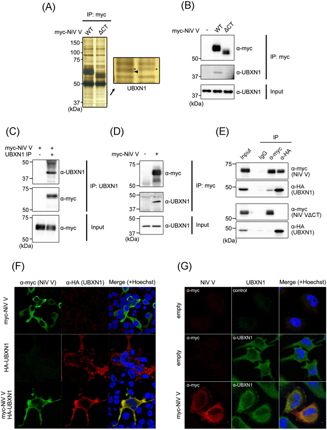
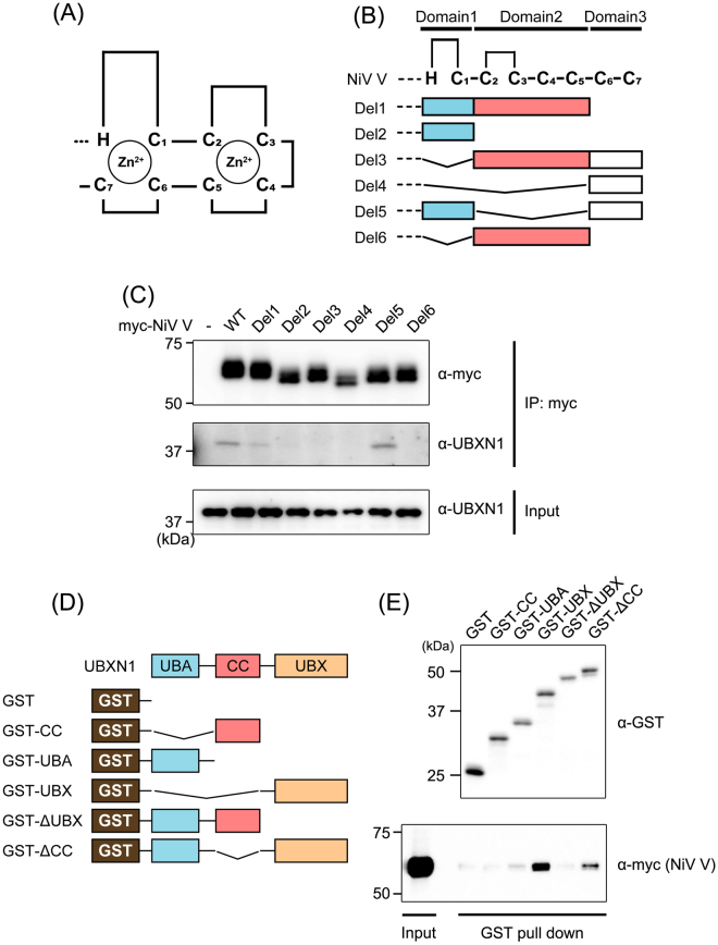
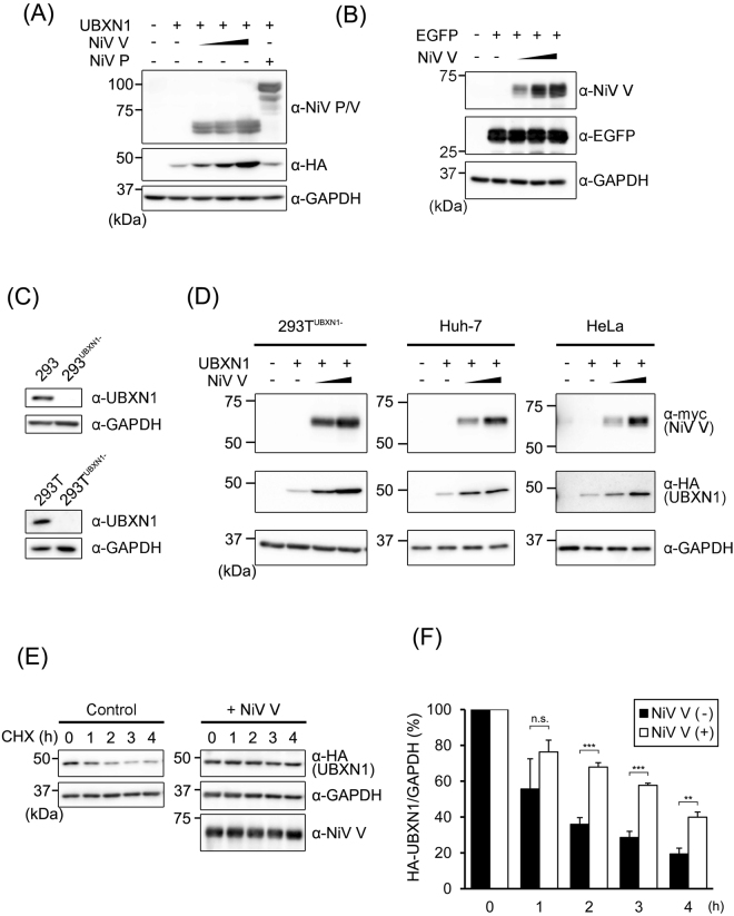
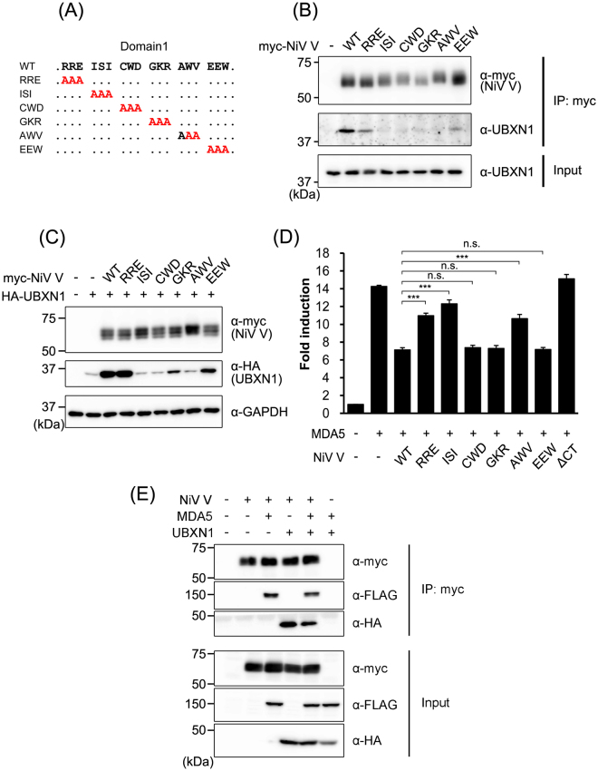
Nipah virus (NiV) is a highly pathogenic paramyxovirus that causes lethal encephalitis in humans. We previously reported that the V protein, one of the three accessory proteins encoded by the P gene, is one of the key determinants of the pathogenesis of NiV in a hamster infection model. Satterfield B.A. et al. have also revealed that V protein is required for the pathogenicity of henipavirus in a ferret infection model. However, the complete functions of NiV V have not been clarified. In this study, we identified UBX domain-containing protein 1 (UBXN1), a negative regulator of RIG-I-like receptor signaling, as a host protein that interacts with NiV V. NiV V interacted with the UBX domain of UBXN1 via its proximal zinc-finger motif in the C-terminal domain. NiV V increased the level of UBXN1 protein by suppressing its proteolysis. Furthermore, NiV V suppressed RIG-I and MDA5-dependent interferon signaling by stabilizing UBXN1 and increasing the interaction between MAVS and UBXN1 in addition to directly interrupting the activation of MDA5. Our results suggest a novel molecular mechanism by which the induction of interferon is potentially suppressed by NiV V protein via UBXN1.


Conflict of interest statement

The authors declare no competing interests.

Figures

Figure 1
Figure 1
Identification of a protein that interacts with NiV V. (A) Myc-tagged NiV V and a mutant lacking the C-terminal domain (ΔCT) were expressed in HEK293T cells. At 48 h posttransfection, an immunoprecipitation assay was performed, and the precipitated proteins were detected with silver staining. The band indicated by the arrowhead was analyzed with mass spectrometry. *Nontargeted bands. (B) The immunoprecipitation assay was performed as described in (A), and the precipitated proteins were detected with western blotting. (C) Myc-tagged NiV V was expressed in HEK293T cells, and at 48 h posttransfection, an immunoprecipitation assay was performed using UBXN1-specific antibody. The precipitated proteins were detected with western blotting. (D) Myc-tagged NiV V expressed in HeLa cells was immunoprecipitated, and the precipitated proteins were detected with western blotting. (E) Myc-tagged NiV V or ΔCT were expressed together with HA-tagged UBXN1 in HEK293T cells, and after 48 h, an immunoprecipitation assay was performed with anti-myc or anti-HA antibody. The precipitated proteins were detected with western blotting. (F) NiV V and HA-tagged UBXN1 were expressed in HEK293T cells, and after 24 h, an indirect immunofluorescence assay was performed. The subcellular localization of NiV V and UBXN1 was observed with confocal microscopy. (G) NiV V was expressed in HeLa cells, and the subcellular localization of NiV V and endogenous UBXN1 was examined by an indirect immunofluorescence assay. The gel and blots presented in (AE) were cropped from different images to improve clarity. Full-length gel and blots are presented in Supplementary Figure S1.
Figure 2
Figure 2
Identification of the binding domains of NiV V and UBXN1. (A,B) Schematic diagrams of the C-terminal domain of V protein (A) and its deletion mutants (B) are shown. (C) Wild-type NiV V and deletion mutants of NiV V were expressed in HEK293T cells, and an immunoprecipitation assay and western blotting were performed as described in Fig. 1B. (D) A schematic diagram of GST-tagged deletion mutants of UBXN1 is shown. (E) The interactions between GST-tagged UBXN1 mutants and myc-tagged NiV V were evaluated with a GST pull-down assay. Pulled-down proteins were detected with western blotting. The blots presented in (C,E) were cropped from different images to improve clarity. Full-length blots are presented in Supplementary Figure S1.
Figure 3
Figure 3
NiV V stabilizes UBXN1. (A) HEK293T cells were transfected with 400 ng of a vector expressing HA-tagged UBXN1 together with various amounts (100 ng, 200 ng, or 400 ng) of vector expressing NiV V or 400 ng NiV P. The total amount of transfected vector was kept constant by the addition of empty vector. After 24 h, the cells were lysed and the proteins were detected with western blotting. (B) HEK293T cells were transfected with 400 ng of a vector expressing for EGFP together with various amounts of vector expressing NiV V as described in (A). The proteins were detected with western blotting. (C) UBXN1 genes in HEK293 and 293 T cells were knocked out by the CRISPR-Cas9 system. The depletion of UBXN1 in 293UBXN1− and 293TUBXN1− cells was verified with western blotting. (D) 293TUBXN1−, Huh-7 and HeLa cells were transfected with 400 ng of a vector expressing HA-tagged UBXN1 together with various amounts (200 ng or 400 ng) of vector expressing NiV V. The total amount of transfected vector was kept constant by the addition of empty vector. After 24 h, the cells were lysed and the proteins were detected with western blotting. (E) HEK293T cells were transfected with 400 ng of a vector expressing for HA-tagged UBXN1 together with 400 ng of a vector expressing NiV V or an empty vector. Then the cells were treated with CHX for 0, 1, 2, 3, or 4 h, and the amount of proteins were evaluated with western blotting. (F) HA-tagged UBXN1 was expressed with or without NiV V in HEK293T cells. Then, the cells were treated with CHX, and the expression amount of HA-UBXN1 and GAPDH was quantitated as described in (E). The CHX assay was repeated three times, and the intensities of the bands were measured and summarized. Error bars indicate standard deviations (N = 3). **P < 0.01, ***P < 0.001, not significant (n.s.) on Student’s t test. The blots presented in (AE) were cropped from different images to improve clarity. Full-length blots are presented in Supplementary Figure S2.
Figure 4
Figure 4
Identification of the domains required to stabilize UBXN1. (A) HEK293T cells were transfected with equal amounts of vector expressing HA-tagged UBXN1 and vectors expressing myc-tagged wild-type NiV V or its deletion mutants. At 48 h posttransfection, the proteins were detected with western blotting. (B) A schematic diagram of the HA-tagged deletion mutants of UBXN1 is shown. (C) HEK293T cells were transfected with vectors expressing HA-tagged deletion mutants of UBXN1 together with a vector expressing NiV V or the empty vector. At 48 h posttransfection, the proteins were detected with western blotting. The blots presented in (A,C) were cropped from different images to improve clarity. Full-length blots are presented in Supplementary Figure S3.
Figure 5
Figure 5
Amino acids in NiV V required for its interaction with UBXN1. (A) Schematic diagrams of the alanine-substitution mutants of NiV V are shown. (B) Myc-tagged wild-type NiV V and its alanine-substitution mutants were expressed in HEK293T cells, and an immunoprecipitation assay and western blotting were performed as described in Fig. 1B. (C) HEK293T cells were transfected with equal amounts of vector expressing HA-tagged UBXN1 and vector expressing myc-tagged wild-type NiV V or its alanine-substitution mutants. At 48 h posttransfection, the proteins were detected with western blotting. (D) HEK293T cells were transfected with an IFNβ reporter vector together with vectors expressing FLAG-tagged MDA5 and wild-type NiV V or its alanine-substitution mutants. The total amount of transfected vector was kept constant by the addition of empty vector. At 24 h posttransfection, a luciferase assay was performed. (E) HEK293T cells were transfected with vectors expressing myc-tagged NiV V, FLAG-tagged MDA5 and HA-tagged UBXN1. At the 48 h posttransfection, an immunoprecipitation assay was performed with anti-myc antibody. The precipitated proteins were detected with western blotting. Error bars indicate standard deviations (N = 3). ***P < 0.001, not significant (n.s.) on Dunnett’s multiple comparison test. The blots presented in (B,C,E) were cropped from different images to improve clarity. Full-length blots are presented in Supplementary Figure S4.
Figure 6
Figure 6
Stabilized UBXN1 suppresses IFN induction. (A) HEK293 cells were transfected with an IFNβ reporter vector together with a vector expressing MDA5 and various amounts of vectors expressing NiV V and UBXN1. The total amount of transfected vector was kept constant by the addition of empty vector. At 24 h posttransfection, a luciferase assay was performed. The values in three data sets, sample No. 1–4, 5–7 and 8–10, were normalized by setting the value of sample No. 2, 5 and 8 to 100% respectively. Samples No. 6, 9 and 7, 10 were statistically compared to No. 3 and 4 respectively. The original data without the normalization were shown in Supplementary Figure S5C. (B) HEK293 cells were transfected with an IFNβ reporter vector together with vectors expressing RIG-IΔ and NiV V with or without a vector expressing UBXN1. The total amount of transfected vector was kept constant by the addition of empty vector. At 24 h posttransfection, a luciferase assay was performed. (C) 293UBXN1− cells were transfected, and the luciferase reporter assay was performed as described in (B). (D) HA-tagged UBXN1 and myc-tagged NiV V were expressed in HEK293T cells. At 48 h posttransfection, endogenous MAVS was immunoprecipitated with a specific antibody, and the precipitated proteins were detected with western blotting. (E) The model of MAVS interference by NiV V suggested by our results is shown. Error bars indicate standard deviations (N = 3). *P < 0.05, **P < 0.01, *** and †††P < 0.001, not significant (n.s.) on Dunnett’s multiple comparison test. The blots presented in (D) were cropped from different images to improve clarity. Full-length blots are presented in Supplementary Figure S4.

References

    1. Chua KB, et al. Nipah virus: a recently emergent deadly paramyxovirus. Science. 2000;288:1432–1435. doi: 10.1126/science.288.5470.1432. - DOI - PubMed
    1. Eaton BT, Broder CC, Middleton D, Wang LF. Hendra and Nipah viruses: different and dangerous. Nat Rev Microbiol. 2006;4:23–35. doi: 10.1038/nrmicro1323. - DOI - PMC - PubMed
    1. Arankalle VA, et al. Genomic characterization of Nipah virus, West Bengal, India. Emerg Infect Dis. 2011;17:907–909. doi: 10.3201/eid1705.100968. - DOI - PMC - PubMed
    1. Chadha MS, et al. Nipah virus-associated encephalitis outbreak, Siliguri, India. Emerg Infect Dis. 2006;12:235–240. doi: 10.3201/eid1202.051247. - DOI - PMC - PubMed
    1. Ching PK, et al. Outbreak of henipavirus infection, Philippines, 2014. Emerg Infect Dis. 2015;21:328–331. doi: 10.3201/eid2102.141433. - DOI - PMC - PubMed

Publication types

MeSH terms