. 2018 May;557(7706):510-515.
doi: 10.1038/s41586-018-0137-8. Epub 2018 May 16.

ANKRD16 prevents neuron loss caused by an editing-defective tRNA synthetase

Affiliations

ANKRD16 prevents neuron loss caused by an editing-defective tRNA synthetase

My-Nuong Vo et al. Nature. 2018 May.

Erratum in

Abstract

Editing domains of aminoacyl tRNA synthetases correct tRNA charging errors to maintain translational fidelity. A mutation in the editing domain of alanyl tRNA synthetase (AlaRS) in Aars sti mutant mice results in an increase in the production of serine-mischarged tRNAAla and the degeneration of cerebellar Purkinje cells. Here, using positional cloning, we identified Ankrd16, a gene that acts epistatically with the Aars sti mutation to attenuate neurodegeneration. ANKRD16, a vertebrate-specific protein that contains ankyrin repeats, binds directly to the catalytic domain of AlaRS. Serine that is misactivated by AlaRS is captured by the lysine side chains of ANKRD16, which prevents the charging of serine adenylates to tRNAAla and precludes serine misincorporation in nascent peptides. The deletion of Ankrd16 in the brains of Aarssti/sti mice causes widespread protein aggregation and neuron loss. These results identify an amino-acid-accepting co-regulator of tRNA synthetase editing as a new layer of the machinery that is essential to the prevention of severe pathologies that arise from defects in editing.


Figures
Extended Data Figure 1
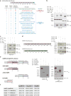
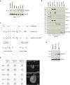
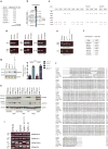
Extended Data Figure 1. Identification of Ankrd16 as a genetic modifier of the B6J.Aarssti/sti mutation
a, 3- to 4-month-old Aarssti/sti mice from crosses to inbred strains. b, Location of Msti relative to microsatellite markers. Centimorgan (cM), megabase pairs (Mbp). c, Nonsynonymous SNPs in Msti candidate genes. The amino acids and position are shown at the top of the table with the B6 residue listed first. d, RT-PCR analysis of Ankrd16 transcripts from cerebellar cDNA prepared from C57BL/6J and CAST/Ei mice. Note, the alternative Ankrd16 transcript containing exon 5’ (see Figure 2c) amplified by primers to exon4/6 or exon 5/7 was present in cDNA from C57BL/6J but not CAST/Ei mice, whereas the alternative transcript detected by exon1/3 primers was present in cDNA from both strains. e, RT-PCR analysis of Ankrd16 transcripts from cerebellar cDNA prepared from C57BL/6J, CAST/Ei, Aarssti/sti, and MstiCAST/CAST; Aarssti/sti mice. Note, the alternative Ankrd16 transcript containing exon 5’ (see Figure 2c) amplified by exon 5/7 or 3/5’ primers was present in cDNA from C57BL/6J and Aarssti/sti, but absent in the presence of CAST/Ei derived Msti (CAST/Ei and MstiCAST/CAST; Aarssti/sti mice). f, Sequence of the SNP-containing region in intron 5 of Ankrd16. Capital letters indicate novel exon 5’ and lower case letters indicate intron 5. The SNP in non-rescuing strains is shown in red. g, Western blotting analysis of ANKRD16 from cerebellar lysates. Note that the expression of ANKRD16 is reduced in C57BL/6J and Aarssti/sti mice relative to mice with CAST/Ei derived Msti (CAST/Ei and MstiCAST/CAST; Aarssti/sti mice) (mean ± SD, n = 3, One-way ANOVA (Tukey correction), **** p ≤ 0.0001). h, Protein levels of AlaRS and ANKRD16 were determined by western blotting using various mouse tissues from C57BL/6J and B6 congenic mice heterozygous for the Msti region (MstiCAST/B6). GAPDH is included as a loading control. Note the increase in ANKRD16 levels in MstiCAST/B6 tissues, whereas AlaRS levels do not change between genotypes. i, PCR results of genomic DNA from B6 transgenic mouse lines Tg25L9-19 and Tg25L9-46, which carry the CAST/Ei BAC. Polymorphic markers, which differentiate between C57BL/6J and CAST/Ei were used as shown. j, Amino acid sequence comparison of ANKRD16 from various species with the C57BL/6J strain shown. Non-synonymous SNPs distinguishing CAST/Ei and C57BL/6J are shown in yellow and serinylated lysines are shown in red.
Extended Data Figure 2
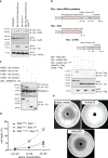
Extended Data Figure 2. Verification of interaction between ANKRD16 and AlaRS in vitro
a, Peptide/spectral counts of proteins co-immunoprecipitated (Co-IP) from transgenic Ankrd16-myc (see cartoon) but not detected in non-transgenic liver tissue. b, HEK293T cells were transiently co-transfected with myc-tagged constructs for mouse Aars, AarsA734E, the Aars aminoacylation domain (AAD), and FLAG-tagged Ankrd16. Co-IP experiments were performed with ANKRD16-FLAG as the bait protein. c, Reciprocal Co-IP experiments were performed by transiently co-transfecting HEK293T cells with HA-tagged constructs for mouse Aars or AarsA734E and the FLAG-tagged Ankrd16. HA-AlaRS proteins were used as bait for pull down. d, Domain structure of mouse ANKRD16 and ANKRD29. HEK293T cells were transiently transfected with FLAG-tagged constructs for mouse Ankrd16, Ankrd29, and the HA-epitope tagged Aars. Co-IP experiments were performed with HA-AlaRS as bait protein. e, Various domain protein products of AlaRS (human) as indicated were bacterially expressed, purified, and incubated with GST-ANKRD16. GST-pull down products and input were immunoblotted with anti-His or -GST antibodies. Asterisks indicate protein degradation products. f, Binding dynamics were determined between mouse wild type or mutant AlaRS and mouse wild type or mutant ANKRD16 using SwitchSENSE (mean ± SD; n = 3).
Extended Data Figure 3
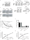
Extended Data Figure 3. Analysis of the effects of ANKRD16 on steps of translation
a, Preassembled ternary complex containing either Ser-tRNAAla or Ala-tRNAAla was mixed with 70S initiation complex (IC) programmed with codon GCU in the A site, aliquots were transferred at various times to quench solution (0.5 M KOH), and products were resolved by eTLC. b, Incubation of full-length AlaRS with preassembled EF-Tu•GTP•Ser-tRNAAla prevents fMet-Ser formation. EF-Tu•GTP•Ser-tRNAAla ternary complex and 70S IC were each preassembled. In reaction scheme 1, AlaRS was incubated with EF-Tu•GTP•Ser-tRNAAla for 15 min, 70S IC was added, and aliquots were removed at various times for eTLC analysis. In contrast (reaction scheme 2), AlaRS was incubated with the 70S IC for 15 min, followed by addition of EF-Tu•GTP•Ser-tRNAAla, and aliquots were removed at various times for eTLC analysis. c, Deacylated tRNAAla was mixed with AlaRS, serine, ATP, and all other components to form ternary complex, aliquots were transferred at various time points to the 70S IC, and dipeptide products were resolved by eTLC. t = 0 (control reactions in the absence of AlaRS); * (oxidized fMet). Note, without alanine supplementation (b and c), trace amounts of fMet-Ala are detected, likely due to AlaRS-bound alanyl-AMP during protein purification. d, Deacylation of [3H]Ser-tRNAAla by mouse wild-type AlaRS or AlaRSA734E in the presence or absence of ANKRD16 (mean ± SD, n = 2, One Phase Decay model [R2AlaRSWT = 0.9892; R2AlaRSA734E = 0.991; R2AlaRSWT/ANKRD16 = 0.9872; R2AlaRSA734E/ANKRD16 = 0.9902; R2ANKRD163xArg = 0.7992]). e, Percentage of EF-Tu retained on filter membrane upon addition of various components as indicated (mean ± SD; n = 2). f, ATP-pyrophosphate exchange by mouse AlaRSA734E in the presence or absence of ANKRD16 (mean ± SD, n = 2, Michaelis-Menten model [R2AlaRSA734E = 0.9763; R2AlaRSA734E/ANKRD16 = 0.9874]). g, Aminoacylation of tRNAAla with alanine by mouse AlaRSA734E in the presence or absence of ANKRD16 (mean ± SD, n = 2, Michaelis-Menten model [R2AlaRSA734E = 0.9879; R2AlaRSA734E/ANKRD16 = 0.9846]).
Extended Data Figure 4
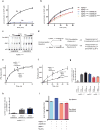
Extended Data Figure 4. Analysis of serinylation of ANKRD16
a, tRNA-independent ATPase activity of mouse wild type AlaRS for serine or alanine (mean ± SD, n = 3, Michaelis-Menten model [R2Ser = 0.9926; R2Ala = 0.8430]). b, tRNA-independent ATPase activity of mouse AlaRSWT or AlaRSA734E for serine in the presence of ANKRD16, ANKRD29, or ANKRD163xArg (mean ± SD, Michaelis-Menten model [R2AlaRSWT = 0.9926, R2AlaRSA734E = 0.9899, R2AlaRSA734E/ANKRD16 = 0.9918, R2AlaRSA734E/ANKRD29 = 0.9939, R2AlaRSA734E/ANKRD163xArg = 0.9841], [AlaRSWT, AlaRSA734E, AlaRSA734E/ANKRD29, n = 3], [AlaRSA734E/ANKRD16, AlaRSA734E/ANKRD163xArg, n = 4]). c, TLC analysis of ATPase activity of AlaRSA734E against serine in the absence and presence of ANKRD16. d, Experimental scheme showing how data were generated for panels e, f, g, and h. e, Acylation reactions with radioactive alanine were performed as above to determine alanine transfer (mean ± SD, n = 2, Michaelis-Menten model [R2AlaRSA734E/tRNA/ANKRD16 = 0.9611, R2AlaRSA734E/tRNA/buffer = 0.9641]). f, Misacylation of tRNAAla with radioactive serine by mouse AlaRSA734E in the presence or absence (buffer) of ANKRD16. After misacylation, reactions were subjected to TCA precipitation of RNA (Ser-tRNAAla) and protein under acidic conditions to maintain Ser-tRNA (mean ± SD, n = 2, Michaelis-Menten model [R2AlaRSA734E/tRNA/ANKRD16 = 0.9577, R2AlaRSA734E/tRNA/buffer = 0.7841]). g, Misacylation reactions using radioactive serine and mouse AlaRSWT or AlaRSA734E were performed in the presence or absence of ANKRD16, ANKRD29, ANKRD163xArg, or tRNAAla. After TCA precipitation under neutral pH conditions, serine was measured (mean ± SD, n = 4). Note, the higher level of TCA-precipitated serine on tRNA or protein in the presence of ANKRD16. h, Acylation reactions using radioactive alanine and mouse AlaRSA734E were performed in the presence or absence of ANKRD16 or tRNAAla. After TCA precipitation under neutral pH conditions, alanine was measured (mean ± SD, n = 4). i, Misacylation reactions using radioactive serine and mouse AlaRSA734E were performed in the presence of ANKRD16 either with or without tRNAAla. Reactions were treated with or without Na2CO3 (final concentration of 0.15 M [alkaline pH]) for 30 minutes, followed by TCA precipitation. Precipitated [3H]-serine-links were measured and plotted as relative level of serinylation (mean, n = 2).
Extended Data Figure 5
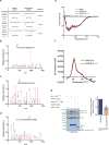
Extended Data Figure 5. Analysis of serinylation of ANKRD16 by mass spectrometry
a, MS/MS spectrum of peptides from ANKRD16. Incorporation of serine onto ANKRD16 was observed when serine was misactivated by mouse AlaRSA734E (+ATP). Misactivation was not observed in the absence of ATP. MS/MS spectrum of peptides from ANKRD16 with serine linked to positions K102 (b), K135 (c) and K165 (d). a ions [a], b ions [b] and y ions [y] are annotated in green, red, and purple, respectively. The triply charged precursor had a mass of 2153.065 Dalton and included carbamidomethyl cysteine. e, Secondary structure analysis of mouse ANKRD16 and ANKRD163xArg. Far-UV circular dichroism (CD) spectra of wild type ANKRD16 (blue) and ANKRD163xArg (red) show highly similar CD spectra (mean ± SD, n = 4). f, Thermal shift analysis of mouse ANKRD16 (blue) and ANKRD163xArg (red) show highly similar thermal stability. g, HEK293T cells were transiently co-transfected with myc-tagged constructs for mouse AarsA734E and FLAG-tagged Ankrd16, Ankrd163xArg, or Ankrd29. Co-IP experiments were performed with FLAG-tagged proteins as the bait protein. Binding affinity was determined by normalizing immunoprecipitation (IP) signal of AlaRSA734E to the input signal, in which the interaction of AlaRSA734E/ANKRD16 was arbitrarily defined as 1 to determine relative binding affinity of AlaRSA734E/ANKRD163xArg (mean ± SD, n = 3).
Extended Data Figure 6
Extended Data Figure 6. Serine-induced cell death in Aarssti/sti fibroblasts
B6.Aarssti/sti embryonic fibroblasts were co-transfected with hrGFP (humanized recombinant GFP, green) and either Ankrd16-Flag or Ankrd163xArg-Flag (n = 4). 12-hours post-transfection, serine was added and cells were cultured for 24 hours prior to staining with propidium iodide (PI) (red) and Hoechst (blue) to determine cell death. Arrowheads represent PI-positive GFP+ cells. Scale bars: 100 µm.
Extended Data Figure 7
Extended Data Figure 7. Analysis of ubiquitous deletion of ANKRD16
a, Protein levels of ANKRD16 were determined by western blotting of tissues from B6 mice homozygous for the Msti region (MstiCAST/CAST). b, Subcellular analysis of ANKRD16 and AlaRS via cell fractionation of brains from B6.MstiCAST/B6 and Ankrd16−/− mice. Cellular fractions were confirmed using antibodies for histone 3 (nuclear marker), GAPDH (cytosolic marker), COX IV (mitochondrial marker), GRP78 (ER marker), and Sec61 beta (rough ER marker). c, To generate a ubiquitous or conditional loss-of-function allele, loxP sites that flank exon 2 of Ankrd16 were inserted by homologous recombination. Removal of exon 2 results in a frame shift and premature stop codon in exon 3. Ankrd16 was ubiquitously deleted by EIIa Cre-mediated removal of exon 2 and the neo cassette. Flp-mediated excision of the neomycin cassette was utilized to generate a conditional loss-of-function Ankrd16 allele. d, Loss of ANKRD16 was verified by western blotting. Protein extracts from Ankrd16−/−, MstiCAST/CAST and C57BL/6J mice were used for comparison purposes and GAPDH was used as a loading control. e, The number of embryos or mice of various genotypes from intercrosses of Aarssti/+; Ankrd16−/− mice over the total number observed. Representative images of E9.5 embryos. Note, Aarssti/sti; Ankrd16−/− embryos are smaller and have failed to turn. Abbreviations: embryonic day (E), postnatal day (P). Scale bars: 500 µm.
Extended Data Figure 8
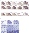
Extended Data Figure 8. Conditional deletion of Ankrd16 accelerates Purkinje cell loss and causes widespread neurodegeneration in the B6.Aarssti/sti cerebellum
a and b, Calbindin D-28 immunohistochemistry of sagittal cerebellar sections. c, Cresyl violet-stained sagittal cerebellar sections. Note, the presence of interneurons in the ML of B6.Aarssti/sti cerebellum despite the thinning of the ML as a consequence of Purkinje cell degeneration. In contrast, loss of Ankrd16 in the B6.Aarssti/sti cerebellum results in degeneration of ML interneurons. Scale bars: 500 µm (a, b, c), 50 µm (c, higher magnification). Abbreviations: postnatal day (P), molecular layer (ML), Purkinje cell layer (PL), granule cell layer (GL).
Extended Data Figure 9
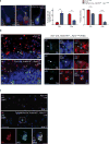
Extended Data Figure 9. Conditional deletion of Ankrd16 accelerates formation of protein aggregates in B6.Aarssti/sti mice
a, Ubiquitin (red), p62 (green), and Calbindin D-28 (blue) immunofluorescence on sagittal cerebellar sections. The percentage of aggregate-positive Purkinje cells and Purkinje cells are shown (mean ± SD, n = 3, Multiple t-tests (Holm-Sidak method), *** p = 0.0002466 (Purkinje cells with aggregates, P21), p = 0.0002336 (Purkinje cells with aggregates, P28), p = 0.0003214 (percent of Purkinje cells, P21), **** p = 7.701787×10⁻⁵ (percent of Purkinje cells, P28)). Note, the percent of Purkinje cells is relative to control C57BL/6J mice. b, Cell type-specific markers (red), and p62 (green) immunofluorescence on sagittal cerebellar sections. Parvalbumin (Parv) was utilized to identify Purkinje cells and interneurons (stellate and basket cells) in the ML. NeuN was utilized to distinguish between granule and Golgi cells in the GL. (En1-Cre; Ankrd16fl/−; Aarssti/sti: Golgi cell [closed arrow head, p62+/NeuN]; granule cell [arrow, p62+/NeuN+]; basket/stellate cell [open arrow head, p62+/Parv+]). c, Ubiquitin (red) and p62 (green) immunofluorescence on sagittal sections of the cortex (layer IV). Scale bars: 10 µm (a), 50 µm (b and c, low magnification) and 10 µm (b and c, higher magnification). Abbreviations: postnatal day (P), molecular layer (ML), Purkinje cell layer (PL), granule cell layer (GL).
Extended Data Figure 10
Extended Data Figure 10. Model for the role of ANKRD16 in translational fidelity
a, A point mutation in the editing domain of AlaRS (A734E) results in editing defects, as indicated by deficits in tRNA-independent ATPase activity, which in turn leads to increased levels of incorrectly aminoacylated Ser-tRNAAla, misincorporation of serine during translation, protein aggregation, and cell death. b, Interaction of ANKRD16 with the aminoacylation domain of editing-deficient AlaRSA734E stimulates tRNA-independent editing and misactivated serines are transferred onto ANKRD16. Mitigation of serine misactivation prevents sti-mediated mistranslation, and thereby prevents protein aggregation and cell death.
Figure 1
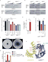
Figure 1. Modifier of sticky (Msti) suppresses Aarssti-mediated neurodegeneration
a, Rotorod analysis of 3-month-old mice (mean ± SD, n = 15 or 16 mice/genotype, Two-way ANOVA (Tukey correction), ** p ≤ 0.01, *** p ≤ 0.001). b, Calbindin D-28 immunohistochemistry. Sections were counterstained with hematoxylin. c, Aggregate-positive cells (mean ± SD, n = 3, Multiple t-tests (Holm-Sidak method), ** p = 0.00148 (6 weeks), p = 0.00498 (12 weeks), **** p = 2.819×10⁻⁶). Scale bars: 500 µm.
Figure 2
Figure 2. Ankrd16 is the modifier of Aarssti/sti
a, Amino acid polymorphisms between C57BL/6J and CAST/Ei in Msti region. b, Alternative splicing of Ankrd16 in the cerebellum. c, An intronic SNP (red) introduces a cryptic exon (exon 5’) in Ankrd6 in non-Msti containing strains. d, Expression of correctly spliced Ankrd16 in the cerebellum (mean ± SEM, n = 3, One-way ANOVA (Tukey correction), * p ≤ 0.05, ** p ≤ 0.01, *** p ≤ 0.001). e, Western blot analysis using cerebellar extracts. f and g, Calbindin D-28 immunohistochemistry. Scale bars: 500 µm.
Figure 3
Figure 3. ANKRD16 interacts with AlaRS and prevents serine-mediated cell death
a, AlaRS/ANKRD16 interaction in the brain. b, Co-IP experiments in HEK293T cells using mouse myc-AlaRS domain constructs. c, Pull down of purified AlaRS, TyrRS, or TrpRS with ANKRD16. d, Cell death of embryonic fibroblasts (mean ± SD, n = 3, Two-way ANOVA (Tukey correction), *** p ≤ 0.001; **** p ≤ 0.0001). e, AarsC666A/Q584H E. coli expressing mouse Ankrd16 or Ankrd29.
Figure 4
Figure 4. ANKRD16 enhances tRNA-independent editing and accepts serine adenylates from AlaRS
a, Mouse AlaRS (±ANKRD16) was added to reactions containing Ser-tRNAAla and alanine. b, Mouse AlaRS (±ANKRD16) was added to reactions containing deacylated tRNAAla, alanine and serine (* oxidized fMet). c, Ser-AMP hydrolysis at 10 min (mean ± SD, n = 3, One-way ANOVA (Tukey correction), *** p ≤ 0.001, **** p ≤ 0.0001). d, Ratio of alanine- or serine-linked ANKRD16/aminoacylated tRNAAla (mean ± SD, n = 3, Two-tailed Student’s t-test, ** p = 0.0011). e, Ser-AMP hydrolysis at 10 min (mean ± SD, n = 3, One-way ANOVA (Tukey correction), *** p ≤ 0.001). Control reactions from panel c (†). f, AarsC666A/Q584H E. coli expressing mouse Ankrd16 or Ankrd163xArg. g, Cell death of embryonic fibroblasts (mean ± SEM, n = 4, One-way ANOVA (Tukey correction), *** p ≤ 0.001, **** p ≤ 0.0001). h, Structural model of ANKRD16 and the aminoacylation domain (AAD) of AlaRS, modified lysines (purple), active site residues (orange), and Ala-AMS (Ala-AMP analog, green).
Figure 5
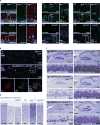
Figure 5. Loss of Ankrd16 in Aarssti/sti mice causes protein aggregation and neurodegeneration
a and b, Immunofluorescence with antibodies to ANKRD16 (green) and calbindin D-28 (red) or ubiquitin (red) and p62 (green). c and d, Cresyl violet staining of hippocampus (c) and cortex (d). Scale bars: 50 µm (a, low magnification), 10 µm (a, higher magnification), 50 µm (b, low magnification), 10 µm (b, higher magnification), 500 µm (c, low magnification), 50 µm (c, higher magnification), 100 µm (d). Postnatal day (P), Cornu Ammonis (CA), Dentate gyrus (DG).

Similar articles

Cited by

References

    1. Guo M, Chong YE, Shapiro R, Beebe K, Yang XL, Schimmel P. Paradox of mistranslation of serine for alanine caused by AlaRS recognition dilemma. Nature. 2009;462:808–812. - PMC - PubMed
    1. Schimmel P. Mistranslation and its control by tRNA synthetases. Philos. Trans. R. Soc. B Biol. Sci. 2011;366:2965–2971. - PMC - PubMed
    1. Ling J, Reynolds N, Ibba M. Aminoacyl-tRNA synthesis and translational quality control. Annu. Rev. Microbiol. 2009;63:61–78. - PubMed
    1. Martinis SA, Boniecki MT. The balance between pre- and post-transfer editing in tRNA synthetases. FEBS Lett. 2010;584:455–459. - PMC - PubMed
    1. Dulic M, Cvetesic N, Perona JJ, Gruic-Sovulj I. Partitioning of tRNA-dependent editing between pre- and post-transfer pathways in class I aminoacyl-tRNA synthetases. J. Biol. Chem. 2010;285:23799–23809. - PMC - PubMed

Publication types