. 2018 May;557(7706):590-594.
doi: 10.1038/s41586-018-0120-4. Epub 2018 May 16.

Structural basis for gating pore current in periodic paralysis

Affiliations

Structural basis for gating pore current in periodic paralysis

Daohua Jiang et al. Nature. 2018 May.

Abstract

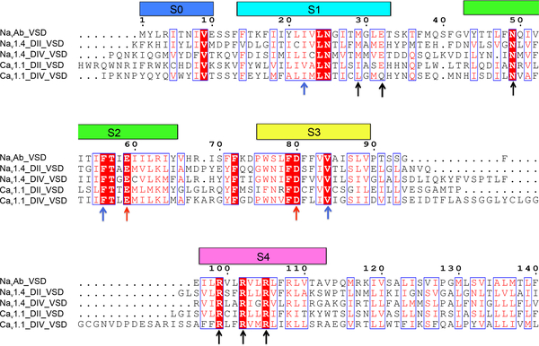
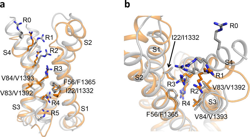
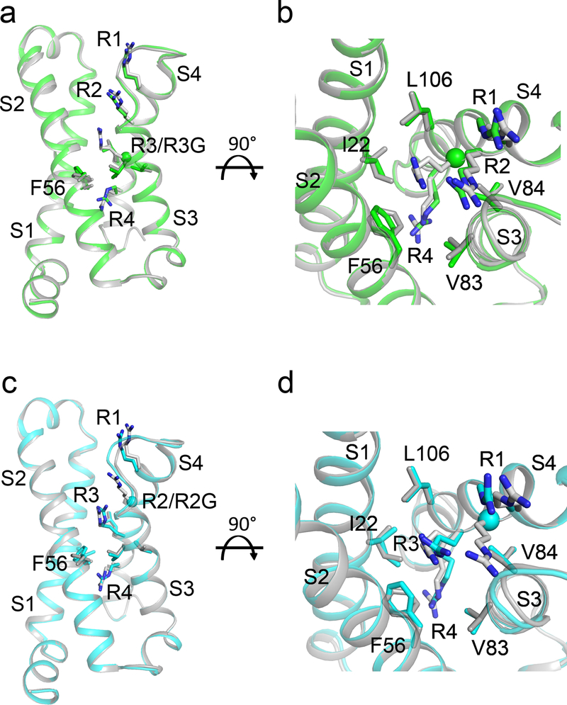
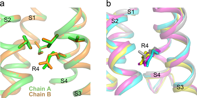
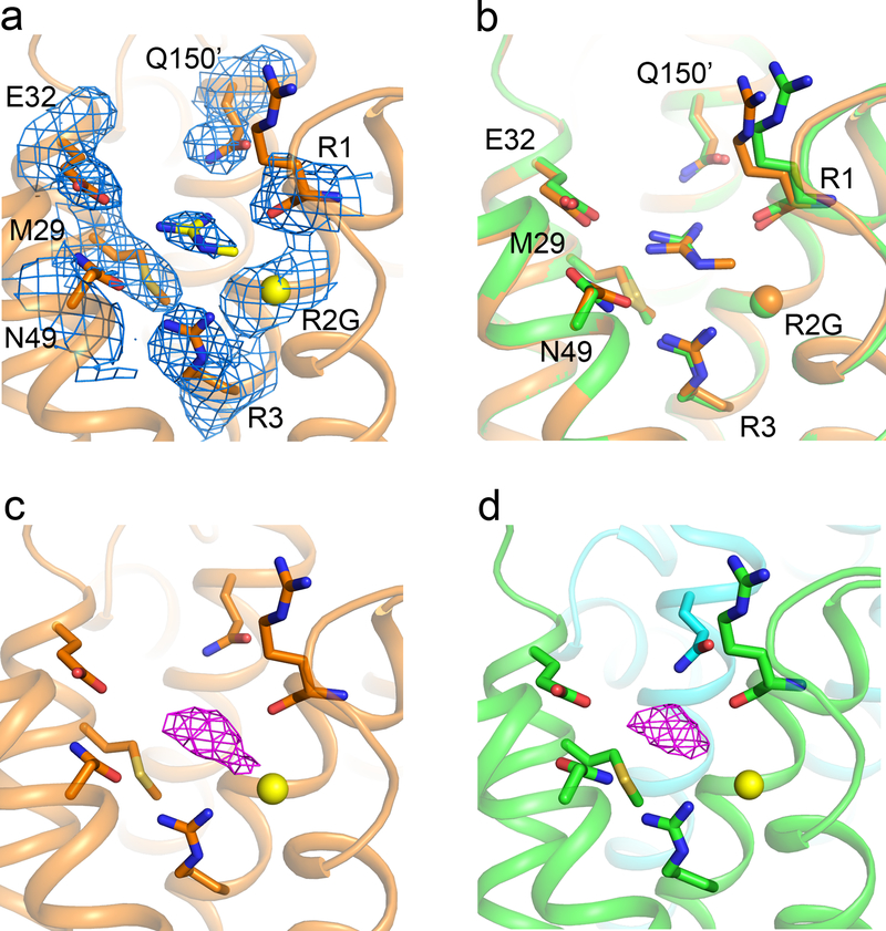
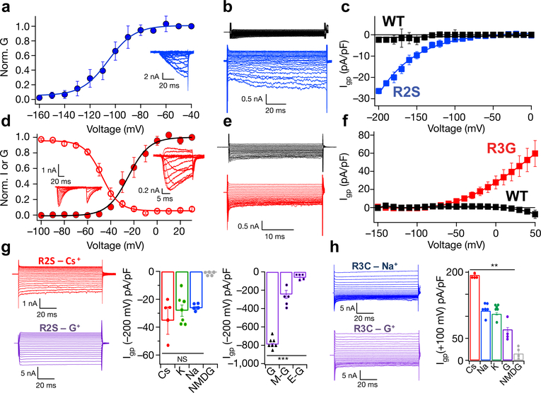
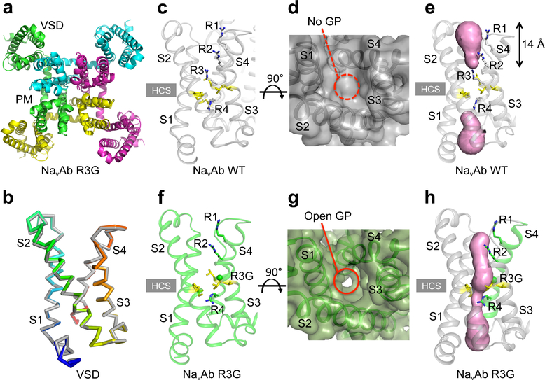
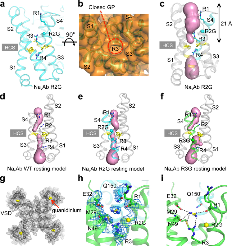
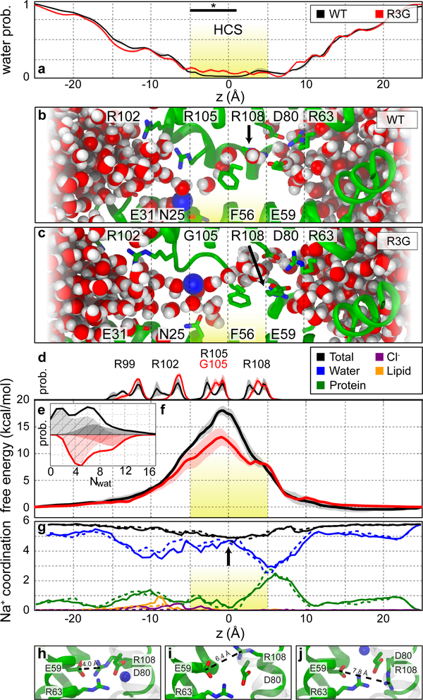
Potassium-sensitive hypokalaemic and normokalaemic periodic paralysis are inherited skeletal muscle diseases characterized by episodes of flaccid muscle weakness1,2. They are caused by single mutations in positively charged residues ('gating charges') in the S4 transmembrane segment of the voltage sensor of the voltage-gated sodium channel Nav1.4 or the calcium channel Cav1.11,2. Mutations of the outermost gating charges (R1 and R2) cause hypokalaemic periodic paralysis1,2 by creating a pathogenic gating pore in the voltage sensor through which cations leak in the resting state3,4. Mutations of the third gating charge (R3) cause normokalaemic periodic paralysis 5 owing to cation leak in both activated and inactivated states 6 . Here we present high-resolution structures of the model bacterial sodium channel NavAb with the analogous gating-charge mutations7,8, which have similar functional effects as in the human channels. The R2G and R3G mutations have no effect on the backbone structures of the voltage sensor, but they create an aqueous cavity near the hydrophobic constriction site that controls gating charge movement through the voltage sensor. The R3G mutation extends the extracellular aqueous cleft through the entire length of the activated voltage sensor, creating an aqueous path through the membrane. Conversely, molecular modelling shows that the R2G mutation creates a continuous aqueous path through the membrane only in the resting state. Crystal structures of NavAb(R2G) in complex with guanidinium define a potential drug target site. Molecular dynamics simulations illustrate the mechanism of Na+ permeation through the mutant gating pore in concert with conformational fluctuations of the gating charge R4. Our results reveal pathogenic mechanisms of periodic paralysis at the atomic level and suggest designs of drugs that may prevent ionic leak and provide symptomatic relief from hypokalaemic and normokalaemic periodic paralysis.


Conflict of interest statement

Author Information. The authors declare no competing financial interests.

Figures

Extended Data Figure 1 |
Extended Data Figure 1 |. Sequence alignment of NavAb VS with human Nav1.4 DII VS, Nav1.4 DIV VS, Cav1.1 DII VS, and Cav1.1 DIV VS.
Colored rectangles represent TM helices. Black arrows indicate residues that form the guanidinium binding site, blue arrows indicate hydrophobic constriction site, and red arrows indicate the conserved intracellular negative cluster.
Extended Data Figure 2 |
Extended Data Figure 2 |. Superposition of NavAb/WT VS and EeNav1.4 DIV VS.
a-b, Comparison of the conformations of NavAb/WT VS (orange) and EeNav1.4 VSDIV (grey) in side view and top view, respectively. Arg sensors and hydrophobic residues in the HCS are labeled and shown side chain in sticks.
Extended Data Figure 3 |
Extended Data Figure 3 |. Superposition of the VS between NavAb/WT and mutants.
a-b, VS structure alignment between NavAb/WT (grey) and NavAb/R3G (green) in side view and top view, respectively. c-d, VS structure alignment between NavAb/WT (grey) and NavAb/R2G (cyan) in side view and top view, respectively. Arg sensors and hydrophobic residues in the HCS are labeled and shown side chain in sticks.
Extended Data Figure 4 |
Extended Data Figure 4 |. R4 side chain conformational changes.
a, Different conformations of the R4 rotamer in NavAb/R3G Chain A (green) and Chain B (orange). b, Different conformations of the R4 rotamer in the four subunits of NaVAb in the slow-inactivated state (PDB code 4EKW).
Extended Data Figure 5 |
Extended Data Figure 5 |. Electron density maps for bound guanidinium and methylguanidinium ions.
a, 2mFo – DFc electron density map (blue mesh) of residues around the methylguanidinium binding site at 1σ. b, Overlay of guanidinium binding site (green) and methylguanidinium binding site (orange). c-d, Simulated annealing map (Fo-Fc) contoured at 3σ for methylguanidinium and guanidinium, respectively.
Extended Data Figure 6 |
Extended Data Figure 6 |. Purification of NavAb/R3G.
a, A representative gel-filtration chromatography of NavAb/R3G, highlighted peak fractions were concentrated for crystallization. b, Concentrated sample was visualized on SDS-PAGE by Coomassie Blue staining.
Figure 1:
Figure 1:. Functional properties of NaVAb/WT, NaVAb/R2S, and NaVAb/R3G.
a, Central pore Na+ currents (inset) and G/V curve for NavAb/R2S during 200-ms depolarizations from −200 mV to the indicated potentials. V1/2= −105 ± 0.6 mV, k=10±0.9 (n=4). b, c, Gating pore Na+ currents and I/V curves for NavAb/R2S (blue) or NavAb/WT (black) during depolarizations from −100 mV to the indicated potentials. n=10. d, Central pore Na+ currents (inset) and G/V curve for NavAb/R3G during depolarizations from-160 mV to the indicated potentials (filled circles; Va=−24.8 ± 1.1 mV, k=9±1 (n=4)). Voltage dependence of steady-state inactivation (open circles) for NavAb/R3G (Vh=−47.7±0.4 mV, k=7.5±0.3 (n=4)). e, f, Gating pore Na+ currents and I/V curves for NaVAb/R3G (red) or NaVAb/WT (black) for voltage steps from 0 mV to the indicated potentials (n=11). g, Gating pore current through NavAb/R2S for Cs+ (n=5), K+ (n=7), Na+ (n=5), N-methyl-D-glucamine (NMDG, n=5), guanidinium (G, n=7), methylguanidinium (M-G, n=5), and ethylguanidinium, (E-G, n=5).. ***, P=0.00029. h, Gating pore current through NavAb/R3C for Cs+ (n=4), Na+ (n=6), K+ (n=6), G (n=4), and NMDG. (n=4). **, P=0.0011. Student’s t-test, two-sided.
Figure 2.
Figure 2.. Structures of the VS of NavAb/WT and NavAb/R3G.
a, Structure of NaVAb/R3G in top view. b, Comparison of the conformations of NaVAb/WT (grey) and NaVAb/R3G (rainbow) VS in side view. c-e, Structures of NavAb/WT VS. (c) Side view highlighting gating charges in sticks. (d) Top view in spacefilling format. (e) MOLE2 analysis of water-accessible space in magenta. f-h, Structures of NavAb/R3G VS. (f) Side view highlighting gating charges. (g) Top view in spacefilling format. (h) MOLE2 analysis of water-filled space in magenta. Green balls in panels f and h indicate the positions of the missing sidechain of R3. In panels d and g, the dotted red line circles the position where the gating pore (GP) would be in the activated state and the solid red line circles the open GP. See Extended Data Table 1 for details.
Figure 3.
Figure 3.. Structure of VS and guanidinium binding site of NaVAb/R2G.
a-c, Structures of the activated VS of NaVAb/R2G. (a) Side view with gating charges highlighted in sticks. (b) Top view in spacefilling format. The dashed red line indicates the position of the closed gating pore (GP) (c) MOLE2 analysis of water-filled space in magenta blobs. d-f. Rosetta structural models of Resting State 2 of the VS were re-optimized with the amino-acid sequence of NaVAb for NaVAb/WT (d), NaVAb/R2G (e), and NaVAb/R3G (f). The perspective is rotated ∼180o around the vertical axis to better illustrate the arginine gating charges in Resting State 2. Green balls represent missing arginine side chains of R2 and R3, respectively. Magenta blobs represent solvent accessible volume modeled with MOLE2. g, Top view of NavAb/R2G with one guanidinium bound to each VS. h, 2mFo – DFc electron density map (blue mesh) of residues around the guanidinium binding site at 1σ. i, Interaction network between guanidinium and amino acids in the VS of NavAb/R2G. Grey dashed lines show interatomic distances shorter than 4 Å. See Extended Data Table 1 for details.
Figure 4.
Figure 4.. R3G mutation lowers the free energy barrier for Na+ conductance.
a, Probability distribution of water along the domain axis for NavAb/WT (black) and NavAb/R3G (red). b and c, Representation of VS from NavAb/WT and NavAb/R3G simulations where Na+ (blue sphere) is restrained at z = −5 Å. The S2 segment (residues 45–65) is omitted for clarity. d, Axial distribution of gating charge Cα for NavAb/WT and NavAb/R3G. The axial position in the crystallographic structure is shown as a vertical line. e, Probability distribution of water in the HCS (−5 Å to 5 Å) across all simulations of WT (black) and R3G (red). The total probability is separated into frames where Na+ occupied the hydrophobic constriction (solid) or was outside this region (cross-hatched). f, Potential of mean force for Na+ conduction within the NavAb/WT (black) and NavAb/R3G (red) pore computed using umbrella sampling. Yellow highlights the HCS. g, Average coordination of Na+ as a function of ionic position along the VS principal axis, for NavAb/WT (solid lines) and NavAb/R3G (dashed lines). The first coordination shell of Na+ is partitioned for coordination to protein (green), water (blue), lipid headgroups (orange), and counterions (purple). h-j, Representative snapshots from NavAb/R3G simulations depicting R4 conformational isomerization. *, P<0.002, n = 60; see Extended Data Table 2 for details.

References

    1. Venance SL et al. The primary periodic paralyses: diagnosis, pathogenesis and treatment. Brain 129, 8–17 (2006). - PubMed
    1. Cannon SC Sodium channelopathies of skeletal muscle. Handb Exp Pharmacol (2017) doi: 10.1007/164_2017_52. [Epub ahead of print]. - DOI - PMC - PubMed
    1. Sokolov S, Scheuer T & Catterall WA Gating pore current in an inherited ion channelopathy. Nature 446, 76–78 (2007). - PubMed
    1. Struyk AF & Cannon SC A Na+ channel mutation linked to hypokalemic periodic paralysis exposes a proton-selective gating pore. J Gen Physiol 130, 11–20 (2007). - PMC - PubMed
    1. Vicart S et al. New mutations of SCN4A cause a potassium-sensitive normokalemic periodic paralysis. Neurology 63, 2120–2127 (2004). - PubMed

Publication types

MeSH terms