. 2018 Mar-Apr;22(2):140-149.
doi: 10.4103/jisp.jisp_5_18.

Evaluation of periosteum eversion and coronally advanced flap techniques in the treatment of isolated Miller's Class I/II gingival recession: A comparative clinical study

Affiliations

Evaluation of periosteum eversion and coronally advanced flap techniques in the treatment of isolated Miller's Class I/II gingival recession: A comparative clinical study

Koel Debnath et al. J Indian Soc Periodontol. 2018 Mar-Apr.

Abstract

Aim: The present investigation aimed to evaluate root coverage (RC) with periosteum eversion technique (PET) using periosteum as a graft and coronally advanced flap (CAF) with platelet-rich fibrin (PRF) membrane as a graft in the treatment of isolated Miller's class I and II gingival recession defects.

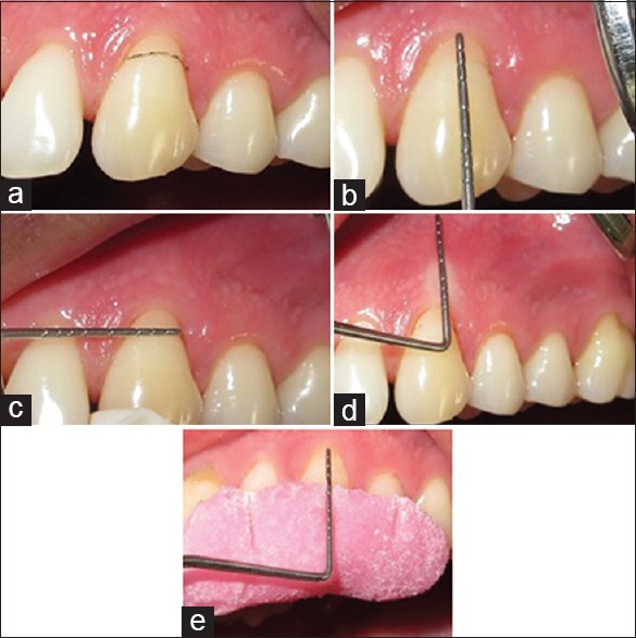
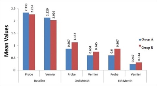
Materials and methods: Thirty sites in 15 participants with Miller's Class I or II gingival recession were randomly treated either with PET using periosteum as graft and CAF + PRF as graft. In a split mouth design, the parameters such as recession depth, recession width at cementoenamel junction, probing depth, periodontal attachment level (PAL), and keratinized gingival width were assessed at baseline, 3 months, and 6 months postoperative follow-up with William's graduated probe and Vernier caliper.

Results: Both the treatment modalities yielded statistically nonsignificant desirable treatment outcomes at both postoperative levels in terms of all the parameters The mean RC with probe method and Vernier method in CAF + PRF was 75.01% and 86.86%, respectively, and PET showed a mean RC of 61.112% and 83.971%, respectively, at 6-month interval period which showed a nonstatistically significant difference.

Conclusion: Both the treatment modalities, i.e., CAF + PRF and PET are essentially and equally effective in the treatment of Miller's Class I or II gingival recession defects.

Keywords: Coronally advanced flap; periosteum eversion technique; platelet-rich fibrin; root coverage.


Conflict of interest statement

There are no conflicts of interest.

Figures

Figure 1
Figure 1
Presurgical measurements of gingival recession defect in relation to 13 in Group A with coronally advanced flap + platelet-rich fibrin membrane by probe method. (a) Preoperative view; (b) recession depth; (c) recession width; (d) width of keratinized tissue; (e) periodontal attachment level with acrylic stent (PAL)
Figure 2
Figure 2
Presurgical measurements of gingival recession defect in relation to 13 in Group A with coronally advanced flap + platelet-rich fibrin membrane by Vernier method. (a) Recession depth; (b) recession width; (c) width of keratinized tissue
Figure 3
Figure 3
Presurgical measurements of gingival recession defect in relation to 23 in Group B with periosteum eversion technique by probe method. (a) Preoperative view; (b) recession depth; (c) recession width; (d) width of keratinized tissue; (e) periodontal attachment level with acrylic stent (PAL)
Figure 4
Figure 4
Presurgical measurements of gingival recession defect in relation to 23 in Group B with periosteum eversion technique by Vernier method: (a) Recession depth; (b) recession width; (c) width of keratinized tissue
Figure 5
Figure 5
Surgical procedure of coronally advanced flap with platelet rich fibrin membrane (a) Placement of horizontal incision joined with vertical incision and sulcular incision was placed; (b)following a full- split- full thickness flap was raised; (c) Platelet rich fibrin (PRF) membrane was placed on the gingival recession defect; (d) direct interrupted suture was placed; (e) tin foil placed and (f) periodontal pack was placed on the surgical site
Figure 6
Figure 6
Preparation of platelet rich fibrin membrane (a) Blood withdrawn from antecubital vein (b) centrifugation in REMI 4c centrifugation machine; (c) centrifuged at 2700 rpm for 12 minutes; (d) the Platelet rich fibrin (PRF) obtained; (e) Platelet rich fibrin PRF gel was squeezed between sterile gauze to form Platelet rich fibrin (PRF) membrane
Figure 7
Figure 7
Surgical procedure of periosteum eversion technique (a) Placement of horizontal incision joined with vertical incision and sulcular incision; (b) a full- split- full thickness flap was raised; (c and d) from the inner surface of flap periosteum was separated and was placed on the gingival recession defect (arrow mark); (e) direct interrupted suture was placed; (f) tin foil placed; (g) periodontal pack was placed
Figure 8
Figure 8
Postsurgical measurements of gingival recession defect in relation to 13 in Group A with coronally advanced flap with platelet-rich fibrin membrane by probe method (a) recession depth; (b) recession width; (c) width of keratinized tissue; (d) periodontal attachment level
Figure 9
Figure 9
Post surgical measurements of gingival recession defects in relation to 13 in group A with coronally advanced flp with platelet rich fibrin membrane by Vernier method (a) recession depth; (b) recession with; (c) width of keratinized tissue
Figure 10
Figure 10
Post surgical measurements of gingival recession defect in relation to 23 in relation to group B with periosteum eversion technique by probe method (a) width of keratinized tissue; (b) recession width; (c) recession depth and (d) periodontal attachment level
Figure 11
Figure 11
Post-surgical measurements of gingival recession defect in relation to 23 in group B with periosteum eversion technique by Vernier method: (a) recession depth (RD); (b) Recession width (RW); (c) Width of keratinized tissue (WKT)
Figure 12
Figure 12
Pre- and postoperative view of Group A with coronally advanced flap with platelet-rich fibrin membrane and Group B with periosteum eversion technique
Figure 13
Figure 13
Comparison of mean values of plaque index in Group A with coronally advanced flap with platelet-rich fibrin membrane and Group B with periosteum eversion technique at baseline, 3rd month, and 6th month
Figure 14
Figure 14
Comparison of mean values of gingival index in Group A with coronally advanced flap with platelet-rich fibrin membrane and Group B with periosteum eversion technique at baseline, 3rd month, and 6th month
Figure 15
Figure 15
Comparison of mean values of recession depth in Group A with coronally advanced flap with platelet rich fibrin membrane and Group B with periosteum eversion technique at baseline, 3rd month, and 6th month
Figure 16
Figure 16
Comparison of mean values of recession width in Group A with coronally advanced flap with platelet-rich fibrin membrane and Group B with periosteum eversion technique at baseline, 3rd month, and 6th month
Figure 17
Figure 17
Comparison of mean values of width of keratinized tissue in Group A with coronally advanced flap with platelet-rich fibrin membrane and Group B with periosteum eversion technique at baseline, 3rd month, and 6th month
Figure 18
Figure 18
Comparison of mean values of probing depth in Group A with coronally advanced flap with platelet-rich fibrin membrane and Group B with periosteum eversion technique at baseline, 3rd month, and 6th month
Figure 19
Figure 19
Comparison of mean values of periodontal attachment level (PAL) in Group A with coronally advanced flap with platelet-rich fibrin membrane and Group B with periosteum eversion technique at baseline, 3rd month, and 6th month
Figure 20
Figure 20
Comparison of mean values of percentage of root coverage in Group A with coronally advanced flap with platelet-rich fibrin membrane and Group B with periosteum eversion technique at 6th month interval period with probe and vernier method of measurement

References

    1. Glossary of Periodontal Terms. 4th ed. Chicago: American Academy of Periodontology; 2001. The American Academy of Periodontology; p. 44.
    1. Roccuzzo M, Bunino M, Needleman I, Sanz M. Periodontal plastic surgery for treatment of localized gingival recessions: A systematic review. J Clin Periodontol. 2002;29(Suppl 3):178–94. - PubMed
    1. Lindhe J, Lang N, Karring T. Mucogingival therapy. Periodontal plastic surgery. In: Ermes E, editor. Clinical Periodontology and Implant Dentistry. 6th ed. Oxford: Blackwell Munksgaard; 2008. pp. 995–1043.
    1. Zucchelli G, De Sanctis M. Treatment of multiple recession-type defects in patients with esthetic demands. J Periodontol. 2000;71:1506–14. - PubMed
    1. Melcher AH. On the repair potential of periodontal tissues. J Periodontol. 1976;47:256–60. - PubMed