Review
. 2018 Jan 31:2018:3804979.
doi: 10.1155/2018/3804979. eCollection 2018.

Free Radical Damage in Ischemia-Reperfusion Injury: An Obstacle in Acute Ischemic Stroke after Revascularization Therapy

Affiliations
Review

Free Radical Damage in Ischemia-Reperfusion Injury: An Obstacle in Acute Ischemic Stroke after Revascularization Therapy

Ming-Shuo Sun et al. Oxid Med Cell Longev. .

Abstract

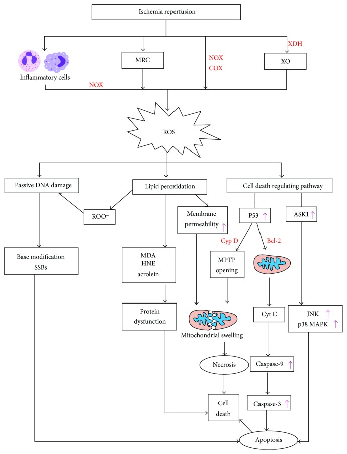
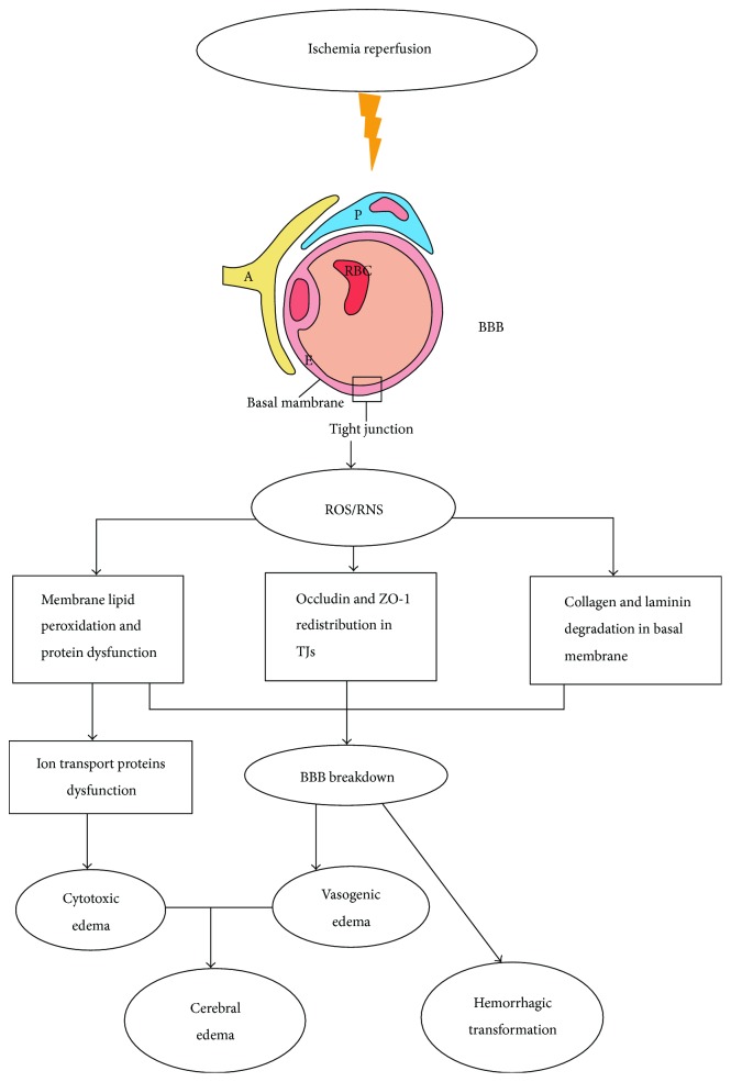
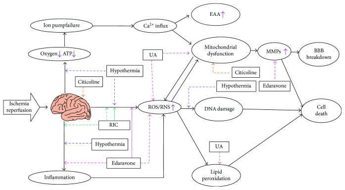
Acute ischemic stroke is a common cause of morbidity and mortality worldwide. Thrombolysis with recombinant tissue plasminogen activator and endovascular thrombectomy are the main revascularization therapies for acute ischemic stroke. However, ischemia-reperfusion injury after revascularization therapy can result in worsening outcomes. Among all possible pathological mechanisms of ischemia-reperfusion injury, free radical damage (mainly oxidative/nitrosative stress injury) has been found to play a key role in the process. Free radicals lead to protein dysfunction, DNA damage, and lipid peroxidation, resulting in cell death. Additionally, free radical damage has a strong connection with inducing hemorrhagic transformation and cerebral edema, which are the major complications of revascularization therapy, and mainly influencing neurological outcomes due to the disruption of the blood-brain barrier. In order to get a better clinical prognosis, more and more studies focus on the pharmaceutical and nonpharmaceutical neuroprotective therapies against free radical damage. This review discusses the pathological mechanisms of free radicals in ischemia-reperfusion injury and adjunctive neuroprotective therapies combined with revascularization therapy against free radical damage.


Figures

Figure 1
Figure 1
ROS damage in ischemia-reperfusion injury. First, ROS-generated pathways: MRC; NOX; COX-2; XO. ROS react with DNA and then cause passive DNA damage leading to base modification and SSBs which induce to apoptosis. Reaction of OH with unsaturated fatty acids generates ROO which may also cause passive DNA damage. The products of lipid peroxidation such as MDA, HNE, and acrolein can lead to protein dysfunction. Besides, lipid peroxidation increases membrane permeability inducing to mitochondrial swelling. P53 activated by ROS can also cause mitochondrial swelling by MPTP opening via reaction of P53 with Cyp D. P53 induces Cyt C released from mitochondria by reacting with Bcl-2 family proteins and subsequently leads to caspase cascade causing apoptosis. What is more, ROS can activate JNK and p38 MAPK pathways which are activated by ASK1 and lead to apoptosis. MRC: mitochondrial respiratory chain; NOX: NADPH oxidases; COX-2: cyclooxygenase-2; XDH: xanthine dehydrogenase; XO: xanthine oxidase; ROS: reactive oxygen species; SSBs: single-strand breaks; ROO: peroxyl radical; MDA: malondialdehyde; HNE: 4-hydroxynonenal; Cyp D: cyclophilin D; Cyt C: cytochrome C; MPTP: mitochondrial permeability transition pore; ASK1: apoptosis signal-regulating kinase 1; JNK: c-Jun NH2-terminal kinase; p38 MAPK: p38 mitogen-activated protein kinase. “↑” demonstrates events that are increased or enhanced.
Figure 2
Figure 2
Complications after revascularization therapy. Severe complications include hemorrhagic transformation and cerebral edema. Hemorrhagic transformation is connected with the increase in the permeability of BBB. H2O2 generated from NADPH oxidase modifies tight junctions by redistribution of occludin and ZO-1. NO/ONOO degrades collagen and laminin in the basal membrane by the activation of MMP pathways. Free radicals also cause lipid peroxidation and protein dysfunction. All these pathophysiologic processes lead to BBB breakdown and subsequently result in hemorrhagic transformation and vasogenic edema. Cytotoxic edema is associated with the dysfunction of the ion transport in the membranes which suffer lipid peroxidation and protein oxidation. BBB: blood-brain barrier; TJs: tight junctions. The meaning of letter in blood-brain barrier: E: endothelial cells; P: pericytes; A: astrocyte end feet.
Figure 3
Figure 3
Adjunctive therapies against free radical damage in ischemia-reperfusion injury. RIC improves cerebral blood flow by increase in the generation of NO and inhibits the activation of NADPH oxidase in neutrophils. Hypothermia decreases the generation of free radicals, inhibits the induction of oxidative DNA lesions, and suppresses immune system. Edaravone eliminates free radicals and enhances the release of NO. Besides, edaravone also inhibits inflammatory responses and MMP cascade. UA can scavenge free radicals and suppress lipid peroxidation. Citicoline plays a key role in maintenance of membrane against free radical damage. Dashed line indicates inhibition, whereas real line indicates enhancement. UA: uric acid; RIC: remote ischemic conditioning; EAA: excitatory amino acid. “↑” demonstrates events that are increased or enhanced.

References

    1. Jauch E. C., Saver J. L., Adams H. P., Jr., et al. Guidelines for the early management of patients with acute ischemic stroke: a guideline for healthcare professionals from the American Heart Association/American Stroke Association. Stroke. 2013;44(3):870–947. doi: 10.1161/STR.0b013e318284056a. - DOI - PubMed
    1. Towfighi A., Saver J. L. Stroke declines from third to fourth leading cause of death in the United States: historical perspective and challenges ahead. Stroke. 2011;42(8):2351–2355. doi: 10.1161/STROKEAHA.111.621904. - DOI - PubMed
    1. Feigin V. L., Forouzanfar M. H., Krishnamurthi R., et al. Global and regional burden of stroke during 1990-2010: findings from the global burden of disease study 2010. The Lancet. 2014;383(9913):245–255. doi: 10.1016/S0140-6736(13)61953-4. - DOI - PMC - PubMed
    1. Ovbiagele B., Goldstein L. B., Higashida R. T., et al. Forecasting the future of stroke in the United States: a policy statement from the American Heart Association and American Stroke Association. Stroke. 2013;44(8):2361–2375. doi: 10.1161/STR.0b013e31829734f2. - DOI - PubMed
    1. Röther J., Ford G. A., Thijs V. N. S. Thrombolytics in acute ischaemic stroke: historical perspective and future opportunities. Cerebrovascular Diseases. 2013;35(4):313–319. doi: 10.1159/000348705. - DOI - PubMed

MeSH terms