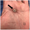
Solitary Palmar Keratoacanthoma: Case Report
- PMID: 29770284
- PMCID: PMC5953505
- DOI: 10.7759/cureus.2331
Solitary Palmar Keratoacanthoma: Case Report
Abstract
Keratoacanthoma (KA) is a squamous neoplasm exhibiting a triphasic growth pattern involving rapid growth, stabilization, and eventual spontaneous resolution. Historically, keratoacanthomas were thought to originate on hair-bearing skin or sun-exposed surfaces. However, recent reports demonstrate that they can occur on the mucous membranes, subungual regions, and palms and soles. We report a 74-year-old man who developed a KA on the left palmar surface after minor trauma, for which he underwent Mohs' micrographic surgery. A literature review for the terms: keratoacanthoma, palm, palmar, volar, plantar, and sole resulted in only four reported cases of solitary or giant KA of the palms and soles; excluding our patient, all of the cases occurred on the plantar foot. A number of reports describe palmar KA in the context of multiple lesions occurring simultaneously. However, to our knowledge, our patient represents the first reported case of a solitary palmar KA in the literature. The features of follicular and non-follicular keratoacanthomas (KAs) and their association with trauma are discussed.
Keywords: keratoacanthoma; mohs micrographic surgery; non-follicular; palmar; trauma.
Conflict of interest statement
The authors have declared that no competing interests exist.
Figures


References
-
- Keratoacanthoma (KA): an update and review. Kwiek B, Schwartz RA. J Am Acad Dermatol. 2016;74:1220–1233. - PubMed
-
- Keratoacanthoma of the foot. Case presentation and review of the literature. Cohn I, Kanat IO. J Am Podiatr Med Assoc. 1985;75:653–655. - PubMed
-
- Giant keratoacanthoma of the plantar foot: a report of two cases. Hale DS, Dockery GL. https://www.ncbi.nlm.nih.gov/pubmed/8318965. J Foot Ankle Surg. 1993;32:75–84. - PubMed
-
- Keratoacanthoma: case presentation and discussion. Strauss H, Potter GK. http://www.ncbi.nlm.nih.gov/pubmed/263082. J Foot Surg. 1979;18:72–74. - PubMed
-
- Multiple palmar keratoacanthomas treated with acitretin. Rosenblum GA. https://www.ncbi.nlm.nih.gov/pubmed/17373152. J Drugs Dermatol. 2006;5:1006–1009. - PubMed
Publication types
LinkOut - more resources
Full Text Sources
Other Literature Sources
