Review
. 2018 Sep;36(9):1329-1340.
doi: 10.1002/stem.2852. Epub 2018 Jul 29.

Concise Review: Current Status of Three-Dimensional Organoids as Preclinical Models

Affiliations
Review

Concise Review: Current Status of Three-Dimensional Organoids as Preclinical Models

Garima Kaushik et al. Stem Cells. 2018 Sep.

Abstract

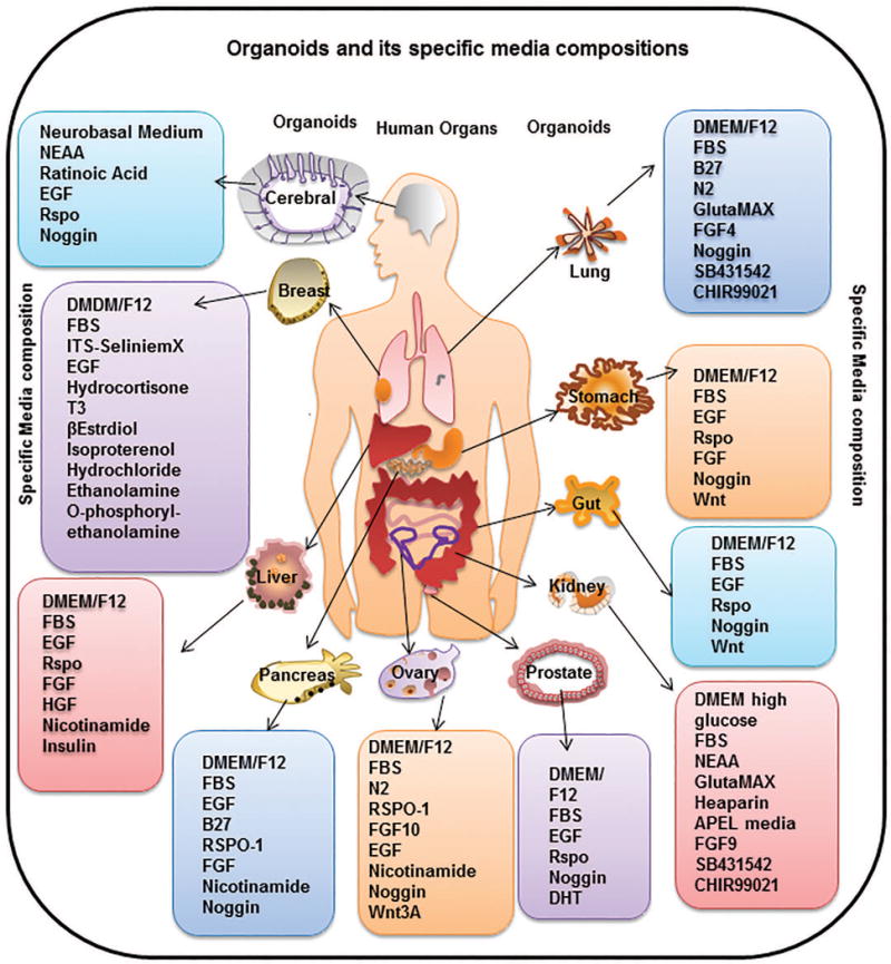
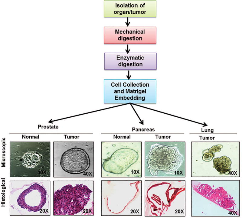
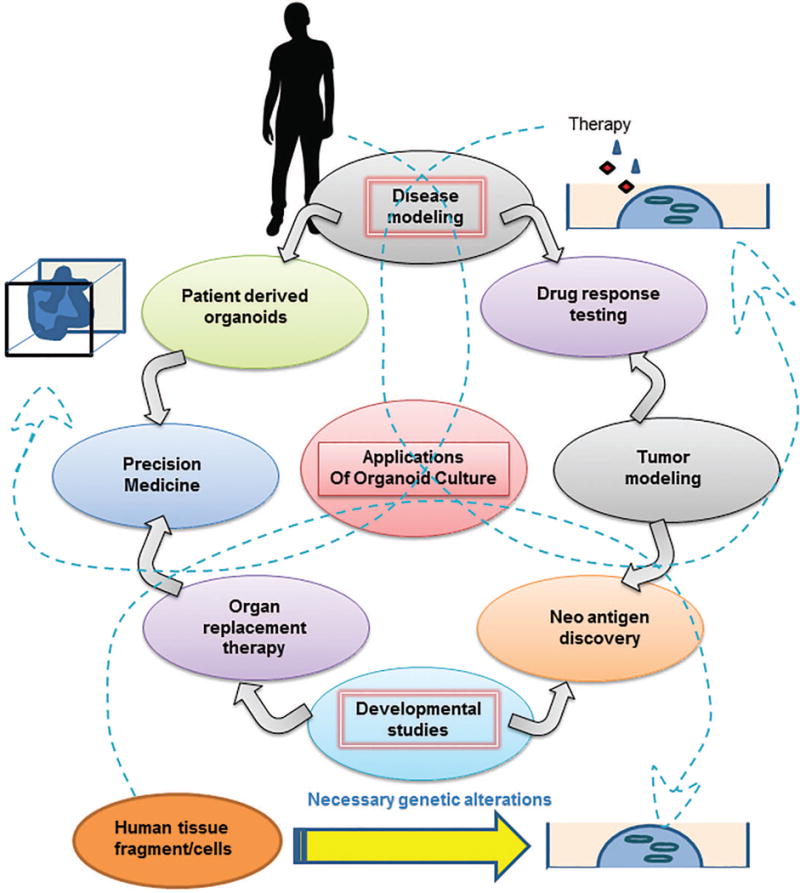
Three-dimensional (3D) cultures use the property of some cells to self-organize in matrices and generate structures that can be programmed to represent an organ or a pathology. Organoid cultures are the 3D cultivation of source tissue (ranging from cells to tissue fragments) in a support matrix and specialized media that nearly resembles the physiological environment. Depending on the source tissue, growth factors, and inhibitors provided, organoids can be programmed to recapitulate the biology of a system and progression of pathology. Organoids are genetically stable, and genetically amenable, making them very suitable tools to study tissue homeostasis and cancer. In this Review, we focus on providing recent technical advances from published literature to efficiently use organoids as a tool for disease modeling and therapeutics. Also, we discuss stem cell biology principles used to generate multiple organoids and their characteristics, with a brief description of methodology. A major theme of this review is to expand organoid applications to the study disease progression and drug response in different cancers. We also discuss shortcomings, limitations, and advantages of developed 3D cultures, with the rationale behind the methodology. Stem Cells 2018;36:1329-1340.

Keywords: Adult stem cells; Differentiation; Experimental models; Induced pluripotent stem cells; Organoids; Stem cell culture; Three-dimensional culture; Tumoroid.


Conflict of interest statement

DISCLOSURE OF POTENTIAL CONFLICT OF INTEREST

The authors indicated no potential conflict of interest.

Figures

Fig. 1
Fig. 1. Representation of the organoids generated, and the media compositions required
These hosts of organoids have been generated from different source materials, including iPSCs, AdSC, embryonic tissues or cells and adult tissue explants. Different media compositions are required for each type of source material used and the type of differentiation to be achieved (organ specific), which is elaborated in detail in the text. Specifically, cerebral organoids need a stepwise incubation of PSC in neural induction media (DMEM-F12, N2 supplement, GlutaMAX supplement, MEM-NEAA, heparin) followed by cerebral induction media (DMEM-F12, Neurobasal medium, N2 supplement, insulin, GlutaMAX supplement, MEM-NEAA, penicillin-streptomycin, 2-mercaptoethanol, B27 supplement). Mammary organoids can be developed from tissue fragments using media composed of DMEM/F12, FBS, ITS Selenite media supplement, FGF2, FGF10 for mouse or EpiCult B medium supplemented with hydrocortisone, insulin, FGF10, HGF for humans. Liver organoids can be generated by mixing tissue fragments in DMEM/F12 media supplemented with FBS, EGF, RSPO1, FGF, HGF, Nicotinamide, and insulin. Pancreatic organoids need a media comprising of DMEM/F12, B27 supplement, Nicotinamide, Noggin, EGF, FGF and RSPO1. Ovarian organoids are generated by seeding fallopian epithelial cells in matrigel with media comprising AdDMEM/F12, Wnt3A, RSPO1, HEPES, GlutaMAX, B27, N2 Supplement, EGF, noggin, FGF10, Nicotinamide, Y-27632, and SB431542. Prostate organoids need a media containing DMEM/F12, B27 Supplement, N-acetylcysteine, EGF, Noggin, RSPO1, A83-01, and DHT. Kidney organoids need a media containing DMEM high glucose, FBS, NEAA, GlutaMAX, Heaparin, APEL media, FGF9, SB431542 and CHIR99021. Gut or intestinal organoids need a media composition of DMEM/F12, FBS, B27, EGF, RSPO1, Noggin and Wnt. Specific cultivation of stomach organoids need media composition same as intestinal organoids with addition of FGF. Lung organoids can be generated and grown in media containing DMEM/F12, FBS, B27, N2 Supplement, GlutaMAX, FGF4, Noggin, SB431542 and CHIR99021. Abbreviation used are: Y-27632:ROCK inhibitor, SB431542:TGF-β R Kinase Inhibitor IV, ITS: Insulin Transferrin-Sodium, NEAA: Non Essential Amino Acid Culture Supplement, EGF: Epidermal Growth Factor, RSPO1: R-spondin-1, Wnt3A: Wingless-Type MMTV Integration Site Family Member 3A, T3: Triiodothyronine, FBS: Fetal Bovine Serum, FGF: Fibroblast Growth Factor, HGF: Hepatocyte Growth factor, DMEM/F12: Dulbecco’s Modified Eagle Medium: Nutrient Mixture F-12, DHT: Dihydrotestosterone, CHIR99021: glycogen synthase kinase 3 inhibitor.
Fig. 2
Fig. 2. General scheme of generating organoids and representative figures of organoids generated by our laboratory
The flowchart represents the scheme of organoid isolation which is modified for each organoid according to the organ or tissue architecture to generate submerged organoids. Briefly, desired source tissue (progenitor cells or tissue fragments) is isolated from host by mincing the organ and then subjecting it to enzymatic digestion. The digestion media composition and the digestion protocol are decided depending on the host tissue. The digestion media usually contains a mixture or Dispase and Collagenase or Collagenase alone and can take from 30 mins to 4-6 hours. Following digestion, the cells are mixed in the matrix (like matrigel or collagen) suitable for the desired organoids. A suitable media is overlaid once the matrix solidifies. Once generated, organoids grow in ductal like morphologies like their human counter parts. Picture panels depict the microscopic pictures of organoids generated in our lab from normal and cancerous prostate and pancreas as well as lung cancer organoids (upper panel) along with hematoxylin and eosin stained sections of the same (lower panel) depicting the difference in organization of cells in each of these organoids. Figure magnifications are mentioned on each of the figures.
Fig. 3
Fig. 3. Potential applications of organoids generated
The figure presents prospective applications of organoid culture tool for advancement of biological research. The arrows in the figure represent the flow of information from tumor modeling, disease modeling and developmental biology studies towards therapeutic interventions. To expand since organoids, represent tissue homeostasis in vitro, they can be used to model pathologies by inducing desired mutations or exposing them to necessary stimulus or pathogens. Following which the pathogenesis and disease development can be studied. Such studies facilitate further research to study drug response or generate organoids directly from patients to device personalized therapeutic strategy as represented by the arrows emerging from disease modeling bubble. Similarly, modeling cancer initiation and progression in organoids can facilitate therapeutic response studies and discovery of new oncogenic proteins or antigens that can be targeted illustrated by arrows emerging from Tumor modeling bubble. Additionally, lineage tracing studies or organ development studies using organoids have immense potential for the field of organ replacement therapy and can help neo-antigen discovery for cancer research (arrows emerging from developmental biology studies bubble). The dashed arrows represent the overlapping domains amongst these applications as explained above.

References

    1. Baker LA, Tiriac H, Clevers H, et al. Modeling pancreatic cancer with organoids. Trends Cancer. 2016;2:176–190. - PMC - PubMed
    1. Campbell JJ, Davidenko N, Caffarel MM, et al. A multifunctional 3D co-culture system for studies of mammary tissue morphogenesis and stem cell biology. PLoS One. 2011;6:e25661. - PMC - PubMed
    1. Dye BR, Hill DR, Ferguson MA, et al. In vitro generation of human pluripotent stem cell derived lung organoids. Elife. 2015;4 - PMC - PubMed
    1. Huch M. Building stomach in a dish. Nat Cell Biol. 2015;17:966–967. - PubMed
    1. Lindborg BA, Brekke JH, Vegoe AL, et al. Rapid Induction of Cerebral Organoids From Human Induced Pluripotent Stem Cells Using a Chemically Defined Hydrogel and Defined Cell Culture Medium. Stem Cells Transl Med. 2016;5:970–979. - PMC - PubMed

Publication types