. 2018 Jun;27(6):967-978.
doi: 10.1177/0963689718759649. Epub 2018 May 17.

Gingival Mesenchymal Stem Cells Outperform Haploidentical Dental Pulp-derived Mesenchymal Stem Cells in Proliferation Rate, Migration Ability, and Angiogenic Potential

Affiliations

Gingival Mesenchymal Stem Cells Outperform Haploidentical Dental Pulp-derived Mesenchymal Stem Cells in Proliferation Rate, Migration Ability, and Angiogenic Potential

Ioannis Angelopoulos et al. Cell Transplant. 2018 Jun.

Abstract

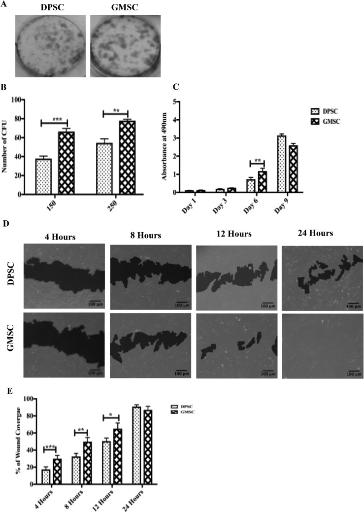
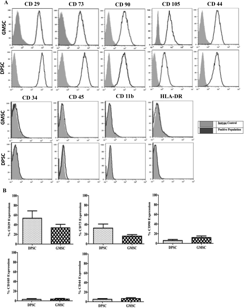
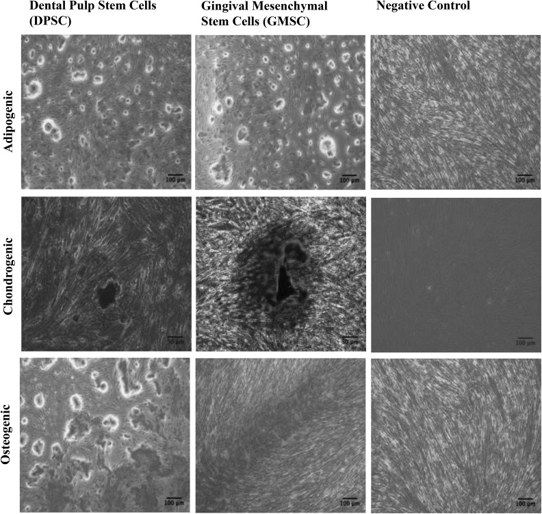
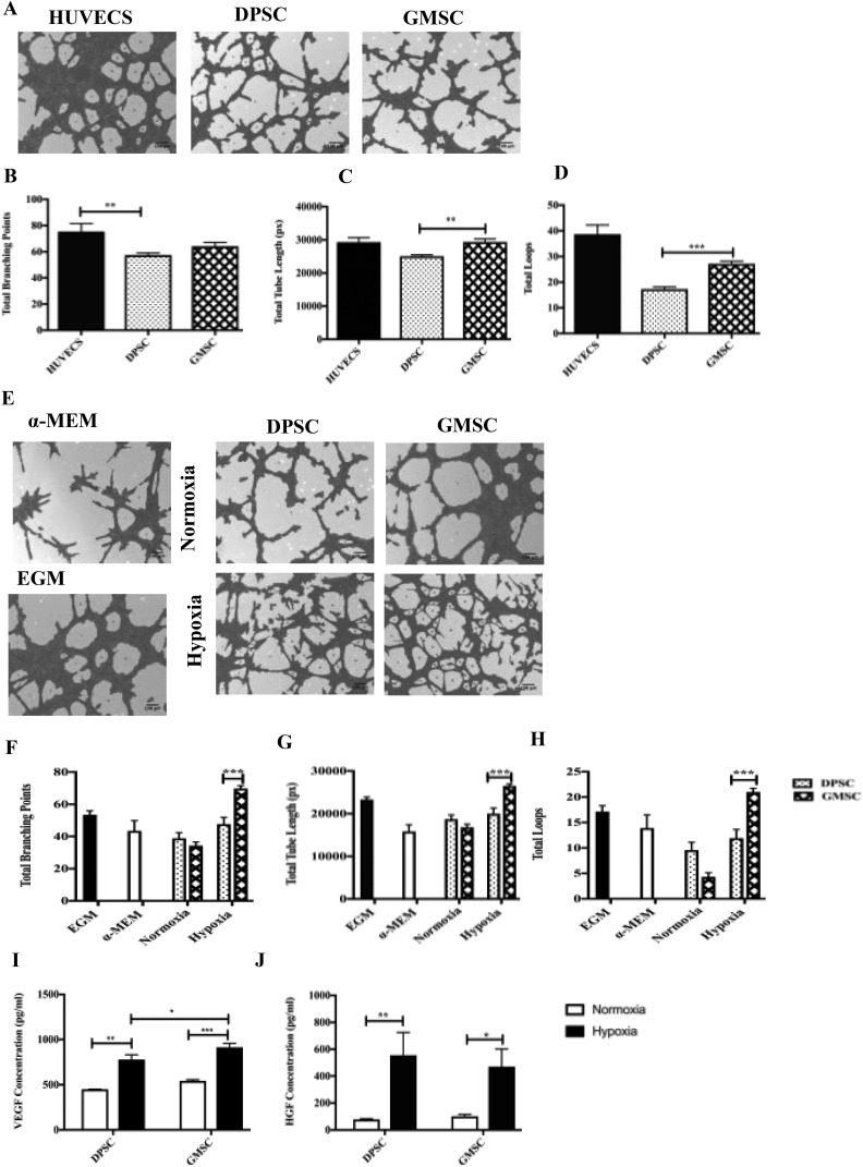
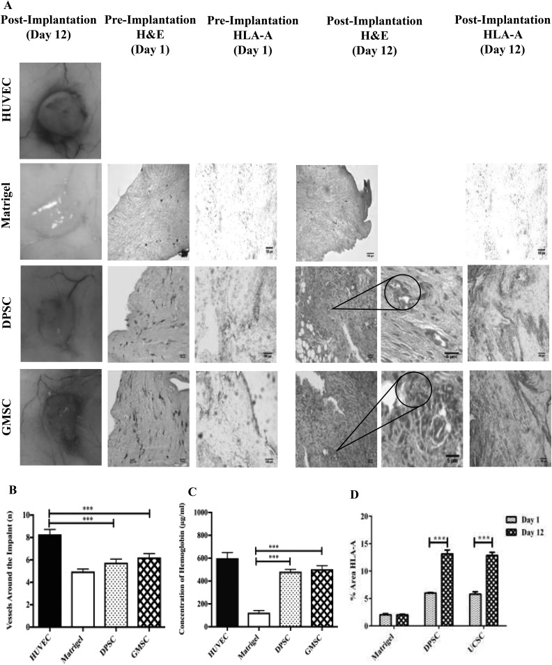
High donor variation makes comparison studies between different dental sources dubious. Dental tissues offer a rare opportunity for comparing the biological characteristics of haploidentical mesenchymal stem cells (MSCs) isolated from the same donor. The objective was to identify the optimal dental source of MSCs through a biological and functional comparison of haploidentical MSCs from gingival (GMSCs) and dental pulp stem cells (DPSCs) focusing mainly on their angiogenic potential. The comparison study included (1) surface markers expression, (2) mesodermal differentiation capacity (chondrogenic, adipogenic, and osteogenic), (3) proliferation, (4) migration potential, (5) ability to form colony units, and (6) angiogenic potential in vitro and in vivo. Comparative analysis showed no difference in the immunophenotypic profile nor for the trilineage differentiation potential. Proliferation of GMSCs was higher than DPSCs at day 6 (2.6-fold higher, P < 0.05). GMSCs showed superior migratory capacity compared to DPSCs at 4, 8, and 12 h (2.1-, 1.5-, and 1.2-fold higher, respectively, P < 0.05). Furthermore, GMSCs formed a higher number of colony units for both cell concentrations (1.7- and 1.4-fold higher for 150 and 250 starting cells, respectively, P < 0.05). GMSCs showed an improved angiogenic capacity compared to DPSCs (total tube lengths 1.17-fold higher and 1.5-fold total loops, P < 0.05). This was correlated with an enhanced release of vascular growth factor under hypoxic conditions. Finally, in the plug transplantation assay evaluating the angiogenesis in vivo, the DPSC and GMSC hemoglobin content was 3.9- and 4-fold higher, respectively, when compared to the control (Matrigel alone). GMSCs were superior to their haploidentical DPSCs in proliferation, migration, and angiogenic potentials. This study positions GMSCs in the forefront of dental cell sources for applications in regenerative medicine.

Keywords: angiogenesis; dental and gingival mesenchymal stem cells; haploidentical; migration.


Conflict of interest statement

Declaration of Conflicting Interests: The author(s) declared the following potential conflicts of interest with respect to the research, authorship, and/or publication of this article: Maroun Khoury is the chief scientific officer of Cells for Cells and REGENERO. The other authors declare no conflicts of interest.

Figures

Fig. 1.
Fig. 1.
Gingival mesenchymal stem cells (GMSCs) and dental pulp stem cells (DPSCs) showed different clonogenic and proliferation potentials. (A) Representative images of colony-forming units (CFUs) stained with crystal violet after 20 d in culture. (B) An increase in the formation of CFUs was observed for both concentrations (150 cells and 250 cells) for GMSCs compared to DPSCs with a P < 0.05. (C) Quantification of cell proliferation between DPSCs and GMSCs incubated at different time points (1, 3, 6, and 9 d). An increase in the proliferation of GMSCs compared to DPSCs was observed between day 6 compared to DPSCs with a P < 0.05. (D) In vitro migration comparison between DPSCs and GMSCs based on a 24-h scratch wound healing assay. (E) GMSCs display a better migratory capacity compared to DPSCs for 4, 8, and 12 h (P < 0.05). At 24 h, no significant change in the proliferation was observed. All data are represented as a mean with the associated standard error of the mean (n = 3) of a minimal 3 donors.
Fig. 2.
Fig. 2.
Gingival mesenchymal stem cells and dental pulp stem cells express common mesenchymal stem cell (MSC) markers. (A) MSCs were stained with labeled monoclonal antibodies against known MSC surface markers (blue) and their respective isotypes (gray), cells were analyzed by flow cytometry. All MSCs were positive for CD29, CD73, CD90, CD105, and CD44 and negative for CD34, CD11b, CD45, and human leukocyte antigen-DR. (B) No significant difference was observed for CD29, CD73, CD90, CD105, and CD44. All data are represented as a mean with the associated standard error of the mean (n = 3) of a minimal 3 donors.
Fig. 3.
Fig. 3.
Dental pulp stem cells and gingival mesenchymal stem cells display similar mesodermal differentiation potential. Images illustrating mesenchymal stem cell trilineage differentiation following incubation with differentiation medium for 30 d and stained with Oil Red O (adipocytes), Alizarin red (osteocytes), and Safranin O (chondrocytes).
Fig. 4.
Fig. 4.
In vitro angiogenesis comparison between dental pulp stem cells (DPSCs) and gingival mesenchymal stem cells (GMSCs) based on a 5-h culture in the Matrigel. (A) Images were analyzed using a Wimasis software. GMSCs were shown a higher potential to form (C) tube-like structure (P < 0.05) and (D) total loops (P < 0.05) in Matrigel-coating cultures compared to DPSC. (B) No statistical difference was observed in the formation of total branching points between DPSCs and GMSCs (b). Human umbilical vein endothelial cells were used as a positive control. All data are represented as a mean with the associated standard error of the mean (n = 3) of a minimal 3 donors. (E) In vitro angiogenesis comparison between DPSC and GMSC-conditioned media (CM) under hypoxic and normoxic conditions. The GMSC-CM under hypoxic conditions were shown a better potential to form (F) total branching points (P < 0.05), (G) total tube length (P < 0.05), and (H) total loops (P < 0.05) conditions compared to supernatant of DPSCs. Endothelial cell growth medium (EGM) and α-minimum essential medium Eagle were used as a control (E). An ELISA was performed to measure (I) protein levels of vascular growth factor (P < 0.05), (J) protein levels of hepatocyte growth factor (P < 0.05). All data are represented as a mean with the associated standard error of the mean (n = 3) of a minimal 3 donors.
Fig. 5.
Fig. 5.
Comparison of the angiogenic potential of dental pulp stem cells (DPSCs) and gingival mesenchymal stem cells (GMSCs) in a mouse plug assay model. In order to determine the angiogenic capacity between DPSCs and GMSCs, a Matrigel plug assay was performed in NOD.Cg-Prkdcscid Il2rgtm1Wjl/SzJ mice. The mice were divided into 4 different groups, namely, human umbilical vein endothelial cells (positive control), Matrigel (negative control), DPSCs, and GMSCs. The different cells (2 × 10⁶) were mixed with a growth factor reduced Matrigel and implanted subcutaneously. At 15-d posttransplantation, the implants were harvested and (A) images were taken, and (B) quantification of the vessels around the implant was performed using the ImageJ software (P < 0.05). (C) Also a quantification of the hemoglobin content (μg/mL) was performed using Drabkin’s reagent at different concentrations (P < 0.05). Histological staining (A) Matrigel implants containing DPSCs or GMSCs were evaluated at 12-d postsubcutaneous implantation in mice. Macroscopic view of explanted Matrigel plugs. Hematoxylin and eosin (H&E)-staining of implants containing DPSCs, GMSCs, or Matrigel alone (control) preimplantation and 12-d post implantation (20× and 40× magnification). H&E-staining showing (40× magnification) high-power view of 1 microvessel containing hematopoietic cells. (A) Human leukocyte antigen (HLA-A) immunostaining revealed the presence of human mesenchymal stem cells within the Matrigel at days 1 and 12. (D) The amount of HLA-A expression was measured using Image J, showing an increase for both DPSCs and GMSCs at day 12 (postimplantation) in comparison to day 1 (preimplantation; P < 0.05).

References

    1. Chen FM, Sun HH, Lu H, Yu Q. Stem cell-delivery therapeutics for periodontal tissue regeneration. Biomaterials. 2012;33(27):6320–6344. - PubMed
    1. Liu Y, Zheng Y, Ding G, Fang D, Zhang C, Bartold PM, Gronthos S, Shi S, Wang S. Periodontal ligament stem cell-mediated treatment for periodontitis in miniature swine. Stem Cells. 2008;26(4):1065–1073. - PMC - PubMed
    1. Feng F, Akiyama K, Liu Y, Yamaza T, Wang TM, Chen JH, Wang BB, Huang GT, Wang S, Shi S. Utility of PDL progenitors for in vivo tissue regeneration: a report of 3 cases. Oral Dis. 2010;16(1):20–28. - PMC - PubMed
    1. Yang Y, Rossi FM, Putnins EE. Periodontal regeneration using engineered bone marrow mesenchymal stromal cells. Biomaterials. 2010;31(33):8574–8582. - PubMed
    1. Tobita M, Uysal AC, Ogawa R, Hyakusoku H, Mizuno H. Periodontal tissue regeneration with adipose-derived stem cells. Tissue Eng Part A. 2008;14(6):945–953. - PubMed

Publication types

LinkOut - more resources