. 2018 Sep;14(3):235-243.
doi: 10.1007/s11302-018-9609-4. Epub 2018 May 16.

Neuronal adenosine A2A receptor overexpression is neuroprotective towards 3-nitropropionic acid-induced striatal toxicity: a rat model of Huntington's disease

Affiliations

Neuronal adenosine A2A receptor overexpression is neuroprotective towards 3-nitropropionic acid-induced striatal toxicity: a rat model of Huntington's disease

Maria Rosaria Domenici et al. Purinergic Signal. 2018 Sep.

Abstract

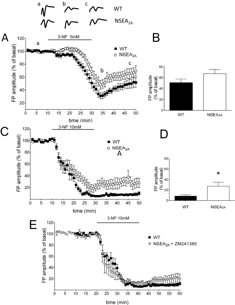
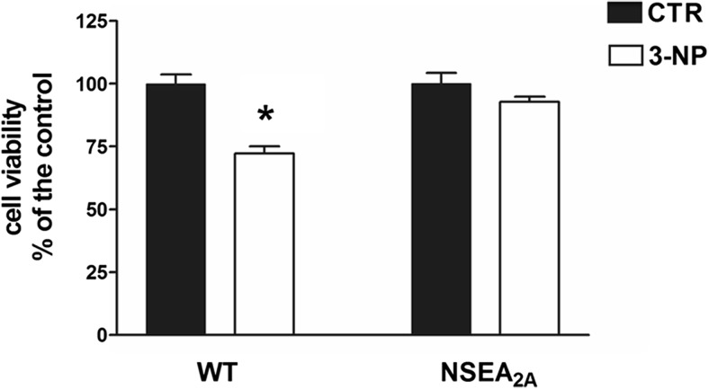
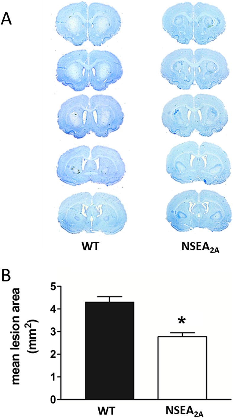
The A2A adenosine receptor (A2AR) is widely distributed on different cellular types in the brain, where it exerts a broad spectrum of pathophysiological functions, and for which a role in different neurodegenerative diseases has been hypothesized or demonstrated. To investigate the role of neuronal A2ARs in neurodegeneration, we evaluated in vitro and in vivo the effect of the neurotoxin 3-nitropropionic acid (3-NP) in a transgenic rat strain overexpressing A2ARs under the control of the neural-specific enolase promoter (NSEA2A rats). We recorded extracellular field potentials (FP) in corticostriatal slice and found that the synaptotoxic effect of 3-NP was significantly reduced in NSEA2A rats compared with wild-type animals (WT). In addition, after exposing corticostriatal slices to 3-NP 10 mM for 2 h, we found that striatal cell viability was significantly higher in NSEA2A rats compared to control rats. These in vitro results were confirmed by in vivo experiments: daily treatment of female rats with 3-NP 10 mg/kg for 8 days induced a selective bilateral lesion in the striatum, which was significantly reduced in NSEA2A compared to WT rats. These results demonstrate that the overexpression of the A2AR selectively at the neuronal level reduced 3-NP-induced neurodegeneration, and suggest an important function of the neuronal A2AR in the modulation of neurodegeneration.

Keywords: 3-Nitropropionic acid; Adenosine A2A receptors; Huntington’s disease; Striatum; Synaptic transmission.


Conflict of interest statement

Conflicts of interest

The authors declare that they have no conflicts of interest.

Ethical approval

All applicable international, national, and/or institutional guidelines for the care and use of animals were followed.

Figures

Fig. 1
Fig. 1
Electrophysiological experiments showing the effect of 3-NP on synaptic transmission in corticostriatal slices. a The concentration of 5 mM 3-NP, applied for 20 min, induced a reduction of synaptic transmission in both WT (n = 9) and NSEA2A (n = 8) rats, that partially recovered 20 min after the washout, with no difference between the two genotypes (b); c slice perfusion with 3-NP 10 mM induced a stronger effect in both WT (n = 8) and NSEA2A (n = 8) and the recovery 20 min after 3-NP washout was significantly higher in NSEA2A than in WT (d); the application of ZM 241385 to corticostriatal slices from NSEA2A rats (1 μM, 10 min before and along with 3-NP, n = 4) worsened 3-NP-induced synaptic depression and, as a result, the effect of 3-NP was comparable to that in WT slices (e). Each point represents the mean of three responses; insets in a show FPs before, during, and after 3-NP application in WT and NSEA2A rats. Horizontal bars indicate the period of drug application. *p ≤ 0.05 vs. WT, Student’s t test
Fig. 2
Fig. 2
Different effect of 3-NP on striatal cell viability in corticostriatal slices from WT and NSEA2A rats. Corticostriatal slices were incubated for 2 h with 3-NP (10 mM) and then cell viability of the striatal area was evaluated by MTT assay. Bar graphs show the mean ± S.E.M. of cell viability in WT (n = 12) and NSEA2A (n = 9) slices with respect to control values. *p ≤ 0.05 vs. CTR, Student’s t test
Fig. 3
Fig. 3
Histological evaluation of 3-NP lesions in WT and NSEA2A rats. a Serial brain sections obtained from WT and NSEA2A rats showing striatal lesions (uncolored areas) induced by i.p. injection of 3-NP. b Bar graphs represent the mean of the lesioned areas (mm2) in WT (n = 6) and NSEA2A (n = 5) rats calculated by Image J software. *p ≤ 0.05 vs. WT, Mann–Whitney U test

References

    1. Fredholm BB, IJzerman AP, Jacobson KA, Klotz KN, Linden J. International Union of Pharmacology. XXV. Nomenclature and classification of adenosine receptors. Pharmacol Rev. 2001;53:527–552. - PMC - PubMed
    1. Fredholm BB, IJzerman AP, Jacobson KA, Linden J, Müller CE. International Union of Basic and Clinical Pharmacology. LXXXI. Nomenclature and classification of adenosine receptors—an update. Pharmacol Rev. 2011;63:1–34. doi: 10.1124/pr.110.003285. - DOI - PMC - PubMed
    1. Svenningsson P, Hall H, Sedvall G, Fredholm BB. Distribution of adenosine receptors in the postmortem human brain: an extended autoradiographic study. Synapse. 1997;27:322–335. doi: 10.1002/(SICI)1098-2396(199712)27:4<322::AID-SYN6>3.0.CO;2-E. - DOI - PubMed
    1. Chen JF, Sonsalla PK, Pedata F, Melani A, Domenici MR, Popoli P, Geiger J, Lopes LV, de Mendonca A. Adenosine A2A receptors and brain injury: broad spectrum of neuroprotection, multifaceted actions and “fine tuning” modulation. Prog Neurobiol. 2007;83:310–331. doi: 10.1016/j.pneurobio.2007.09.002. - DOI - PubMed
    1. Gomes CV, Kaster MP, Tome AR, Agostinho PM, Cunha RA. Adenosine receptors and brain diseases: neuroprotection and neurodegeneration. Biochim Biophys Acta. 2011;1808:1380–1399. doi: 10.1016/j.bbamem.2010.12.001. - DOI - PubMed