

Visual impairment and progressive phthisis bulbi caused by recessive pathogenic variant in MARK3
- PMID: 29771303
- PMCID: PMC6048992
- DOI: 10.1093/hmg/ddy180
Visual impairment and progressive phthisis bulbi caused by recessive pathogenic variant in MARK3
Abstract
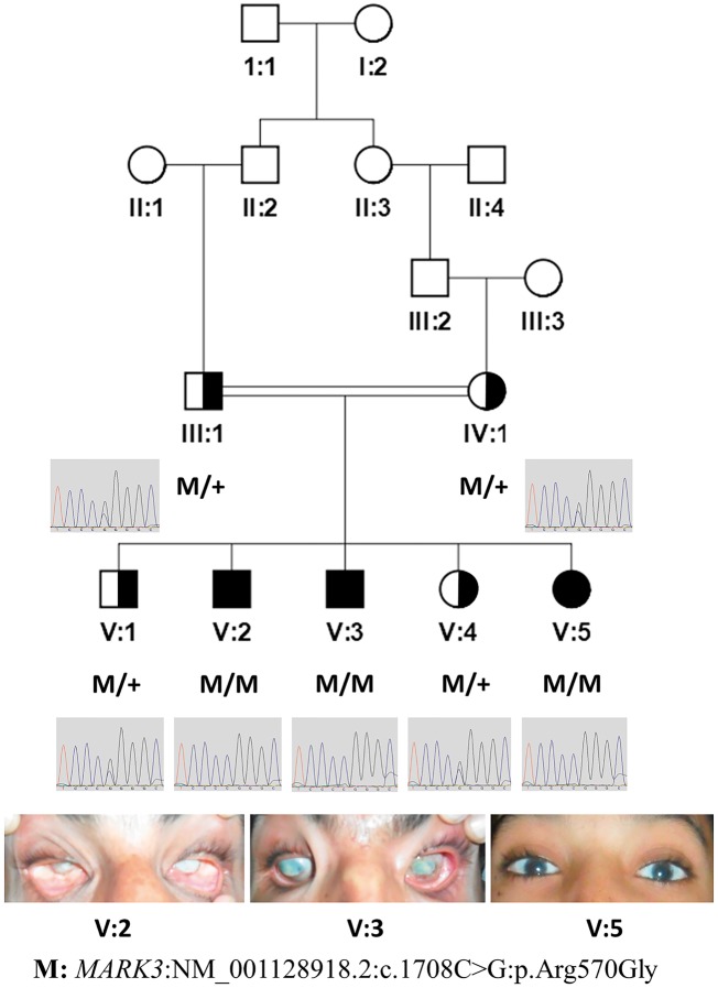
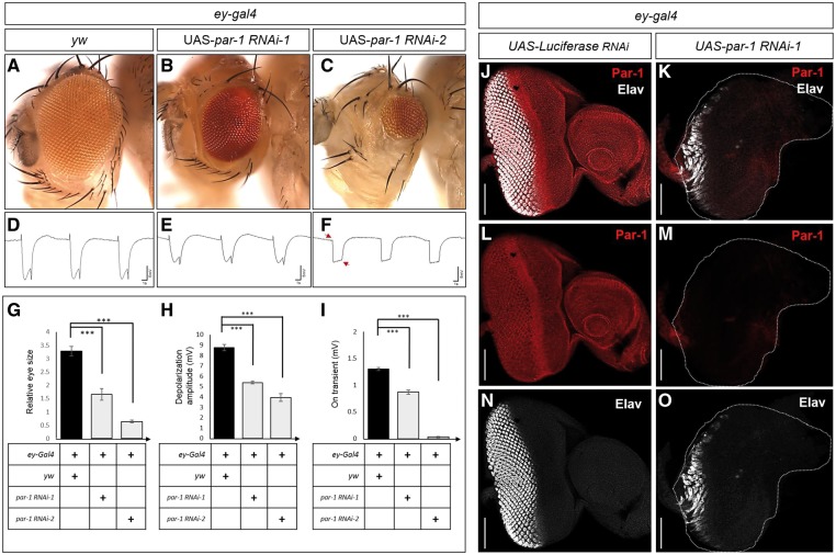
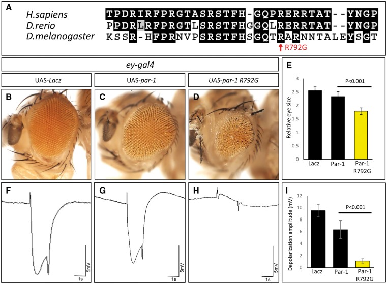
Developmental eye defects often severely reduce vision. Despite extensive efforts, for a substantial fraction of these cases the molecular causes are unknown. Recessive eye disorders are frequent in consanguineous populations and such large families with multiple affected individuals provide an opportunity to identify recessive causative genes. We studied a Pakistani consanguineous family with three affected individuals with congenital vision loss and progressive eye degeneration. The family was analyzed by exome sequencing of one affected individual and genotyping of all family members. We have identified a non-synonymous homozygous variant (NM_001128918.2: c.1708C > G: p.Arg570Gly) in the MARK3 gene as the likely cause of the phenotype. Given that MARK3 is highly conserved in flies (I: 55%; S: 67%) we knocked down the MARK3 homologue, par-1, in the eye during development. This leads to a significant reduction in eye size, a severe loss of photoreceptors and loss of vision based on electroretinogram (ERG) recordings. Expression of the par-1 p.Arg792Gly mutation (equivalent to the MARK3 variant found in patients) in developing fly eyes also induces loss of eye tissue and reduces the ERG signals. The data in flies and human indicate that the MARK3 variant corresponds to a loss of function. We conclude that the identified mutation in MARK3 establishes a new gene-disease link, since it likely causes structural abnormalities during eye development and visual impairment in humans, and that the function of MARK3/par-1 is evolutionarily conserved in eye development.
© The Author(s) 2018. Published by Oxford University Press. All rights reserved. For permissions, please email: journals.permissions@oup.com.
Figures
Similar articles
-
Homozygous Variant in ARL3 Causes Autosomal Recessive Cone Rod Dystrophy.Invest Ophthalmol Vis Sci. 2019 Nov 1;60(14):4811-4819. doi: 10.1167/iovs.19-27263. Invest Ophthalmol Vis Sci. 2019. PMID: 31743939 Free PMC article.
-
A mutation in DOP1B identified as a probable cause for autosomal recessive Peters anomaly in a consanguineous family.Mol Vis. 2020 Nov 25;26:757-765. eCollection 2020. Mol Vis. 2020. PMID: 33273802 Free PMC article.
-
Whole exome sequencing identified a novel missense alteration in CC2D2A causing Joubert syndrome 9 in a Pakhtun family.J Gene Med. 2021 Jan;23(1):e3279. doi: 10.1002/jgm.3279. Epub 2020 Oct 27. J Gene Med. 2021. PMID: 32989887
-
Identification of a novel truncating variant in AHI1 gene and a brief review on mutations spectrum.Mol Biol Rep. 2021 Jun;48(6):5339-5345. doi: 10.1007/s11033-021-06508-5. Epub 2021 Jun 30. Mol Biol Rep. 2021. PMID: 34191236 Review.
-
MARK3 kinase: Regulation and physiologic roles.Cell Signal. 2023 Mar;103:110578. doi: 10.1016/j.cellsig.2022.110578. Epub 2022 Dec 26. Cell Signal. 2023. PMID: 36581219 Review.
Cited by
-
Comparative Genomics Sheds Light on the Convergent Evolution of Miniaturized Wasps.Mol Biol Evol. 2021 Dec 9;38(12):5539-5554. doi: 10.1093/molbev/msab273. Mol Biol Evol. 2021. PMID: 34515790 Free PMC article.
-
Bi-allelic Variants in DYNC1I2 Cause Syndromic Microcephaly with Intellectual Disability, Cerebral Malformations, and Dysmorphic Facial Features.Am J Hum Genet. 2019 Jun 6;104(6):1073-1087. doi: 10.1016/j.ajhg.2019.04.002. Epub 2019 May 9. Am J Hum Genet. 2019. PMID: 31079899 Free PMC article.
-
Integrating non-mammalian model organisms in the diagnosis of rare genetic diseases in humans.Nat Rev Genet. 2024 Jan;25(1):46-60. doi: 10.1038/s41576-023-00633-6. Epub 2023 Jul 25. Nat Rev Genet. 2024. PMID: 37491400 Review.
-
Variants in CAPZA2, a member of an F-actin capping complex, cause intellectual disability and developmental delay.Hum Mol Genet. 2020 Jun 3;29(9):1537-1546. doi: 10.1093/hmg/ddaa078. Hum Mol Genet. 2020. PMID: 32338762 Free PMC article.
-
The fruit fly at the interface of diagnosis and pathogenic mechanisms of rare and common human diseases.Hum Mol Genet. 2019 Nov 21;28(R2):R207-R214. doi: 10.1093/hmg/ddz135. Hum Mol Genet. 2019. PMID: 31227826 Free PMC article.
References
-
- Graw J. (2003) The genetic and molecular basis of congenital eye defects. Nat. Rev. Genet., 4, 876–888. - PubMed
-
- Antonarakis S.E., Beckmann J.S. (2006) Mendelian disorders deserve more attention. Nat. Rev. Genet., 7, 277–282. - PubMed
-
- Waryah A.M., Narsani A.K., Sheikh S.A., Shaikh H., Shahani M.Y. (2013) The novel heterozygous Thr377Arg MYOC mutation causes severe Juvenile Open Angle Glaucoma in a large Pakistani family. Gene, 528, 356–359. - PubMed
Publication types
MeSH terms
Substances
Grants and funding
LinkOut - more resources
Full Text Sources
Other Literature Sources
Medical
Molecular Biology Databases
