. 2018 Sep 1;125(3):687-696.
doi: 10.1152/japplphysiol.00895.2017. Epub 2018 May 17.

Paced breathing and phrenic nerve responses evoked by epidural stimulation following complete high cervical spinal cord injury in rats

Affiliations

Paced breathing and phrenic nerve responses evoked by epidural stimulation following complete high cervical spinal cord injury in rats

Tatiana Bezdudnaya et al. J Appl Physiol (1985). .

Abstract

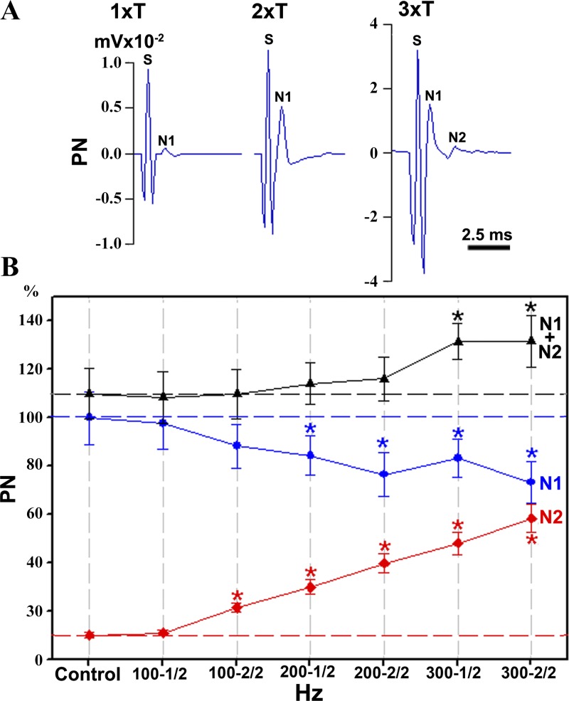
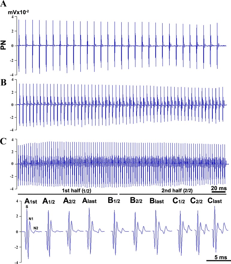
Spinal cord injury (SCI) at the level of cervical segments often results in life-threatening respiratory complications and requires long-term mechanical ventilator assistance. Thus restoring diaphragm activity and regaining voluntary control of breathing are the primary clinical goals for patients with respiratory dysfunction following cervical SCI. Epidural stimulation (EDS) is a promising strategy that has been explored extensively for nonrespiratory functions and to a limited extent within the respiratory system. The goal of the present study is to assess the potential for EDS at the location of the phrenic nucleus (C3-C5) innervating the diaphragm: the main inspiratory muscle following complete C1 cervical transection. To avoid the suppressive effect of anesthesia, all experiments were performed in decerebrate, C1 cervical transection, unanesthetized, nonparalyzed ( n = 13) and paralyzed ( n = 7) animals. Our results show that C4 segment was the most responsive to EDS and required the lowest threshold of current intensity, affecting tracheal pressure and phrenic nerve responses. High-frequency (200-300 Hz) EDS applied over C4 segment (C4-EDS) was able to maintain breathing with normal end-tidal CO2 level and raise blood pressure. In addition, 100-300 Hz of C4-EDS showed time- and frequency-dependent changes (short-term facilitation) of evoked phrenic nerve responses that may serve as a target mechanism for pacing of phrenic motor circuits. The present work provides the first report of successful EDS at the level of phrenic nucleus in a complete SCI animal model and offers insight into the potential therapeutic application in patients with high cervical SCI. NEW & NOTEWORTHY The present work offers the first demonstration of successful life-supporting breathing paced by epidural stimulation (EDS) at the level of the phrenic nucleus, following a complete spinal cord injury in unanesthetized, decerebrate rats. Moreover, our experiments showed time- and frequency-dependent changes of evoked phrenic nerve activity during EDS that may serve as a target mechanism for pacing spinal phrenic motor networks.

Keywords: breathing; complete spinal cord injury; epidural stimulation; phrenic nerve; rats.


Conflict of interest statement

No conflicts of interest, financial or otherwise, are declared by the authors.

Figures

Fig. 1.
Fig. 1.
Schematic epidural stimulation (EDS) electrode location and identification of threshold EDS currents. A: schematic diaphragm of EDS electrode location near the ventral and dorsolateral surface of C4 cervical segment. Phrenic motor nucleus and phrenic nerve (PN; red) are shown relative to the electrodes for recording of PN activity (and input to operational amplifier; black). The details of stimulation paradigm are shown at the bottom. B: an example of “no-inflation” test performed before EDS to confirm completeness of C1-transected rats (there were no signs of spontaneous breathing). C: an example of detecting the minimal EDS current affecting tracheal pressure (TP; marked with black dots) (see also statistics graph in Fig. 2A). B and C: traces from the top to bottom indicate blood pressure (BP in mmHg; red), end-tidal carbon dioxide concentration (CO2 in %; green), and TP (in cmH2O; blue). The bottom black trace in C indicates the current of EDS (µA) applied to C4 cervical segment.
Fig. 2.
Fig. 2.
Statistical graphs of thresholds (A) and successful vs. unsuccessful (B) epidural stimulation (EDS)-paced breathing in C1-transected rats. A: average minimal thresholds of EDS current affecting tracheal pressure (see Fig. 1C), applied with different pulse lengths (0.1, 0.2, and 0.5 ms) to C3–C5 segments (C3, C4, and C5), are shown with relative significant differences (horizontal lines) and corresponding P values. The mean (±SD) threshold values are shown inside the bars. B: the differences between proportions of successful and unsuccessful C4-EDS trials are indicated with corresponding P values. The proportions (ratio between successful EDS and all EDS trials) are shown inside the bars. Current intensity of EDS in thresholds applied with different frequencies (100, 200, and 300 Hz) are as follows: 1.5× threshold (1.5T), 2× threshold (2T), and 3× threshold (3T).
Fig. 3.
Fig. 3.
A: an example of the stable breathing during high-frequency (300 Hz) C4-epidural stimulation (EDS) pacing with 3× thresholds of current intensity. Black arrows indicate turning assisted ventilation off or on (up and down arrows, respectively). B1B3: highlighted segments demonstrate stable changes of paced breathing in expanded time scale during EDS with respiratory flow (cyan) and tidal volume (Tv; purple). Labels and legends for three top traces in A and B1B3 are as follows (from top to bottom): blood pressure (BP in mmHg; red), end-tidal carbon dioxide concentration (CO2 in %; green), tracheal pressure (TP in cmH2O; blue) was recorded before and after C4-EDS, respiratory flow in milliliters per sample rate (Flow, ml/sample rate; blue) was recorded during C4-EDS. The bottom black trace on in A and B1B3 indicates the current of EDS (µA) applied to C4 cervical segment.
Fig. 4.
Fig. 4.
A: an example of ineffective breathing during high-frequency (300 Hz) C4-epidural stimulation (EDS) pacing with 1.5× threshold current intensity. A series of EDS pulses (0.2 ms, 300 Hz, 1.5× threshold, train duration 0.33 s, 1 Hz) was applied at C4 level. Black arrows indicate turning assisted ventilation off or on (up and down arrows, respectively). B1B3: highlighted segments demonstrate unstable (declined) paced breathing in expanded time scale during EDS with respiratory flow (cyan) and tidal volume (Tv; purple). Labels and legends for three top traces in A and B1–B3 are as follows (from top to bottom): blood pressure (BP in mmHg; red), end-tidal carbon dioxide concentration (CO2 in %; green), tracheal pressure (TP in cmH2O; blue) was recorded before and after C4-EDS, respiratory flow in milliliters per sample rate (Flow, ml/sample rate, blue) was recorded during C4-EDS. The bottom black trace in A and B1B3 indicates the current of EDS (µA) applied to C4 cervical segment. Note decrease in respiratory flow and Tv in the middle part and the end of EDS pacing.
Fig. 5.
Fig. 5.
The responses of phrenic nerve to single pulse and train of epidural stimulation (EDS) applied to C4 segment with different frequencies. A: phrenic nerve (PN) responses (in mV, mV × 10−2) to single pulse (0.2 ms) of EDS with different current intensity (1–3× thresholds, 1 × T to 3 × T). S, N1, and N2 are abbreviations used for marking the stimulus artifact (S) and the first (N1) and second (N2) phrenic nerve components, respectively. Phrenic nerve response to 2 × T to 3 × T of current intensity has a high-amplitude stimulus artifact that partially overlaps the onset of N1 component. This overlap is absent at 1 × T of current intensity. B: the graph of phrenic nerve components (%, for numbers, see Table 1) changes in response to 3 × T of EDS-train stimulation at different frequencies (blue circles, N1; red diamonds, N2; black triangles, N1 + N2 components). x-Axis (100–1/2 to 300–2/2) shows stimulus-triggered averaging of phrenic nerve components during the first (1/1) and second (2/2) half of EDS-train stimulation for 100, 200, and 300 Hz. Control (100%) corresponds to the amplitude of N1 component in the first response of phrenic nerve during stimulations (blue dashed line). Relative controls for N2 and sum of components (N1 + N2) are shown in red and black dashed lines, respectively. *Significant differences of component amplitude means were estimated relative to their controls (see Table 1 for P values).
Fig. 6.
Fig. 6.
An example of phrenic nerve component changes in response to different frequencies of C4-epidural stimulation (EDS). AC: phrenic nerve (PN) responses (in mV, mV × 10−2) to C4-EDS at different frequencies (A, 100 Hz; B, 200 Hz; and C: 300 Hz) with 3× threshold current intensity (0.33-s train length, 0.2-ms pulse duration). Bottom: A1/2 to C1/2 and A2/2 to C2/2 are stimulus-triggered averaging of phrenic nerve responses during first and second half period of stimulation at different frequencies (each half is marked with solid line below C). A1st to C1st and Alast to Clast are the first and last phrenic nerve responses at the beginning and the end of each frequency train. S, N1, and N2 are abbreviations used for marking the stimulus artifact (S) and the first (N1) and second (N2) phrenic nerve components, respectively.

Similar articles

Cited by

References

    1. Agostoni E, Mognoni P, Torri G, Saracino F. Relation between changes of the rib cage circumference and lung volume. J Appl Physiol 20: 1179–1186, 1965. doi:10.1152/jappl.1965.20.6.1179. - DOI
    1. Alves-Neto O, Tavares P, Rocco PRM, Zin WA. Respiratory mechanics and morphometric changes during anesthesia with ketamine in normal rats. Braz J Med Biol Res 34: 1217–1223, 2001. doi:10.1590/S0100-879X2001000900016. - DOI - PubMed
    1. Bezdudnaya T, Hormigo KM, Marchenko V, Lane MA. Spontaneous respiratory plasticity following unilateral high cervical spinal cord injury in behaving rats. Exp Neurol 305: 56–65, 2018. doi:10.1016/j.expneurol.2018.03.014. - DOI - PMC - PubMed
    1. Betz AL, Coester HC. Effect of steroids on edema and sodium uptake of the brain during focal ischemia in rats. Stroke 21: 1199–1204, 1990. doi:10.1161/01.STR.21.8.1199. - DOI - PubMed
    1. Connelly CA, Otto-Smith MR, Feldman JL. Blockade of NMDA receptor-channels by MK-801 alters breathing in adult rats. Brain Res 596: 99–110, 1992. doi:10.1016/0006-8993(92)91537-O. - DOI - PubMed

Publication types

LinkOut - more resources