

Brillouin microscopy: assessing ocular tissue biomechanics
- PMID: 29771749
- PMCID: PMC6012042
- DOI: 10.1097/ICU.0000000000000489
Brillouin microscopy: assessing ocular tissue biomechanics
Abstract
Purpose of review: Assessment of corneal biomechanics has been an unmet clinical need in ophthalmology for many years. Many researchers and clinicians have identified corneal biomechanics as source of variability in refractive procedures and one of the main factors in keratoconus. However, it has been difficult to accurately characterize corneal biomechanics in patients. The recent development of Brillouin light scattering microscopy heightens the promise of bringing biomechanics into the clinic. The aim of this review is to overview the progress and discuss prospective applications of this new technology.
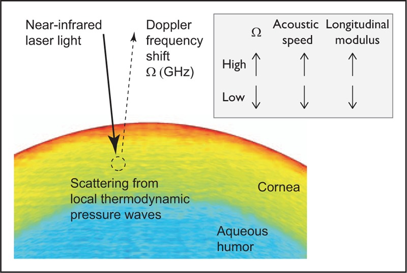
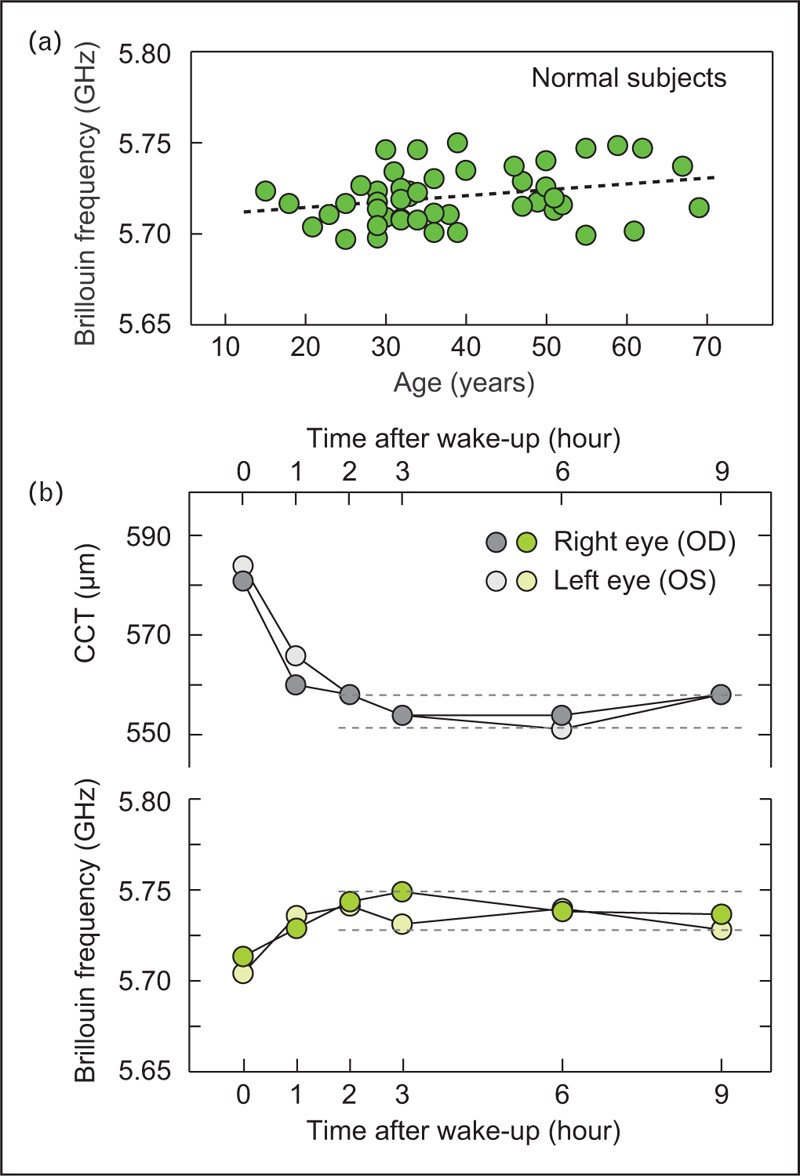
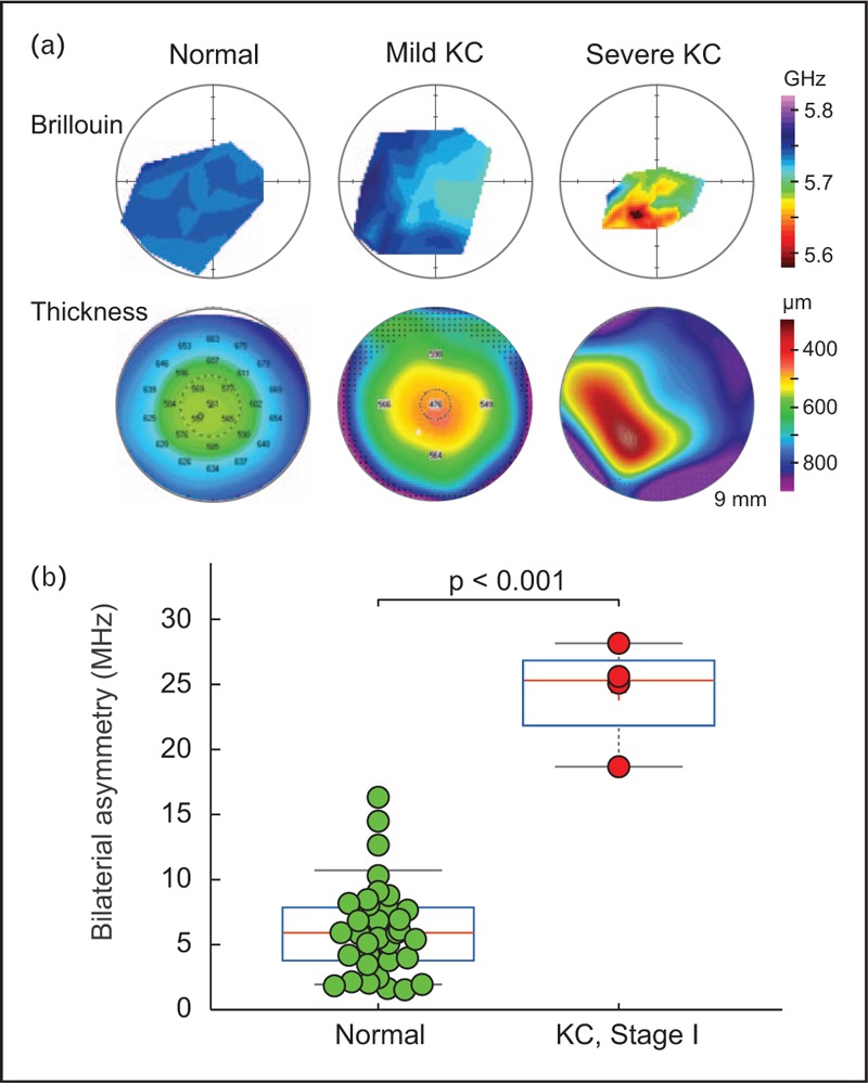
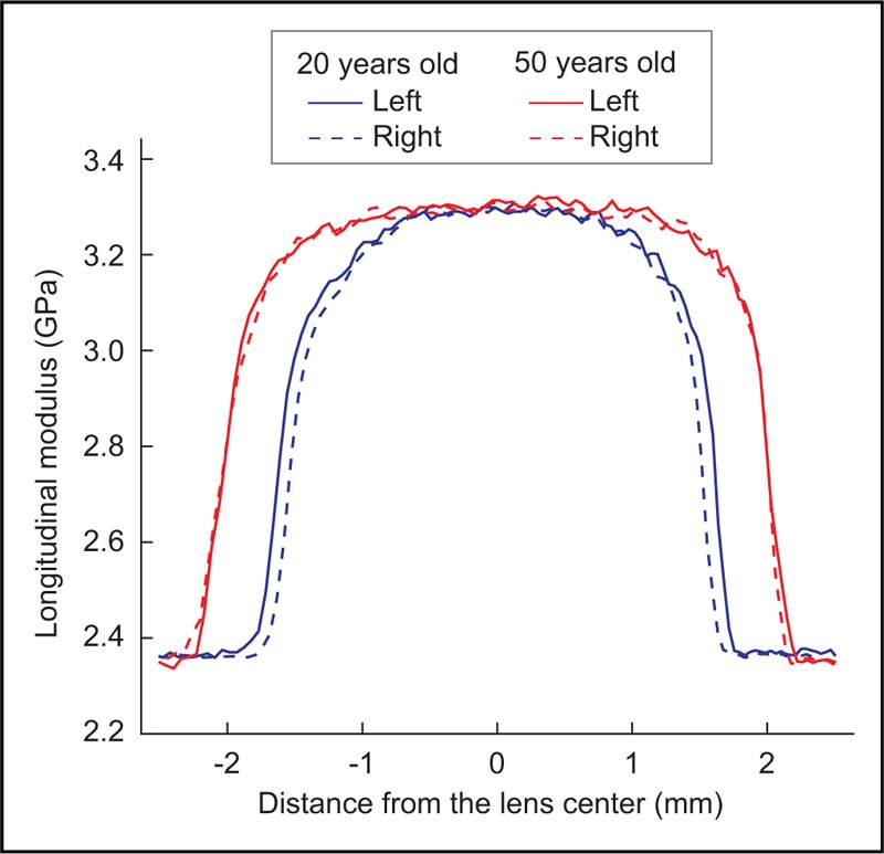
Recent findings: Brillouin microscopy uses a low-power near-infrared laser beam to determine longitudinal modulus or mechanical compressibility of tissue by analyzing the return signal spectrum. Human clinical studies have demonstrated significant difference in the elastic properties of normal corneas versus corneas diagnosed with mild and severe keratoconus. Clinical data have also shown biomechanical changes after corneal cross-linking treatment of keratoconus patients. Brillouin measurements of the crystalline lens and sclera have also been demonstrated.
Summary: Brillouin microscopy is a promising technology under commercial development at present. The technique enables physicians to characterize the biomechanical properties of ocular tissues.
Figures
Similar articles
-
Biomechanical characterization of keratoconus corneas ex vivo with Brillouin microscopy.Invest Ophthalmol Vis Sci. 2014 Jun 17;55(7):4490-5. doi: 10.1167/iovs.14-14450. Invest Ophthalmol Vis Sci. 2014. PMID: 24938517 Free PMC article.
-
Brillouin Microscopy: An Emerging Tool for Biomechanical Analysis in Ophthalmology.J Refract Surg. 2025 Jul;41(7):e731-e746. doi: 10.3928/1081597X-20250513-02. Epub 2025 Jul 1. J Refract Surg. 2025. PMID: 40626435 Review.
-
Brillouin Spectroscopy in Ophthalmology.Klin Monbl Augenheilkd. 2023 Jun;240(6):779-782. doi: 10.1055/a-2085-5738. Epub 2023 May 4. Klin Monbl Augenheilkd. 2023. PMID: 37142238 Review. English, German.
-
Brillouin optical microscopy for corneal biomechanics.Invest Ophthalmol Vis Sci. 2012 Jan 20;53(1):185-90. doi: 10.1167/iovs.11-8281. Invest Ophthalmol Vis Sci. 2012. PMID: 22159012 Free PMC article.
-
Comparison of corneal biomechanical properties following penetrating keratoplasty and deep anterior lamellar keratoplasty for keratoconus.Clin Exp Ophthalmol. 2020 Mar;48(2):174-182. doi: 10.1111/ceo.13677. Epub 2019 Nov 27. Clin Exp Ophthalmol. 2020. PMID: 31705767
Cited by
-
In Vivo Evaluation of Corneal Biomechanics Following Cross-Linking Surgeries Using Optical Coherence Elastography in a Rabbit Model of Keratoconus.Transl Vis Sci Technol. 2024 Feb 1;13(2):15. doi: 10.1167/tvst.13.2.15. Transl Vis Sci Technol. 2024. PMID: 38376862 Free PMC article.
-
Brillouin microscopy: an emerging tool for mechanobiology.Nat Methods. 2019 Oct;16(10):969-977. doi: 10.1038/s41592-019-0543-3. Epub 2019 Sep 23. Nat Methods. 2019. PMID: 31548707 Review.
-
Ultrahigh-sensitive optical coherence elastography.Light Sci Appl. 2020 Apr 13;9:58. doi: 10.1038/s41377-020-0297-9. eCollection 2020. Light Sci Appl. 2020. PMID: 32337022 Free PMC article.
-
Stress-Strain Index Map: A New Way to Represent Corneal Material Stiffness.Front Bioeng Biotechnol. 2021 Mar 11;9:640434. doi: 10.3389/fbioe.2021.640434. eCollection 2021. Front Bioeng Biotechnol. 2021. PMID: 33777912 Free PMC article.
-
Viscoelasticity in 3D Cell Culture and Regenerative Medicine: Measurement Techniques and Biological Relevance.ACS Mater Au. 2024 Jun 18;4(4):354-384. doi: 10.1021/acsmaterialsau.3c00038. eCollection 2024 Jul 10. ACS Mater Au. 2024. PMID: 39006396 Free PMC article. Review.
References
-
- Kotecha A. What biomechanical properties of the cornea are relevant for the clinician? Surv Ophthalmol 2007; 52:S109–S114. - PubMed
-
- Roberts C. The cornea is not a piece of plastic. J Refract Surg 2000; 16:407–413. - PubMed
-
- Ortiz D, Pinero D, Shabayek M, et al. Corneal biomechanical properties in normal, postlaser in situ keratomileusis, and keratoconic eyes. J Cataract Refract Surg 2007; 33:1371–1375. - PubMed
-
- Touboul D, Roberts C, Kérautret J, et al. Correlation between corneal hysteresis intraocular pressure, and corneal central pachymetry. J Cataract Refract Surg 2008; 34:616–622. - PubMed
-
- Shah S, Laiquzzaman M, Bhojwani R, et al. Assessment of the biomechanical properties of the cornea with the ocular response analyzer in normal and keratoconic eyes. Invest Ophthalmol Vis Sci 2007; 48:3026–3031. - PubMed
Publication types
MeSH terms
Grants and funding
LinkOut - more resources
Full Text Sources
Other Literature Sources
Research Materials
