. 2018 May 17;13(5):e0197618.
doi: 10.1371/journal.pone.0197618. eCollection 2018.

Another cat and mouse game: Deciphering the evolution of the SCGB superfamily and exploring the molecular similarity of major cat allergen Fel d 1 and mouse ABP using computational approaches

Affiliations

Another cat and mouse game: Deciphering the evolution of the SCGB superfamily and exploring the molecular similarity of major cat allergen Fel d 1 and mouse ABP using computational approaches

Rajesh Durairaj et al. PLoS One. .

Abstract

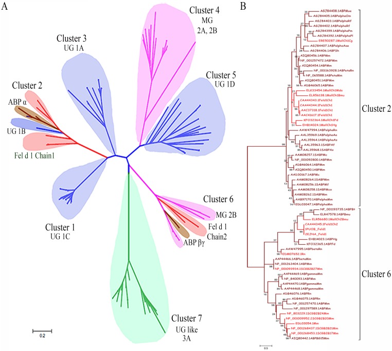
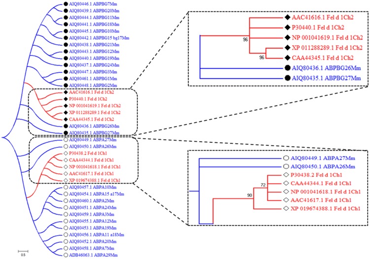
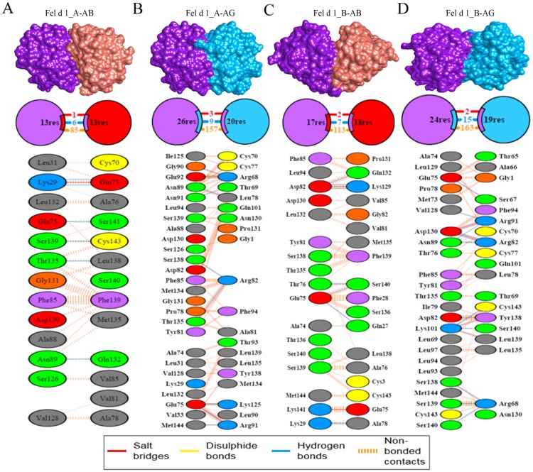
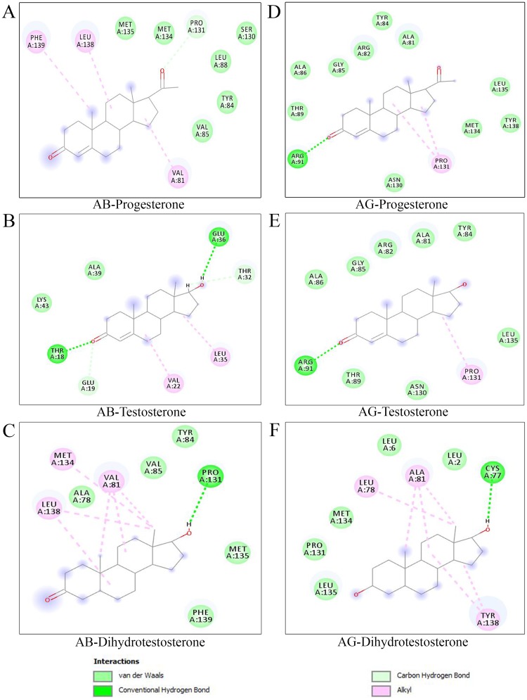
The mammalian secretoglobin (SCGB) superfamily contains functionally diverse members, among which the major cat allergen Fel d 1 and mouse salivary androgen-binding protein (ABP) display similar subunits. We searched for molecular similarities between Fel d 1 and ABP to examine the possibility that they play similar roles. We aimed to i) cluster the evolutionary relationships of the SCGB superfamily; ii) identify divergence patterns, structural overlap, and protein-protein docking between Fel d 1 and ABP dimers; and iii) explore the residual interaction between ABP dimers and steroid binding in chemical communication using computational approaches. We also report that the evolutionary tree of the SCGB superfamily comprises seven unique palm-like clusters, showing the evolutionary pattern and divergence time tree of Fel d 1 with 28 ABP paralogs. Three ABP subunits (A27, BG27, and BG26) share phylogenetic relationships with Fel d 1 chains. The Fel d 1 and ABP subunits show similarities in terms of sequence conservation, identical motifs and binding site clefts. Topologically equivalent positions were visualized through superimposition of ABP A27:BG27 (AB) and ABP A27:BG26 (AG) dimers on a heterodimeric Fel d 1 model. In docking, Fel d 1-ABP dimers exhibit the maximum surface binding ability of AG compared with that of AB dimers and the several polar interactions between ABP dimers with steroids. Hence, cat Fel d 1 is an ABP-like molecule in which monomeric chains 1 and 2 are the equivalent of the ABPA and ABPBG monomers, respectively. These findings suggest that the biological and molecular function of Fel d 1 is similar to that of ABP in chemical communication, possibly via pheromone and/or steroid binding.


Conflict of interest statement

The authors have declared that no competing interests exist.

Figures

Fig 1
Fig 1. Computational methodology.
Graphical representation of the sequence and structural annotation methods were employed for the SCGB dataset.
Fig 2
Fig 2. Dataset collection.
All sequence in the SCGB protein dataset (969) sequences were separated by subtypes and classified by organism. Rodents and non-human primates exhibit more SCGBs than other mammals. The numbers of Fel d 1 chains and ABP subunit sequences are greater in cats and rodents, respectively, compared with those in other mammals. UG and related sequences are present in all species.
Fig 3
Fig 3. Construction of the phylogenetic tree and clustering branching pattern of Fel d 1 and ABP.
(A). The radial evolutionary tree was generated using selective members of the SCGB superfamily proteins from different organisms. The UG, MG, UG like, Fel d 1/Mall, and ABP are represented as blue, pink, green, red, and brown, respectively, using a subtree coloring scheme, which provides the distribution of distantly related SCGB superfamily members. The SCGB divergence tree was estimated using a scale bar of 0.2 amino acid substitution matrices per site. (B). Phylogenetic tree of clusters 2 and 6. The dendrogram of Fel d 1/Mall-ABP showed clustering pattern of chains 1 and 2 of Fel d 1. Fel d 1/Mall-ABP was estimated using a scale bar of 0.5 amino acid substitution matrices per site and the BS values indicated on the tree branches.
Fig 4
Fig 4. Evolutionary pattern of Fel d 1 and ABP subunits.
The phylogenetic tree shows the evolutionary pattern of Fel d 1 and 28 paralogs. Chains 1 and 2 of Fel d 1 cluster are enlarged, and the arrangement of coclusters with ABP paralogs is shown. Chain 1 of Fel d 1 or ABPA is depicted as an open diamond or open circle, respectively. Similarly, the chain 2 of Fel d 1 or ABPBG is depicted as a closed diamond or closed circle respectively. The Fel d 1-ABP paralogs were estimated using a scale bar of 0.5 amino acid substitution matrices per site.
Fig 5
Fig 5. Illustration of sequence similarity and putative sites of Fel d 1 and ABP using MSA.
Multiple functional site information is described in the MSA with a residue scale. The Fel d 1 chains 1 and 2 sequences were aligned with various subunits of ABP sequences in MSA. In general, identical and conserved residues are indicated with differently colored dotted boxes: black for chain 1 and blue for chain 2. The cysteine-rich residues are indicated with blue stars, and the unique conserved residue, Tyr21, is denoted with an orange star. The calcium-binding sites are identified with red triangles in subunits A and B of Fel d 1. UG signature and motif sites are indicated with blue, red, and black underlining, respectively. The similarity of the functional sites from pheromaxein is indicated with yellow arrows. N-glycosylation (-NATE-) and N-myristylation sites in chain 2 are indicated with red and dark blue boxes, respectively.
Fig 6
Fig 6. Structural superimposition of subunit A of Fel d 1 with AB and AG dimers.
(A) Overlap of the AB (red) structural model with Fel d 1 (purple). (B) Overlap of the AG (light blue) structural model with Fel d 1 (purple). The pipes and plank and molecular surface views represent good superimposition of both proteins. Projections of the superimposition and topologically equivalent positions are shown in dotted boxes. The black and yellow arrows denote the surface cavity in the superimposed structures.
Fig 7
Fig 7. Representation of the surface binding site cleft.
(A) The surface and molecular bubble mesh view of the binding site cleft is shown for subunits A (purple) and B (pink) of Fel d 1. (B) The surface and molecular bubble mesh view of the binding site cleft is shown for subunits A of Fel d 1 (purple) and AB dimer (red). (C) The surface and molecular bubble mesh view of binding site cleft were showed for subunit A of Fel d 1 (purple) and AG dimer (light blue). The surface cleft located between two proteins is shown in green in all figures. The arrows represent the surface patches of proteins near the dimer interface.
Fig 8
Fig 8. Fel d 1-ABP dimers similarity.
(A). Molecular surface view and residue contacts between subunit A of Fel d 1 with the AB dimer. (B). Subunit A of Fel d 1 with the AG dimer. (C). Subunit B of Fel d 1 with the AB dimer. (D). Subunit B of Fel d 1 with AG. All the structural contacts exhibit three different contacts (salt bridges, H-bonds and nonbonded contacts), which are represented with different colors, as indicated in the legend.
Fig 9
Fig 9. Molecular docking of ABP dimers with steroids.
The 2D-residue map of steroid interaction with ABP dimers; the residues within 4.0 Å of the steroids are shown. (A-C). Residue interactions of progesterone (P), testosterone (T), and dihydrotestosterone (DHT) with the AB dimer. (D-F). Residue interactions of progesterone, testosterone, and dihydrotestosterone with the AG dimer. The protein-ligand interactions are depicted as Van der Waals, conventional hydrogen bonds, and alkyl bonds.

Similar articles

Cited by

References

    1. Mukherjee AB, Chilton BS. The uteroglobin/Clara cell protein family. Ann N Y Acad Sci. 2000;923: 1–356. - PubMed
    1. Daniel JC. Discovery and perspectives from the blastokinin era. Ann N Y Acad Sci. 2000;923: 1–8. doi: 10.1111/j.1749-6632.2000.tb05515.x . - DOI - PubMed
    1. Beier HM. The discovery of uteroglobin and its significance for reproductive biology and endocrinology. Ann N Y Acad Sci. 2000;923: 9–24. doi: 10.1111/j.1749-6632.2000.tb05516.x . - DOI - PubMed
    1. Niimi T, Keck-Waggoner CL, Popescu NC, Zhou Y, Levitt RC, Kimura S. UGRP1, a uteroglobin/Clara cell secretory protein-related protein, is a novel lung-enriched downstream target gene for the T/EBP/NKX2.1 homeodomain transcription factor. Mol Endocrinol. 2001;15: 2021–2036. doi: 10.1210/mend.15.11.0728 . - DOI - PubMed
    1. Watson MA, Fleming TP. Mammaglobin, a mammary-specific member of the uteroglobin gene family, is overexpressed in human breast cancer. Cancer Res. 1996;56: 860–865. . - PubMed

Publication types

MeSH terms

LinkOut - more resources