. 2018 May 17;14(5):e1007017.
doi: 10.1371/journal.ppat.1007017. eCollection 2018 May.

Induction of influenza-specific local CD8 T-cells in the respiratory tract after aerosol delivery of vaccine antigen or virus in the Babraham inbred pig

Affiliations

Induction of influenza-specific local CD8 T-cells in the respiratory tract after aerosol delivery of vaccine antigen or virus in the Babraham inbred pig

Katie Tungatt et al. PLoS Pathog. .

Abstract

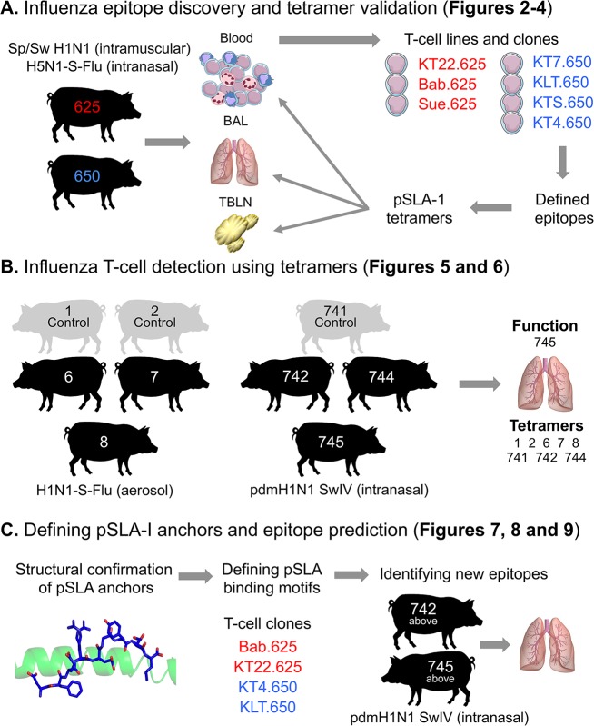
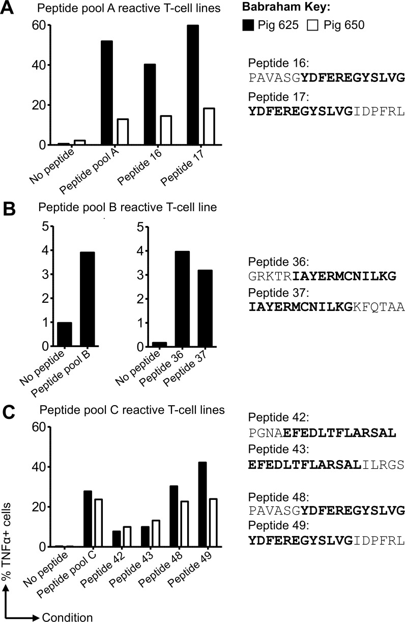
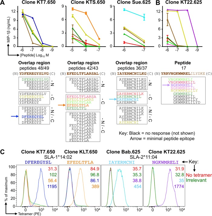
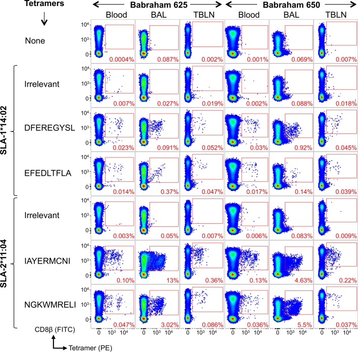
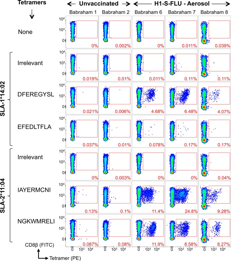
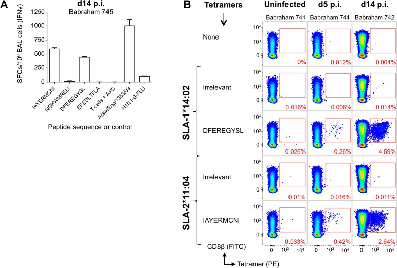
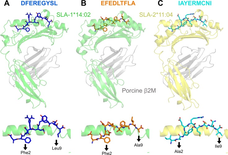
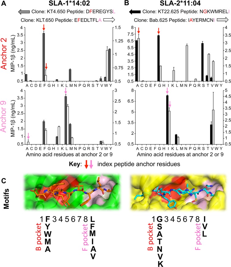
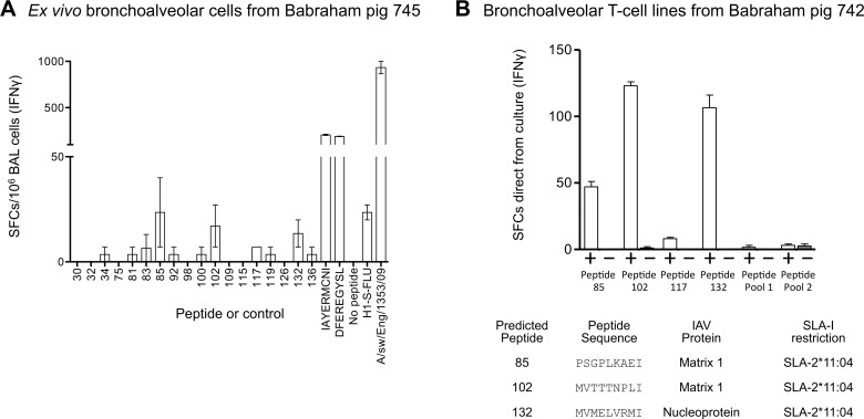
There is increasing evidence that induction of local immune responses is a key component of effective vaccines. For respiratory pathogens, for example tuberculosis and influenza, aerosol delivery is being actively explored as a method to administer vaccine antigens. Current animal models used to study respiratory pathogens suffer from anatomical disparity with humans. The pig is a natural and important host of influenza viruses and is physiologically more comparable to humans than other animal models in terms of size, respiratory tract biology and volume. It may also be an important vector in the birds to human infection cycle. A major drawback of the current pig model is the inability to analyze antigen-specific CD8+ T-cell responses, which are critical to respiratory immunity. Here we address this knowledge gap using an established in-bred pig model with a high degree of genetic identity between individuals, including the MHC (Swine Leukocyte Antigen (SLA)) locus. We developed a toolset that included long-term in vitro pig T-cell culture and cloning and identification of novel immunodominant influenza-derived T-cell epitopes. We also generated structures of the two SLA class I molecules found in these animals presenting the immunodominant epitopes. These structures allowed definition of the primary anchor points for epitopes in the SLA binding groove and established SLA binding motifs that were used to successfully predict other influenza-derived peptide sequences capable of stimulating T-cells. Peptide-SLA tetramers were constructed and used to track influenza-specific T-cells ex vivo in blood, the lungs and draining lymph nodes. Aerosol immunization with attenuated single cycle influenza viruses (S-FLU) induced large numbers of CD8+ T-cells specific for conserved NP peptides in the respiratory tract. Collectively, these data substantially increase the utility of pigs as an effective model for studying protective local cellular immunity against respiratory pathogens.


Conflict of interest statement

AMT is named on a European patent (publication no. EP2758525 A2, published July 30, 2014) concerning the use of S-FLU as a vaccine. AKS. is an inventor of patent WO 2010032022, “Use of protein kinase inhibitor to detect immune cells, such as T-cells. The other authors have no financial conflicts of interest.

Figures

Fig 1
Fig 1. Study overview.
The inbred Babraham pig was used throughout this study for vaccination and infection, with each pig assigned an identifying number, shown here within each silhouette. (A) Pigs 625 (red) and 650 (blue) were vaccinated intranasally and intramuscularly as depicted. Blood, bronchoalveolar lavage (BAL) and tracheobronchial lymph nodes (TBLNs) were harvested, with peripheral blood mononuclear cells (PBMCs) purified from blood and single suspensions from BAL and TBLNs generated for experiments. Overlapping peptides from the NP of PR8 were used to create T-cell lines (Fig 2) and T-cell clones (named and shown in red or blue text) (Fig 3). The red clones came from pig 625 (red) and the blue from pig 650 (blue). The clones were used to define minimal NP peptides, which were subsequently refolded with SLA-1*14:02 or SLA-2*11:04 to create pSLA-I tetramers. The tetramers were used to stain the clones (Fig 3) and harvested tissues from pigs 625 and 650 (Fig 4). (B) The BAL from pigs vaccinated or infected intranasally with influenza, as shown, were stained with the tetramers from A (Figs 5 and 6). The BAL from pig 745 was used for ex vivo ELISPOTS. (C) SLA-1*14:02 or SLA-2*11:04 were refolded with the epitopes defined in A to confirm peptide anchor residues (Fig 7). T-cell clones from A were used to define a SLA-1*14:02 or SLA-2*11:04 peptide anchor binding motif (Fig 8), which were then used to predict other influenza epitopes, tested using BAL from the two pigs shown (Fig 9).
Fig 2
Fig 2. Generation of influenza-specific CD8β T-cell lines from Babraham pigs simultaneously immunized with H5N1-S-FLU and Sp/Sw H1N1.
CD8β T-cells purified from the peripheral blood mononuclear cells of Babraham pigs 625 (filled bars) and 650 (open bars) were stimulated in vitro with pools (four pools: A, B, C and D) of overlapping peptides (81 in total, S2 Table) from the NP of S-FLU (PR8). Two weeks post stimulation the cells were tested for reactivity towards individual peptides from each pool. Intracellular cytokine staining for TNF was performed following a 5 h incuabtion with no peptide (DMSO control) or peptides (2 μM). All associated flow cytometry data is shown in S2 Fig (pig 625) and S3 Fig (pig 650) with the percentage of TNF producing cells displayed here. (A) T-cell lines generated against pool A mapped to individual peptides 16 and 17 (sequences shown with overlap region in bold). (B) Reactivity for pool B for pig 625 (left graph) had been seen in previous experiments with indications for reactivity towards peptides 36 and 37, thus lines were successfully generated against individual peptides 36 and 37 (right graph). (C) Lines responsive to pool C mapped to individual peptide 42, 43, 48 and 49. T-cell clones were grown directly from these lines by limiting dilution or first by enriching for peptide specific T-cells using a TAPI-0 assay (S1 Fig) and flow cytometry.
Fig 3
Fig 3. Minimal epitope identification and pSLA tetramer staining of influenza-specific T-cell clones.
CD8β clones were grown from T-cell lines generated from Babraham pigs 625 and 650 using overlapping peptides from the NP of S-FLU (PR8) (Fig 2). The .650 or .625 indicates the pig the clones were grown from. (A) Clones KT7.650, KTS.650 and Sue.625 responded to the overlapping peptide regions shown in brown text below each graph. Clones were incubated with decreasing concentrations (upper panel) of truncated versions of the respective peptide (sequences shown in the lower panel). Truncations were performed at the amino (N) and/or carboxyl (C) terminal ends of each peptide as indicated (lower panel). MIP-1β ELISAs were performed to assess T-cell activation after overnight incubation. Peptides that did not elicit a repsonse are shown in black and are not displayed on the graphs. The minimal peptide epitope is indicated by the arrow. (B) Using a similar approach as in (A). In order to induce maximal activation and define the minimal epitope, clone KT22.625 (right) required the isoleucine from peptide 17, which was not present in the overlap region of 16/17. (C) The minimal epitopes defined in (A and B) were refolded with both SLA-1*14:02 and SLA-2*11:04, with successful refolding determining restriction. pSLA-I tetramers were assembled and used to stain clones with respective peptide specificity, as shown. Irrelevant tetramers: SLA-1*14:02-AFAAAAAAL and SLA-2*11:04-AGAAAAAAI.
Fig 4
Fig 4. Nuceloprotein pSLA-I tetramer staining of tissues from influenza vaccinated Babraham Pigs.
Babraham pig 625 (left panel of 21 plots) and 650 (right panel of 21 plots) received H5N1 S-FLU intranasally and inactivated H1N1 virus [A/Swine/Spain/SF11131/2007] with montanide adjuvant intramuscularly, followed by a boost at day 25 using the same preparation. Pigs were culled at day 38 (day 13 post boost) and blood, bronchoalveolar lavage (BAL) and tracheobronchial lymph nodes (TBLNs) harvested and frozen as single cell suspensions. Tetramer staining was performed on thawed cells from the blood, BAL and TBLN using a no tetramer control, and staining with Irrelevant and nucleoprotein peptide tetramers. The sequences for the nucleoprotein peptides are shown. Irrelevant tetramers: SLA-1*14:02-AFAAAAAAL, SLA-2*11:04-AGAAAAAAI (pig 625) and SLA-2*11:04-GAGGGGGGI (pig 650). Gating strategy: lymphocytes, single cells, viability (Vividneg) CD3+ CD14neg then CD8β+ CD4+ and displayed as CD8β versus tetramer (S1 Fig).
Fig 5
Fig 5. Nuceloprotein pSLA-I tetramer staining of bronchoalveolar lavage samples from Babraham pigs vaccinated with H1N1 S-FLU.
Babraham pigs were either left unvaccinated (1 and 2) or received H1N1 S-FLU via aerosol administration (6, 7, 8). H1N1 S-FLU vaccinated animals received a boost at day 28 with the same vaccine. Animals were culled and bronchoalveolar lavage harvested at day 57. Nucleoprotein and irrelevant peptide SLA-I tetramer staining was performed on thawed bronchoalveolar lavage samples and the percentage of tetramer+ cells of CD8β+ cells displayed in red. The sequences of the nucleoprotein peptides are shown. Irrelevant tetramers: SLA-1*14:02-AFAAAAAAL and SLA-2*11:04-GAGGGGGGI. Gating strategy: lymphocytes, single cells, viability (Vividneg) CD3+ CD14neg then CD8β+ CD4+ and displayed as CD8β versus tetramer (S1 Fig).
Fig 6
Fig 6. Identification of influenza-specific T-cells in the bronchoalveolar lavage from Babraham pigs infected with pandemic H1N1 swine influenza virus.
Babraham pigs were either left uninfected (741) or infected intranasally with pandemic H1N1 [A/sw/Eng/1353/09] (742, 744, 745). The pigs were culled on day 0 (741), 5 (744) or 14 (742 and 745) post infection. (A) 200,000 bronchoalveolar lavage cells from pig 745 (infected, day 14 cull) were incubated alone, with 10-5M peptide, or virus for 16–18 h. A Babraham kidney cell line was included in each well (15,000 per well) to act as antigen presenting cells. All conditions were performed in duplicate and spot forming cells (SFCs) detected by IFNγ ELISPOT and displayed as mean +SEM and scaled (X5) to 106 BAL cells. (B) Irrelevant and nucleoprotein peptide-SLA-1*14:02 and SLA-2*11:04 tetramer staining was performed on thawed bronchoalveolar lavage samples and the percentage of tetramer+ cells of CD8β cells displayed in red. Nucleoprotein peptide sequences are shown. Irrelevant tetramers were SLA-1*14:02-AFAAAAAAL and SLA-2*11:04-AGAAAAAAI. Gating strategy: lymphocytes, single cells, viability (Vividneg) CD3+ CD14neg then CD8β+ CD4+ and displayed as CD8β versus tetramer (S1 Fig).
Fig 7
Fig 7. Structural overview of influenza peptides bound to SLA-1*14:02 and SLA-2*11:04.
(A) Upper panel: SLA-1*14:02 (α1, α2 and α3 domains in green) with nucleoprotein peptide DFEREGYSL (blue) and porcine β2M (grey). Lower panel: position of DFEREGYSL within the SLA-1*14:02 binding groove and amino (position 2) and carboxyl (position 9) terminus anchor residues of the peptide are shown below the black arrows. (B) Same figure layout as for (A). SLA-1*14:02 (α1, α2 and α3 domains in green) with nucleoprotein peptide EFEDLTFLA (orange) and porcine β2M (grey). (C) Same figure layout as for (A). SLA-2*11:04 (α1, α2 and α3 domains in yellow) with nucleoprotein peptide IAYERMCNI (cyan) and porcine β2M (grey).
Fig 8
Fig 8. Peptide-SLA anchor residue preferences and proposed binding motifs for SLA-1*14:02 and SLA-2*11:04.
(A) SLA-1*14:02 restricted, nucleoprotein peptide specific CD8 clones grown from Babraham pig 650 were used to define the peptide binding motif for SLA-1*14:02. Clone KT4.650 (left axis) recognizes index peptide DFEREGYSL and clone KLT.650 (right axis) index peptide EFEDLTFLA. Each of the proteogenic amino acids residues was tested at positions 2 (upper graph) and 9 (lower graph) by substitution of the index peptides. For example: DFEREGYSL index peptide and anchor 2 variants: DA/C/D/E/G/H/I/K/L/M/N/P/Q/R/S/T/V/W/Y/EREGYSL (each residue in bold tested in turn). The corresponding clone was used in peptide titration assays and ELISAs were performed to determine MIP-1β release, with data displayed for 10−7 M peptide. The limit of maximal detection of MIP-1β release was ~10 ng/mL, data below 0.5 ng/mL has been omitted for clarity, and mean + SEM shown. (B) As for (A), but using the SLA-1*11:04 restricted, nucleoprotein peptide specific clones KT22.625 (left axis, index peptide NGKWMRELI) and Bab.625 (right axis, index peptide IAYERMCNI) grown from Babraham pig 625 to define the peptide binding motif for SLA-2*11:04. Data displayed for 10−8 M peptide. (C) Binding pocket composition and proposed binding motif for SLA-1*14:02 and SLA-2*11:04 determined from the data in panels A and B. SLA-1*14:02 (green) with EFEDLTFLA (orange sticks) and SLA-2*11:04 (yellow) with IAYERMCNI (cyan sticks). Double conformers have been removed for visual clarity. B pocket is shown in red and the F pocket in pink.
Fig 9
Fig 9. Verification of predicted influenza T-cell epitopes in bronchoalveolar from Babraham pigs infected with pandemic H1N1 swine influenza.
Babraham pigs 742 and 745 were experimentally infected intranasally with H1N1 [A/sw/Eng/1353/09] and culled on day 14 post infection. Based on the binding motifs of SLA-1*14:02 and SLA-2*11:04 (Fig 8), predicted epitopes from matrix proteins 1 and 2, nucleoprotein, and polymerase basic proteins 1 and 2 (S6 Table) were tested as pooled peptides using BAL cells from pig 745 and IFNγ ELISpots (S7 Fig). Individual peptides highlighted from this process were then tested: (A) BAL cells from pig 745 were thawed and 200,000 used per well for IFNγ ELISPOT. 18 individual peptides (numbered on the x-axis) were used alongside our validated nucleoprotein epitopes, IAYERMCNI and DFEREGYSL. A no peptide and viral controls were included, and conditions performed in duplicate, scaled (X5) to 106 spot forming cells (SFCs) and with the mean displayed +SEM. Babraham kidney cells were used in every well (15,000 per well) to act as antigen presenting cells. (B) BAL cells from pig 742 were cultured in the presence of the peptides from (A) to create six T-cell lines (labelled on the x-axis). Peptides 85, 102, 117 and 132 were used individually as they gave relatively more SFCs for the ELISPOT in A. The remaining peptides were assembled in to two pools; pool 1 consisted of peptides 30, 32, 34, 75, 81, 83, 92; and pool 2 contained peptides 98, 100, 109, 115, 126, 136 (sequences in S6 Table). After two weeks, the lines were taken straight from culture for IFNγ ELISPOT and incubated with 10−5 M of the peptide(s) used to generate the line (+) or with no peptide (−). The actual number of SFCs is displayed as mean +SEM. The table shows the peptide sequence, the protein of origin and SLA-I restriction of the three epitopes that gave robust responses.

Similar articles

Cited by

References

    1. Molinari N-AM, Ortega-Sanchez IR, Messonnier ML, Thompson WW, Wortley PM, Weintraub E, et al. The annual impact of seasonal influenza in the US: Measuring disease burden and costs. Vaccine. 2007;25: 5086–5096. doi: 10.1016/j.vaccine.2007.03.046 - DOI - PubMed
    1. Tchilian E, Holzer B. Harnessing Local Immunity for an Effective Universal Swine Influenza Vaccine. Viruses. Multidisciplinary Digital Publishing Institute; 2017;9: 98 doi: 10.3390/v9050098 - DOI - PMC - PubMed
    1. Paules CI, Marston HD, Eisinger RW, Baltimore D, Fauci AS. The Pathway to a Universal Influenza Vaccine. Immunity. 2017. pp. 599–603. doi: 10.1016/j.immuni.2017.09.007 - DOI - PubMed
    1. Schulman JL, Kilbourne ED. Induction of Partial Specific Heterotypic immunity in Mice by a Single Infection with Influenza A virus. J Bacteriol. American Society for Microbiology (ASM); 1965;89: 170–4. - PMC - PubMed
    1. McMichael AJ, Gotch FM, Noble GR, Beare PAS. Cytotoxic T-Cell Immunity to Influenza. N Engl J Med. 1983;309: 13–17. doi: 10.1056/NEJM198307073090103 - DOI - PubMed

Publication types

MeSH terms