Review
. 2018 May 16;98(4):687-705.
doi: 10.1016/j.neuron.2018.03.045.

The Neural Basis of Timing: Distributed Mechanisms for Diverse Functions

Affiliations
Review

The Neural Basis of Timing: Distributed Mechanisms for Diverse Functions

Joseph J Paton et al. Neuron. .

Abstract

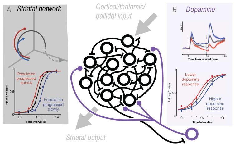
Timing is critical to most forms of learning, behavior, and sensory-motor processing. Converging evidence supports the notion that, precisely because of its importance across a wide range of brain functions, timing relies on intrinsic and general properties of neurons and neural circuits; that is, the brain uses its natural cellular and network dynamics to solve a diversity of temporal computations. Many circuits have been shown to encode elapsed time in dynamically changing patterns of neural activity-so-called population clocks. But temporal processing encompasses a wide range of different computations, and just as there are different circuits and mechanisms underlying computations about space, there are a multitude of circuits and mechanisms underlying the ability to tell time and generate temporal patterns.


Figures

Figure 1
Figure 1. Taxonomy of timing tasks
The continuum along at least two task dimensions are likely to be important for understanding the neural basis of timing: sensory versus motor, and interval versus pattern timing. Some tasks (Interval Timing) require the discrimination (Sensory Timing) or production (Motor Timing) of simple durations or intervals (or anticipation of an external event). Other tasks (Pattern Timing) require the discrimination or production of complex temporal or spatiotemporal patterns—such as deciphering Morse code signals (Sensory timing) or tapping a complex temporal pattern (Motor Timing). Upper left: adapted from Gouvêa et al, 2015. Lower left: adapted from Kawai et al, 2015.
Figure 2
Figure 2. Example of interval tuned neurons
A. Voltage traces from a neuron in the midbrain of an electric fish to trains of electrical pulses presented at intervals of 100 (left), 50 (center) and 10 ms (right). The rows represent three separate repetitions of each train. This neuron was tuned to pulses delivered at intervals of 50 ms (right). Adapted from Carlson (2009). B. Rastergram of a neuron from rat auditory cortex in response to five different stimuli, each composed of a 200 ms 3 kHz tone followed by a 50 ms 7 kHz (CF) tone with different stimulus-onset asynchronies. Numbers represent the facilitation index. Rats were trained to detect an interval of 100 ms between both tones (red arrow), and this was the spatiotemporal pattern that elicited the maximal response across the population (right). Adapted from Zhou et al. (2010). C. Model of how STP can generate an interval selective neuron in a disynaptic circuit composed of an excitatory (blue) and inhibitory (red) neuron. Left, the input to both neurons exhibits paired-pulse facilitation. By adjusting the weights onto both the Ex and Inh neurons it is possible to create an Ex neuron that functions as a 50, 100, or 200 ms detector. Modified from Buonomano (2000).
Figure 3
Figure 3. Midbrain dopamine neurons and striatal dynamics may interact to regulate timing
A. The speed with which striatal ensembles traverse neural space (top panel) predicts duration judgments (lower panel) in an interval discrimination task. Colored schematic trajectories in top panel depict a quickly (red) or slowly (blue) evolving ensemble activity pattern during interval presentation in a space defined by the firing of simultaneously recorded striatal neurons. Psychometric curves for trials segregated on the basis of whether activity proceeded quickly or slowly during interval presentation. Adapted from Gouvêa et al. 2015. B. Calcium signals collected from dopamine neurons in the SNc exhibited trial-to-trial variability during interval presentations (top panel) that predicted the timing judgments of mice during the same interval discrimination task used during the data collected in A (adapted from Soares et al, 2016). Given the dense innervation of striatal networks (in black, center) by nigro-striatal dopamine neurons (in purple, center) and the fact that SNc dopamine neurons receive significant input from striatum, these data support a hypothesis where the two brain areas reciprocally influence each other’s timing functions.
Figure 4
Figure 4. Examples of experimentally observed neural responses (left) and simulated models of timing (right)
A. Two ramping MFC neurons recorded during trials in which the animal anticipated reward availability at 3 or 12 seconds. From Emmons et. al. (2017). B. Model of an integrator that generates ramping, and that can be rescaled to time different durations by changing the magnitude of the input. Adapted from Balci and Simen (2016). C. Example of the sequential activation of neurons in area HVC. Each line represents a burst in a neuron (neurons that bursted more than once are represented in different lines). The pattern drives the timing of the zebra finch song. Adapted from Lynch et al. (2016). D. Schematic of a simple feedforward network (a synfire chain) that can implement a sparse population clock. E. Example of the trial-averaged activity in simultaneously recorded OFC neurons in response to an olfactory cue (blue bar) that predicts a delayed reward (red arrowhead). Cells are sorted according to the time of the peak firing rate. From Bakhurin et al. (2017). F. Simulation of a firing-rate based RNN that generates a complex population clock. Units are sorted according to the time of peak activity after the end of the input (blue bar). Adapted from Laje and Buonomano (2013).

References

    1. Abeles M. Local cortical circuits: an electrohysiological study. Berlin: Springer; 1982.
    1. Aschoff J. On the perception of time during prolonged temporal isolation. Hum Neurobiol. 1985;4:41–52. - PubMed
    1. Aubie B, Becker S, Faure PA. Computational Models of Millisecond Level Duration Tuning in Neural Circuits. J Neurosci. 2009;29:9255–9270. - PMC - PubMed
    1. Aubie B, Sayegh R, Faure PA. Duration Tuning across Vertebrates. The Journal of Neuroscience. 2012;32:6373–6390. - PMC - PubMed
    1. Baker CA, Carlson BA. Short-Term Depression, Temporal Summation, and Onset Inhibition Shape Interval Tuning in Midbrain Neurons. The Journal of Neuroscience. 2014;34:14272–14287. - PMC - PubMed

Publication types