Review
. 2018 May 17;19(5):1496.
doi: 10.3390/ijms19051496.

Oxidative Stress in Preeclampsia and Placental Diseases

Affiliations
Review

Oxidative Stress in Preeclampsia and Placental Diseases

Rajaa Aouache et al. Int J Mol Sci. .

Abstract

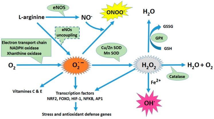
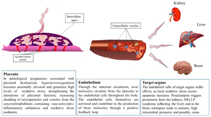
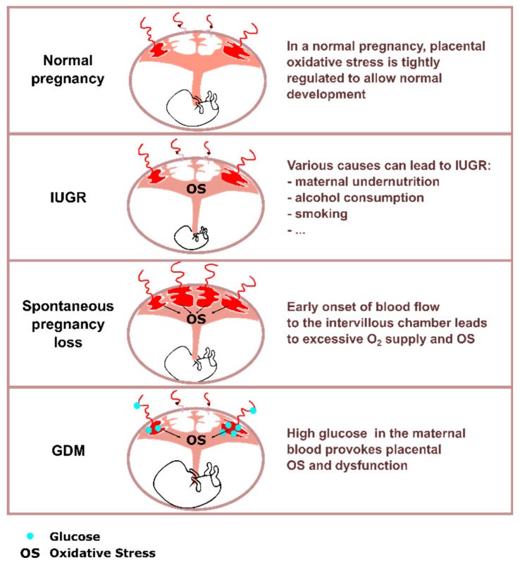
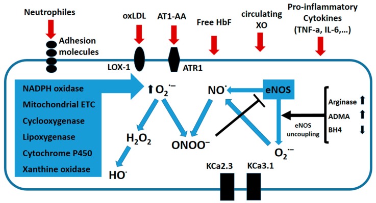
Preeclampsia is a persistent hypertensive gestational disease characterized by high blood pressure and proteinuria, which presents from the second trimester of pregnancy. At the cellular level, preeclampsia has largely been associated with the release of free radicals by the placenta. Placenta-borne oxidative and nitrosative stresses are even sometimes considered as the major molecular determinants of the maternal disease. In this review, we present the recent literature evaluating free radical production in both normal and pathological placentas (including preeclampsia and other major pregnancy diseases), in humans and animal models. We then assess the putative effects of these free radicals on the placenta and maternal endothelium. This analysis was conducted with regard to recent papers and possible therapeutic avenues.

Keywords: oxidative stress; placenta; preeclampsia; pregnancy; vascular endothelium.


Conflict of interest statement

All authors declare no conflict of interest.

Figures

Figure 1
Figure 1
Oxidative stress plays a central role in the physiopathology of preeclampsia. Schematic of the principal sources of oxidative stress and its antioxidant mechanisms. The mitochondria, endoplasmic reticulum (ER), and nuclear membrane produce anions as a byproduct of the auto-oxidation of electron transport chain (ETC) components. ROS are also produced as a consequence of arachidonic acid metabolism by Cyclooxygenase 2 (COX-2), Lipoxygenases, Xanthine Oxydase (XO), and Cytochrome P450. NADPH oxidases (NOX) are another significant source of ROS. NOX generates superoxide (O2•−) by transferring electrons from NADPH inside the cell across its membrane and coupling them to O2. eNO synthase (eNOS) can generate O2•− and H2O2, specifically when the concentrations of its substrate, l-arginine, or its cofactor, tetrahydrobiopterin (BH4), are low. When intracellular ROS production increases (especially O2•− ions), •NO may react with ROS to form peroxynitrite (ONOO). Superoxide is rapidly dismutated to H2O2 by superoxide dismutase (SOD). H2O2 can be transformed into H2O and O2 by catalase and glutathione peroxidase (GPX). However, in the presence of Fe2+, and through Fenton’s reaction, H2O2 can generate the highly reactive radical hydroxyl (OH).
Figure 2
Figure 2
The placenta is the interface between mother and fetus. The placenta plays an indispensable and multifunctional role as the interface between the two adjoined organisms, regulating immunological dialogue and tolerance, nutrient and gas exchange, and producing hormones essential for pregnancy. The first trimester of gestation in humans is characterized by general hypoxia in the intervillous space, as many maternal spiral arteries are plugged with extravillous trophoblast cells, preventing maternal red blood cells from passing into this space. After 12–14 weeks of normal gestation, these placental extravillous trophoblasts colonize the maternal spiraled arteries as far as the proximal third of the myometrium. This innervation leads to the arteries to relax and lose contractile properties, which increases blood flow and raises oxygen partial pressure, thus reversing the previously hypoxic environment. Therefore, oxidative stress normally occurs in the healthy placenta and may in fact be important for its organogenesis. However, in cases of incomplete placentation, this stress occurs at an excessive level and leads to an elevated release of placental debris and vesicles into the maternal circulation. These extracellular vesicles carry active molecules (proteins of microRNA) generated by stressed placental cells. Once in the blood, they meet maternal endothelial cells and potentially transfer their contents, leading to transcriptome alterations and inflammation. Eventually, the endothelium of maternal organs is affected. In the case of preeclampsia, this most heavily occurs in the kidney, liver, and brain.
Figure 3
Figure 3
Schematic representation of first trimester placentas in IUGR, GDM, and spontaneous pregnancy loss. Oxidative stress (OS) is a key characteristic in various placental pathologies. Although the causes of IUGR can be numerous, placental oxidative stress is recurrently found. In the case of spontaneous pregnancy loss, initial onset of the blood flow in the intervillous chamber happens earlier and is less organized than in normal pregnancies, which leads to an increase in oxidative stress in the placenta. In gestational diabetes, variations in maternal glycaemia participate in placental oxidative stress induction. In all the pathologies mentioned, placental oxidative stress leads to various dysfunctions. Key: glucose (blue dots), oxidative stress (OS), Intra-Uterine Growth Restriction (IUGR), and Gestational Diabetes Mellitus (GDM).
Figure 4
Figure 4
Principal mediators and sources of oxidative stress in the preeclamptic endothelium. Circulating factors in the blood of preeclamptic women can act on endothelial cells (ECs) to induce oxidative stress. These include reactive oxygen species (ROS) produced by neutrophils (oxLDL), agonist autoantibodies against angiotensin receptors (AT1-AA), free fetal hemoglobin (HbF), circulating Xanthine oxidase (XO), and cytokines (i.e., TNF-α). In ECs, several enzymatic systems including the electron transport chain, NADPH oxidases, and cyclooxigenases can produce superoxide (O2•−). Under certain circumstances, this can lead to increased Arginase II expression, increased asymmetric dimethyl arginine (ADMA), and the loss of the cofactor tetrahydrobiopterin (BH4), and endothelial nitric oxide synthase (eNOS) synthase can become uncoupled. Instead of •NO, uncoupled eNOS produces (O2•−). Nitric oxide can then react with O2•− to produce peroxynitrite (ONOO), a powerful oxidant whose nitrate proteins can induce DNA damage. Additionally, ONOO can inhibit eNOS activity. Superoxide scavenging of •NO impairs endothelium-dependent vasodilation. ROS can also down regulate the calcium-activated potassium channels KCa2.3 and KCa3.1, which are important electrical triggers of vasodilation. Figure adapted from [105].

Similar articles

Cited by

References

    1. Steegers E.A., von Dadelszen P., Duvekot J.J., Pijnenborg R. Pre-eclampsia. Lancet. 2010;376:631–644. doi: 10.1016/S0140-6736(10)60279-6. - DOI - PubMed
    1. Zabul P., Wozniak M., Slominski A.T., Preis K., Gorska M., Korozan M., Wieruszewski J., Zmijewski M.A., Zabul E., Tuckey R., et al. A proposed molecular mechanism of high-dose vitamin d3 supplementation in prevention and treatment of preeclampsia. Int. J. Mol. Sci. 2015;16:13043–13064. doi: 10.3390/ijms160613043. - DOI - PMC - PubMed
    1. Ghosh G., Grewal J., Mannisto T., Mendola P., Chen Z., Xie Y., Laughon S.K. Racial/ethnic differences in pregnancy-related hypertensive disease in nulliparous women. Ethn. Dis. 2014;24:283–289. - PMC - PubMed
    1. Hod T., Cerdeira A.S., Karumanchi S.A. Molecular mechanisms of preeclampsia. Cold Spring Harb. Perspect. Med. 2015;5:a023473. doi: 10.1101/cshperspect.a023473. - DOI - PMC - PubMed
    1. Goulopoulou S., Davidge S.T. Molecular mechanisms of maternal vascular dysfunction in preeclampsia. Trends Mol. Med. 2015;21:88–97. doi: 10.1016/j.molmed.2014.11.009. - DOI - PubMed

LinkOut - more resources