. 2018 May 17;19(5):1497.
doi: 10.3390/ijms19051497.

Tau Fibril Formation in Cultured Cells Compatible with a Mouse Model of Tauopathy

Affiliations

Tau Fibril Formation in Cultured Cells Compatible with a Mouse Model of Tauopathy

Gen Matsumoto et al. Int J Mol Sci. .

Abstract

Neurofibrillary tangles composed of hyperphosphorylated tau protein are primarily neuropathological features of a number of neurodegenerative diseases collectively termed tauopathy. To understand the mechanisms underlying the cause of tauopathy, precise cellular and animal models are required. Recent data suggest that the transient introduction of exogenous tau can accelerate the development of tauopathy in the brains of non-transgenic and transgenic mice expressing wild-type human tau. However, the transmission mechanism leading to tauopathy is not fully understood. In this study, we developed cultured-cell models of tauopathy representing a human tauopathy. Neuro2a (N2a) cells containing propagative tau filaments were generated by introducing purified tau fibrils. These cell lines expressed full-length (2N4R) human tau and the green fluorescent protein (GFP)-fused repeat domain of tau with P301L mutation. Immunocytochemistry and super-resolution microscopic imaging revealed that tau inclusions exhibited filamentous morphology and were composed of both full-length and repeat domain fragment tau. Live-cell imaging analysis revealed that filamentous tau inclusions are transmitted to daughter cells, resulting in yeast-prion-like propagation. By a standard method of tau preparation, both full-length tau and repeat domain fragments were recovered in sarkosyl insoluble fraction. Hyperphosphorylation of full-length tau was confirmed by the immunoreactivity of phospho-Tau antibodies and mobility shifts by sodium dodecyl sulfate-polyacrylamide gel electrophoresis (SDS-PAGE). These properties were similar to the biochemical features of P301L mutated human tau in a mouse model of tauopathy. In addition, filamentous tau aggregates in cells barely co-localized with ubiquitins, suggesting that most tau aggregates were excluded from protein degradation systems, and thus propagated to daughter cells. The present cellular model of tauopathy will provide an advantage for dissecting the mechanisms of tau aggregation and degradation and be a powerful tool for drug screening to prevent tauopathy.

Keywords: cellular model; sarkosyl insoluble tau; super-resolution microscopy; tauopathy.


Conflict of interest statement

The authors have no conflicts of interest to report.

Figures
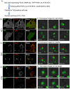
Figure 1
Figure 1
Stable Neuro2a cell lines expressing the human 2N4R tau isoform and repeat domain fragment with the P301L mutation. (A) Schematic representation of cloning for tau fibril cell lines. To generate cells with Tau inclusions, recombinant K18 tau fibrils were transduced into 4C1 cells by lipofectamine 3000. Single cells with GFP-positive inclusion were cloned for generating stable cell lines; (BD) GFP fluorescence imaging and immunocytochemistry of stable Neuro2a cell lines ((B), 4C1 cell; (C), D1C cell; (D), F1B cell). Methanol-formaldehyde fixed cells were immunolabeled with the Tau12 or AT8 antibody. GFP signal and immunoreactivity were captured by fluorescence microscopy with green and red filters, respectively. Co-localization with GFP and immunoreactivity was indicated by a yellow signal in merged images; (EG) Time-lapse images of the F1C cell during cell division (E), stable period (F), and cell death (G). GFP fluorescence was captured by an incubator microscope. GFP inclusions were transmitted from mother to daughter cells within an hour (E). Representative images showed a long living inclusion of more than 20 h ((F); red arrow indicates a stable inclusion). The other representative images show a bursting GFP inclusion during cell death (G).
Figure 2
Figure 2
Biochemical characterization of tau protein in cells and mouse brains. (A) Cellular fractionation protocol for detecting tau protein in TBS-extractable (S1) and sarkosyl-insoluble fractions (P3) from 4C1, D1C, and F1B cells; (B) 2.5 μL of each sample from fractionated cell lysate was loaded on gel and SDS-PAGE was performed. Subsequently, western blots of S1 and P3 fractions with Tau12, Tau46, PHF1, TauRD, and β-actin antibodies were carried out. All cell lines (4C1, D1C, and F1B) expressed both human full-length tau (65–72 kDa) and GFP-K18 (43 kDa, purple arrowhead) recognized by Tau12 and TauRD antibodies, respectively. Both full-length and GFP-K18 tau were recovered in P3 fractions of D1C and F1B cells, but not in the P3 fraction of the 4C1 cell. High molecular weight aggregates (blue arrowheads) also appeared in stacking gel of P3 fractions of D1c and F1B cells. Hyperphosphorylated tau (72 kDa, red arrowheads) was detected in S1 and P3 fractions of D1C and F1B cells; (C) Western blots for detecting TBS-extractable tau in rTg4510 mice. A certain amount of S1 fraction (loading sample containing 0.01 mg wet-weight of brain) from 2-, 5.9-, 6.7-, 8-, and 11-month-old rTg4510 mice was separated by SDS-PAGE, and then western blotting with Tau12, Tau46, pS396, PHF1, and β-actin antibodies was conducted. Green arrowhead indicates hyperphosphorylated 64 kDa tau. Mobility shift of full-length 0N4R tau (50–60 kDa to 64 kDa) was clearly observed from 5.9- to 6.7-month-old rTg4510 mice; (D) Western blots for detecting sarkosyl-insoluble tau in rTg4510 mice. A certain amount of P3 fraction (loading sample containing 0.5 mg wet-weight of brain) from the above-mentioned mice was separated by SDS-PAGE, and then western blotting with E1, Tau46, and PHF1 antibodies was conducted. Green arrowhead indicated hyperphosphorylated 64 kDa tau.
Figure 2
Figure 2
Biochemical characterization of tau protein in cells and mouse brains. (A) Cellular fractionation protocol for detecting tau protein in TBS-extractable (S1) and sarkosyl-insoluble fractions (P3) from 4C1, D1C, and F1B cells; (B) 2.5 μL of each sample from fractionated cell lysate was loaded on gel and SDS-PAGE was performed. Subsequently, western blots of S1 and P3 fractions with Tau12, Tau46, PHF1, TauRD, and β-actin antibodies were carried out. All cell lines (4C1, D1C, and F1B) expressed both human full-length tau (65–72 kDa) and GFP-K18 (43 kDa, purple arrowhead) recognized by Tau12 and TauRD antibodies, respectively. Both full-length and GFP-K18 tau were recovered in P3 fractions of D1C and F1B cells, but not in the P3 fraction of the 4C1 cell. High molecular weight aggregates (blue arrowheads) also appeared in stacking gel of P3 fractions of D1c and F1B cells. Hyperphosphorylated tau (72 kDa, red arrowheads) was detected in S1 and P3 fractions of D1C and F1B cells; (C) Western blots for detecting TBS-extractable tau in rTg4510 mice. A certain amount of S1 fraction (loading sample containing 0.01 mg wet-weight of brain) from 2-, 5.9-, 6.7-, 8-, and 11-month-old rTg4510 mice was separated by SDS-PAGE, and then western blotting with Tau12, Tau46, pS396, PHF1, and β-actin antibodies was conducted. Green arrowhead indicates hyperphosphorylated 64 kDa tau. Mobility shift of full-length 0N4R tau (50–60 kDa to 64 kDa) was clearly observed from 5.9- to 6.7-month-old rTg4510 mice; (D) Western blots for detecting sarkosyl-insoluble tau in rTg4510 mice. A certain amount of P3 fraction (loading sample containing 0.5 mg wet-weight of brain) from the above-mentioned mice was separated by SDS-PAGE, and then western blotting with E1, Tau46, and PHF1 antibodies was conducted. Green arrowhead indicated hyperphosphorylated 64 kDa tau.
Figure 3
Figure 3
Visualization of cellular tau aggregates in tau fibril cell lines. (A,B) D1C (A) and F1B (B) cells labeled by the PBB5 ligand were captured with GFP and Cy3 filters to detect GFP and PBB5 fluorescence, respectively. Both cell lines showed co-localization of GFP and PBB5 signals, indicating the existence of tau fibrils composed of β-sheets in both cell lines. Bars = 20 μm. Right end panels showed high-magnified images detecting GFP (green) and PBB5 (purple) signals. Bars = 1 μm; (C) F1B cell was immunostained with Tau12 and p62 antibodies following formaldehyde fixation, and visualized by super-resolution structured illumination microscopy (SR-SIM). Whole cell (upper panel) and magnified (lower panel) images are shown. Tau12-positive tau (red channel) represents full-length 2N4R tau. Both full-length and GFP-K18 tau were co-localized and formed fibular aggregates. Co-localization with p62 and tau aggregates (arrows) was partial. Bar for whole cell = 5 μm. Bar for magnified image = 1 μm; (D) F1B cell was immunostained with AT8 and p62 antibodies followed by formaldehyde fixation, and visualized by super-resolution microscopy. Whole cell (upper panel) and magnified (lower panel) images are shown. AT8 signal was not completely co-labeled with GFP, suggesting that phosphorylated tau was a part of tau fibrils. On the other hand, AT8-positive fibrils tended to co-localize with p62.
Figure 3
Figure 3
Visualization of cellular tau aggregates in tau fibril cell lines. (A,B) D1C (A) and F1B (B) cells labeled by the PBB5 ligand were captured with GFP and Cy3 filters to detect GFP and PBB5 fluorescence, respectively. Both cell lines showed co-localization of GFP and PBB5 signals, indicating the existence of tau fibrils composed of β-sheets in both cell lines. Bars = 20 μm. Right end panels showed high-magnified images detecting GFP (green) and PBB5 (purple) signals. Bars = 1 μm; (C) F1B cell was immunostained with Tau12 and p62 antibodies following formaldehyde fixation, and visualized by super-resolution structured illumination microscopy (SR-SIM). Whole cell (upper panel) and magnified (lower panel) images are shown. Tau12-positive tau (red channel) represents full-length 2N4R tau. Both full-length and GFP-K18 tau were co-localized and formed fibular aggregates. Co-localization with p62 and tau aggregates (arrows) was partial. Bar for whole cell = 5 μm. Bar for magnified image = 1 μm; (D) F1B cell was immunostained with AT8 and p62 antibodies followed by formaldehyde fixation, and visualized by super-resolution microscopy. Whole cell (upper panel) and magnified (lower panel) images are shown. AT8 signal was not completely co-labeled with GFP, suggesting that phosphorylated tau was a part of tau fibrils. On the other hand, AT8-positive fibrils tended to co-localize with p62.
Figure 4
Figure 4
Visualization of polyubiquitinated tau by super-resolution microscopy. (AC) Cell was immunostained with multiubiquitin (FK2) and p62 (p62c) antibodies following formaldehyde fixation, and was visualized by super-resolution microscopy (SR-SIM). Whole cell (A), magnified (B), and super-magnified (C) images are shown. Polyubiquitin signals were detected in p62 bodies, but only a little in tau fibrils (shown in B). FK2-positive patch (arrow in C) was observed in tau fibrils (merged image of C). Bar for super-magnified image = 200 nm.
Figure 4
Figure 4
Visualization of polyubiquitinated tau by super-resolution microscopy. (AC) Cell was immunostained with multiubiquitin (FK2) and p62 (p62c) antibodies following formaldehyde fixation, and was visualized by super-resolution microscopy (SR-SIM). Whole cell (A), magnified (B), and super-magnified (C) images are shown. Polyubiquitin signals were detected in p62 bodies, but only a little in tau fibrils (shown in B). FK2-positive patch (arrow in C) was observed in tau fibrils (merged image of C). Bar for super-magnified image = 200 nm.

Similar articles

Cited by

References

    1. Lee V.M., Goedert M., Trojanowski J.Q. Neurodegenerative tauopathies. Annu. Rev. Neurosci. 2001;24:1121–1159. doi: 10.1146/annurev.neuro.24.1.1121. - DOI - PubMed
    1. Iqbal K., Liu F., Gong C.X. Tau and neurodegenerative disease: The story so far. Nat. Rev. Neurol. 2016;12:15–27. doi: 10.1038/nrneurol.2015.225. - DOI - PubMed
    1. Goedert M., Jakes R., Spillantini M.G., Hasegawa M., Smith M.J., Crowther R.A. Assembly of microtubule-associated protein tau into alzheimer-like filaments induced by sulphated glycosaminoglycans. Nature. 1996;383:550–553. doi: 10.1038/383550a0. - DOI - PubMed
    1. Perez M., Valpuesta J.M., Medina M., Montejo de Garcini E., Avila J. Polymerization of tau into filaments in the presence of heparin: The minimal sequence required for tau-tau interaction. J. Neurochem. 1996;67:1183–1190. doi: 10.1046/j.1471-4159.1996.67031183.x. - DOI - PubMed
    1. DeTure M., Ko L.W., Easson C., Yen S.H. Tau assembly in inducible transfectants expressing wild-type or ftdp-17 tau. Am. J. Pathol. 2002;161:1711–1722. doi: 10.1016/S0002-9440(10)64448-3. - DOI - PMC - PubMed

MeSH terms