. 2018 Dec;56(1):32-42.
doi: 10.1080/13880209.2017.1412468.

The suppressive effect of the three-herb extract mixture on vascular and liver inflammation in atherogenic diet with high fructose-fed mice

Affiliations

The suppressive effect of the three-herb extract mixture on vascular and liver inflammation in atherogenic diet with high fructose-fed mice

Hae Seong Song et al. Pharm Biol. 2018 Dec.

Abstract

Context: Cynanchum wilfordii (Maximowicz) Hemsley (Apocynaceae), Arctium lappa L. var. rubescens Frivald (Asteraceae) and Dioscorea opposite Thunb (Dioscoreaceae) root extracts have been widely used as an alternative for intervening obesity.

Objectives: The synergistic effect of three-herb mixture of C. wilfordii, A. lappa and D. opposita was determined on aortic and liver inflammatory responses.

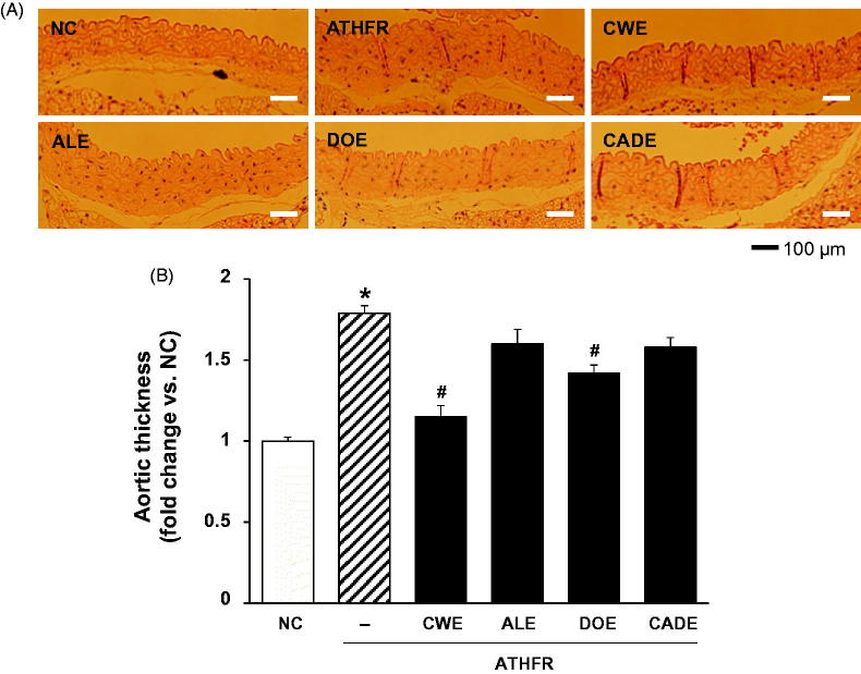
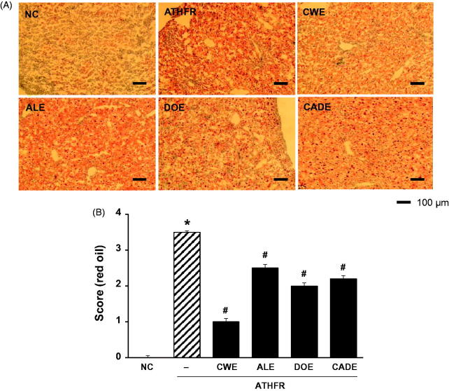
Materials and methods: CWE, ALE and DOE were prepared from the root of C. wilfordii, A. lappa and D. opposite by 70% ethanol extraction, respectively. CADE was prepared using a powder mixture of 2 CWE:1 ALE:1 DOE. C57BL/6 mice were fed an atherogenic diet combined with 10% fructose (ATHFR) in the presence of 200 mg/kg/day CWE, ALE, DOE or CADE for 8 weeks (each group, n = 6). Biochemical profiles, protein expression of vascular cell adhesion molecule-1 (VCAM-1) on the aorta and liver were determined.

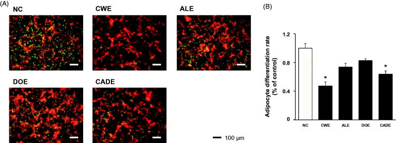
Results: CADE could significantly suppress the protein expression of VCAM-1 in both the aorta and liver (80% reduction) compared to ATHFR-fed mice. Impairment of liver function was significantly ameliorated by CADE supplement, as determined by GOT (60% reduction) and GPT (51% reduction) levels.

Conclusions: CADE should be considered when developing medications to suppress the vascular and liver inflammatory responses for individuals who are either non-responsive or resistant to lipid-lowering drugs.

Keywords: Inflammation; P-selectin; fat accumulation; lipid-lowering; obesity; synergistic effect; vascular cell adhesion molecule-1.


Figures

Figure 1.
Figure 1.
The suppressive effects of CWE, ALE, DOE and CADE on TNF-α induced mRNA expression level of VCAM-1 (A) and ICAM-1 (B). HASMC cells were treated with 20 μg/mL of CWE, ALE, DOE and CADE in the presence of TNF-α (10 ng/mL) for 12 h. The mRNA expression levels of VCAM-1 and ICAM-1 were determined by RT-PCR. Data are mean ± SEM. *p < 0.05, significantly different from the untreated group, #p < 0.05, significantly different from the TNF-α group. ATHFR: an atherogenic diet plus 10% fructose; CWE: C. wilfordii extract; ALE: A. lappa L. extract; DOE: D. opposita extract; CADE: mixture of CWE:ALE:DOE at a ratio of 2:1:1; NC: untreated control.
Figure 2.
Figure 2.
The suppressive effects of CWE, ALE, DOE and CADE on adipogenesis in 3T3-L1 cells. 3T3-L1 preadipocytes were differentiated in the presence or absence of CWE, ALE, DOE and CADE (200 μg/mL). After staining with AdipoRedTM, the lipid droplets were quantified by the intensities of fluorescence. Small dot indicates a lipid droplet. Original magnification, ×100. Data are mean ± SEM. *p < 0.05, significantly different from the untreated group. CWE: C. wilfordii extract; ALE: A. lappa L. extract; DOE: D. opposita extract; CADE: mixture of CWE:ALE:DOE at ratio of 2:1:1; NC: untreated control.
Figure 3.
Figure 3.
Changes in body weight and both food and water intake rate. Mice were fed a normal chow diet or an ATHFR diet in the presence or absence of CWE, ALE, DOE and CADE for 8 weeks. Body weight and food and water intake were measured weekly. Data are mean ± SEM (each group, n = 6). ATHFR: an atherogenic diet plus 10% fructose; CWE: C. wilfordii extract; ALE: A. lappa L. extract; DOE: D. opposita extract; CADE: mixture of CWE:ALE:DOE at ratio of 2:1:1; NC: untreated control.
Figure 4.
Figure 4.
Changes in the weight of liver, kidney, spleen and thymus. Mice were fed with normal chow diet and ATHFR diet in the presence or absence of CWE, ALE, DOE and CADE for 8 weeks. At the end of experiment, the weights of liver, kidney, spleen and thymus were measured. Data are mean ± SEM (each group, n = 6). Omega-3 and simvastatin were used as controls for nutritional alternative and lipid-lowering drug, respectively. *p < 0.05, significantly different from the NC group, #p < 0.05, significantly different from the ATHFR group. ATHFR: an atherogenic diet plus 10% fructose; CWE: C. wilfordii extract; ALE: A. lappa L. extract; DOE: D. opposita extract; CADE: mixture of CWE:ALE:DOE at a ratio of 2:1:1; NC: untreated control.
Figure 5.
Figure 5.
Effect of CWE, ALE, DOE and CADE on the status of lipid profiles. Mice were fed with normal chow diet or ATHFR diet in the presence or absence of CWE, ALE, DOE and CADE for 8 weeks. At the end of experiment, blood specimens were collected the serum level of total-cholesterol (A), HDL-cholesterol (B), LDL-cholesterol (C), and triglyceride levels (D) were measured using commercially available assay kits. Data are mean ± SEM (each group, n = 6). Omega-3 and simvastatin were used as controls for nutritional alternative and lipid-lowering drug, respectively. ATHFR: an atherogenic diet plus 10% fructose; CWE: C. wilfordii extract; ALE: A. lappa L. extract; DOE: D. opposita extract; CADE: mixture of CWE:ALE:DOE at a ratio of 2:1:1; NC: untreated control.
Figure 6.
Figure 6.
Effect of CWE, ALE, DOE and CADE on the thickening of the aorta in ATHFR diet-fed mice. Mice were fed with a normal chow diet or ATHFR diet in the presence or absence of CWE, ALE, DOE and CADE for 8 weeks (each n = 6). At the end of the experiment, aorta tissues were collected and stained with H/E staining. Data are mean ± SEM (each group, n = 6). Omega-3 and simvastatin were used as controls for nutritional alternative and lipid-lowering drug, respectively. Original magnification, ×100. *p < 0.05, significantly different from the NC group, #p < 0.05, significantly different from the ATHFR group. ATHFR: an atherogenic diet plus 10% fructose; CWE: C. wilfordii extract; ALE: A. lappa L. extract; DOE: D. opposita extract; CADE: mixture of CWE:ALE:DOE at a ratio of 2:1:1; NC: untreated control.
Figure 7.
Figure 7.
Effect of CWE, ALE, DOE and CADE on protein expression levels of VCAM-1 and P-selectin in the aorta from ATHFR diet-fed mice. Mice were fed with normal chow diet or ATHFR diet in the presence or absence of CWE, ALE, DOE and CADE for 8 weeks (each n = 6). At the end of experiment, aorta tissues were collected, and the protein expression levels of VCAM-1 (A) and P-selectin (B) were detected by immunohistochemistry. Omega-3 and simvastatin were used as controls for nutritional alternative and lipid-lowering drug, respectively. Original magnification, ×200. Bar indicates 100 μm. ATHFR: an atherogenic diet plus 10% fructose; CWE: C. wilfordii extract; ALE: A. lappa L. extract; DOE: D. opposita extract; CADE: mixture of CWE:ALE:DOE at a ratio of 2:1:1; NC: untreated control.
Figure 8.
Figure 8.
Effect of CWE, ALE, DOE and CADE on enzymatic activities of GOT and GPT in ATHFR diet-fed mice. Mice were fed with normal chow diet or an ATHFR diet in the presence or absence of CWE, ALE, DOE and CADE for 8 weeks (each n = 6). At the end of the experiment, both GOT and GPT levels were determined using a colorimetric method. Omega-3 and simvastatin were used as controls for nutritional alternative and lipid-lowering drug, respectively. *p < 0.05, significantly different from the NC group, #p < 0.05, significantly different from the ATHFR group. ATHFR: an atherogenic diet plus 10% fructose; CWE: C. wilfordii extract; ALE: A. lappa L. extract; DOE: D. opposita extract; CADE: mixture of CWE:ALE:DOE at a ratio of 2:1:1; NC: untreated control.
Figure 9.
Figure 9.
Effect of CWE, ALE, DOE and CADE on lipid contents in livers from ATHFR diet-fed mice. Mice were fed with normal chow diet or an ATHFR diet in the presence or absence of CWE, ALE, DOE and CADE for 8 weeks (each n = 6). At the end of the experiment, liver specimens were collected, and frozen liver sections were stained with H&E staining. Omega-3 and simvastatin were used as controls of nutritional alternative and lipid-lowering drug, respectively. Original magnification, ×200. Bar indicates 100 μm. *p < 0.05, significantly different from the NC group, #p < 0.05, significantly different from the ATHFR group. ATHFR: an atherogenic diet plus 10% fructose; CWE: C. wilfordii extract; ALE: A. lappa L. extract; DOE: D. opposita extract; CADE: mixture of CWE:ALE:DOE with ratio of 2:1:1; NC: untreated control.
Figure 10.
Figure 10.
Effect of CWE, ALE, DOE and CADE on fat accumulation in livers from ATHFR diet-fed mice. Mice were fed with a normal chow diet or an ATHFR diet in the presence or absence of CWE, ALE, DOE or CADE for 8 weeks (each n = 6). At the end of the experiment, liver specimens were collected, and frozen liver sections were stained with H&E staining. Omega-3 and simvastatin were used as controls of nutritional alternative and lipid-lowering drug, respectively. Original magnification, ×100. Bar indicates 100 μm. *p < 0.05, significantly different from the NC group, #p < 0.05, significantly different from the ATHFR group. ATHFR: an atherogenic diet plus 10% fructose; CWE: C. wilfordii extract; ALE: A. lappa L. extract; DOE: D. opposita extract; CADE: mixture of CWE:ALE:DOE at a ratio of 2:1:1; NC: untreated control.
Figure 11.
Figure 11.
Effect of CWE, ALE, DOE and CADE on protein expression of VCAM-1 in livers from ATHFR diet-fed mice. Mice were fed with a normal chow diet or an ATHFR diet in the presence or absence of CWE, ALE, DOE or CADE for 8 weeks (each n = 6). At the end of the experiment, liver specimens were collected, and protein expression levels of VCAM-1 were detected in the frozen liver sections using immunohistochemical staining. Original magnification, ×100. Bar indicates 100 μm. *p < 0.05, significantly different from the NC group, #p < 0.05, significantly different from the ATHFR group. ATHFR: an atherogenic diet plus 10% fructose; CWE: C. wilfordii extract; ALE: A. lappa L. extract; DOE: D. opposita extract; CADE: mixture of CWE:ALE:DOE at a ratio of 2:1:1; NC: untreated control.

Similar articles

Cited by

References

    1. Aeberli I, Zimmermann MB, Molinari L, Lehmann R, l’Allemand D, Spinas GA, Berneis K.. 2007. Fructose intake is a predictor of LDL particle size in overweight school children. Am J Clin Nutr. 86:1174–1178. - PubMed
    1. Ali AT, Hochfeld WE, Myburgh R, Pepper MS.. 2013. Adipocyte and adipogenesis. Eur J Cell Biol. 92:229–236. - PubMed
    1. wCho Y-M, Song H-S, Jang S-A, Park D-W, Shin YS, Jeong YJ, Kang SC.. 2016. Suppression of VCAM-1 expression in human aortic smooth muscle cells treated with ethanol extracts of Cynanchum wilfordii radix, Arctium lappa L., and Dioscorea opposita. Korean J Plant Resour. 29:525–531.
    1. da Silva AM, Correa CL, Neves RH, Machado-Silva JR.. 2012. A high-fat diet associated with acute Schistosomiasis mansoni causes disorganization in splenic architecture in mice. Exp Parasitol. 132:193–199. - PubMed
    1. Dai S, McNeill JH.. 1995. Fructose-induced hypertension in rats is concentration- and duration-dependent. J Pharmacol Toxicol Methods. 33:101–107. - PubMed

MeSH terms

LinkOut - more resources