. 2018 Dec;48(3):513-522.
doi: 10.1016/j.semarthrit.2018.03.016. Epub 2018 Mar 28.

Medications received by patients with juvenile dermatomyositis

Affiliations

Medications received by patients with juvenile dermatomyositis

Takayuki Kishi et al. Semin Arthritis Rheum. 2018 Dec.

Abstract

Objective: Few controlled studies are available to guide treatment decisions in juvenile dermatomyositis (JDM). This study evaluated therapies received, changes of treatment over time, and factors associated with medication choices in JDM.

Methods: We performed a retrospective analysis of the number and type of therapies and duration of treatment for 320 patients with JDM enrolled in a North American registry. Kaplan-Meier and logistic regression analysis were used to assess the association of demographic and clinical features and autoantibodies with medication usage.

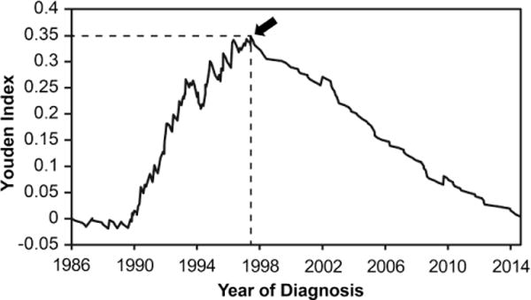
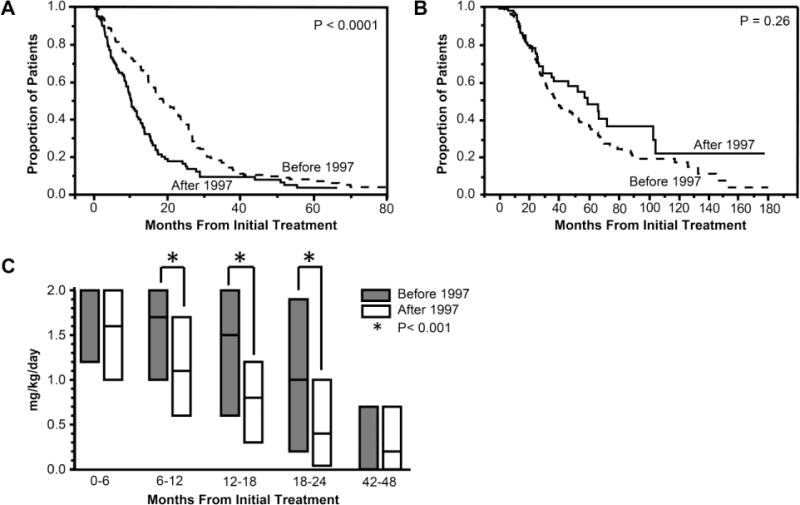
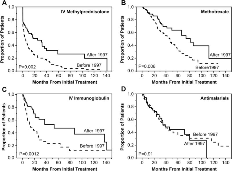
Results: High-dose oral prednisone was the primary therapy given to 99% of patients. 1997 was determined to be a threshold year for increasing usage of medications other than prednisone. The median time to half the initial oral prednisone dose was shorter in patients diagnosed after vs. before 1997 (10 vs. 19 months, P < 0.01). Patients received intravenous methylprednisolone (IVMP), methotrexate, intravenous immunoglobulin, antimalarial drugs, and combination therapy more frequently when diagnosed after 1997. IVMP was frequently received by patients with severe illness onset, anti-p155/140 (anti-TIF1) and anti-MJ (anti-NXP2) autoantibodies. Treatment with methotrexate was associated with older age at diagnosis and anti-MJ autoantibodies, while antimalarial therapy was associated with anti-p155/140 autoantibodies and mild onset severity.

Conclusion: Oral prednisone has been the mainstay of therapy in JDM, and prednisone was reduced faster in patients diagnosed after 1997 when there was also an increase in other medications. Specific medications received by patients with JDM correlated with year and age at diagnosis, myositis autoantibodies, onset severity, and illness features.

Keywords: Juvenile dermatomyositis; Medications; Methotrexate; Myositis autoantibodies; Prednisone; Treatment.


Conflict of interest statement

Disclosures of conflict of interest: None

Figures

Figure 1
Figure 1
Plot of year of diagnosis versus Youden index. Youden index is defined as (Sensitivity-(1-Specificity)) for major drug usage other than prednisone, and is calculated by Receiver Operating Characteristic (ROC) curve analysis. The sensitivity and specificity of major drug usage beyond oral prednisone was calculated for each year after diagnosis. The arrow indicates the maximum point of the Youden index (0.348). This corresponds to the threshold date for major medication usage beyond oral prednisone, which was July 11, 1997.
Figure 2
Figure 2
A, Kaplan-Meier analysis plot showing time to half of initial dose of oral prednisone in juvenile dermatomyositis patients diagnosed before 1997 versus after 1997. The Y axis shows the proportion of patients who were still receiving more than 50% of the initial dose of prednisone at any point in time. X axis shows months from initial treatment. B, Kaplan-Meier analysis plot showing time to final discontinuation of oral prednisone in juvenile dermatomyositis patients diagnosed before 1997 versus after 1997. The Y axis shows the proportion of patients who were still receiving prednisone at any point in time. X axis shows months from initial treatment. C, Median daily maximum dose of oral prednisone in each 6 month treatment period. The Y axis shows the median daily maximum dose of oral prednisone. X axis shows the months from initial treatment. The bars show the median prednisone dose with 25% and 75%. For 0–6 months, the median dose of prednisone before 1997 is the same as the 75%, and for 42–48 months, the median dose of prednisone before 1997 is the same as the 25%. The median daily maximum dose for patients diagnosed before 1997 and after 1997 was 1.7 [IQR 1.0–2.0] vs. 1.1 [IQR 0.6–1.7] mg/kg per day for 6–12 months, 1.5 [0.6–2.0] vs. 0.8 [0.3–1.2] mg/kg per day for 12–18 months, and 1.0 [0.2–1.9] vs. 0.4 [0.04–1.0] mg/kg per day for 18–24 months.
Figure 3
Figure 3
Kaplan-Meier analysis plots showing time to discontinuation of each medication. The Y axis shows the proportion of patients who were still receiving each medication at any point in time. The X axis shows the months from initial treatment. A, Intravenous methylprednisolone (IVMP). The median time to discontinuation of IVMP was 5 months from the initiation of IVMP treatment in patients diagnosed before 1997 vs. 25 months in patients diagnosed after 1997. B, Methotrexate. The median time to discontinuation of methotrexate was 46 months from methotrexate treatment initiation in patients diagnosed before 1997 vs. 81 months in patients diagnosed after 1997. C, Intravenous immunoglobulin (IVIG). The median time to discontinuation of IVIG was 13 months after IVIG treatment initiation in patients diagnosed before 1997 vs. 53 months in patients diagnosed after 1997. D, Antimalarial drugs. The median time to discontinuation of antimalarial drugs was 37 months after initiation of antimalarial drugs in patients diagnosed before 1997 vs. 39 months in patients diagnosed after 1997.

References

    1. Feldman BM, Rider LG, Reed AM, Pachman LM. Juvenile dermatomyositis and other idiopathic inflammatory myopathies of childhood. Lancet. 2008;371:2201–12. - PubMed
    1. Rider LG, Nistala K. The juvenile idiopathic inflammatory myopathies: pathogenesis, clinical and autoantibody phenotypes, and outcomes. J Intern Med. 2016;280:24–38. - PMC - PubMed
    1. Rider LG, Katz JD, Jones OY. Developments in the classification and treatment of the juvenile idiopathic inflammatory myopathies. Rheum Dis Clin North Am. 2013;39:877–904. - PMC - PubMed
    1. Tansley SL, Simou S, Shaddick G, Betteridge ZE, Almeida B, Gunawardena H, et al. Autoantibodies in juvenile-onset myositis: Their diagnostic value and associated clinical phenotype in a large UK cohort. J Autoimmun. 2017;84:55–64. - PMC - PubMed
    1. Bowyer SL, Blane CE, Sullivan DB, Cassidy JT. Childhood dermatomyositis: factors predicting functional outcome and development of dystrophic calcification. J Pediatr. 1983;103:882–8. - PubMed

Publication types