. 2018 May 1;32(9-10):658-669.
doi: 10.1101/gad.313460.118. Epub 2018 May 17.

PRDX1 and MTH1 cooperate to prevent ROS-mediated inhibition of telomerase

Affiliations

PRDX1 and MTH1 cooperate to prevent ROS-mediated inhibition of telomerase

Wareed Ahmed et al. Genes Dev. .

Abstract

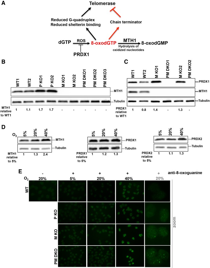
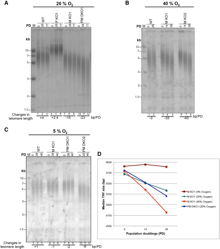
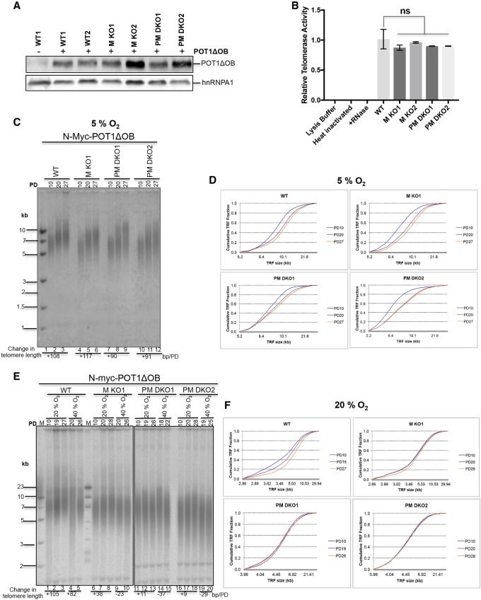
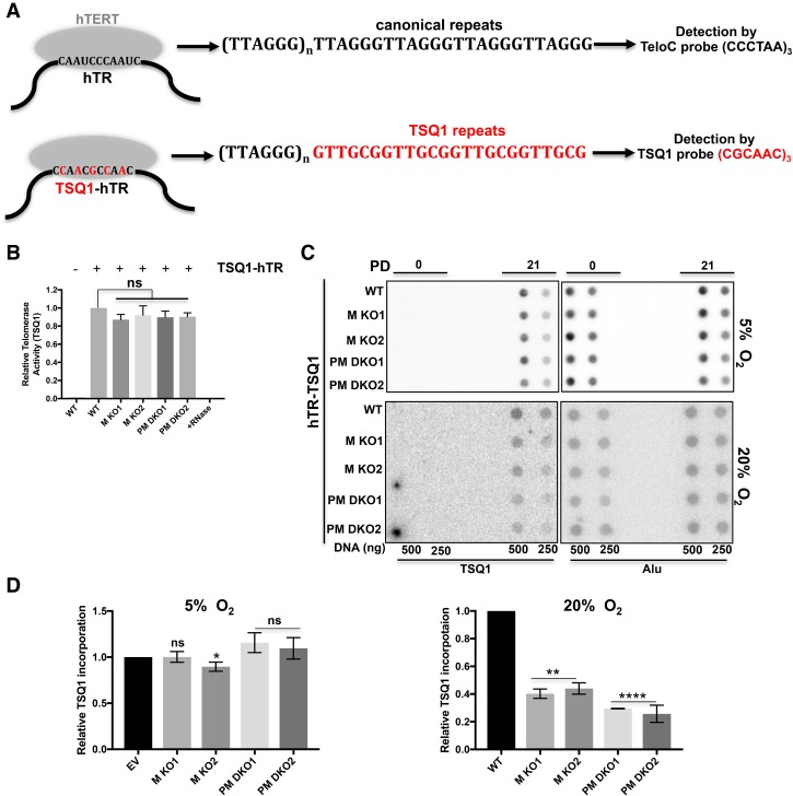
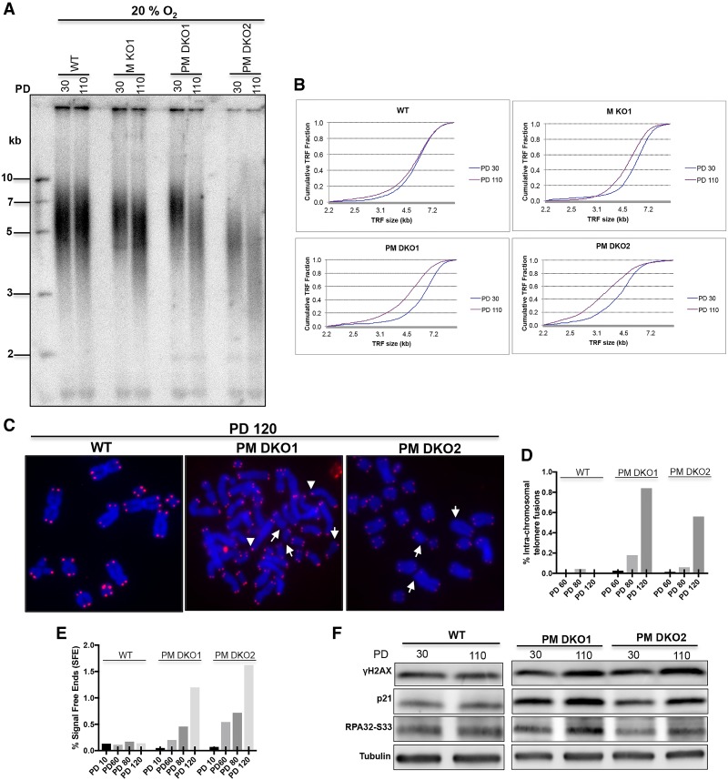
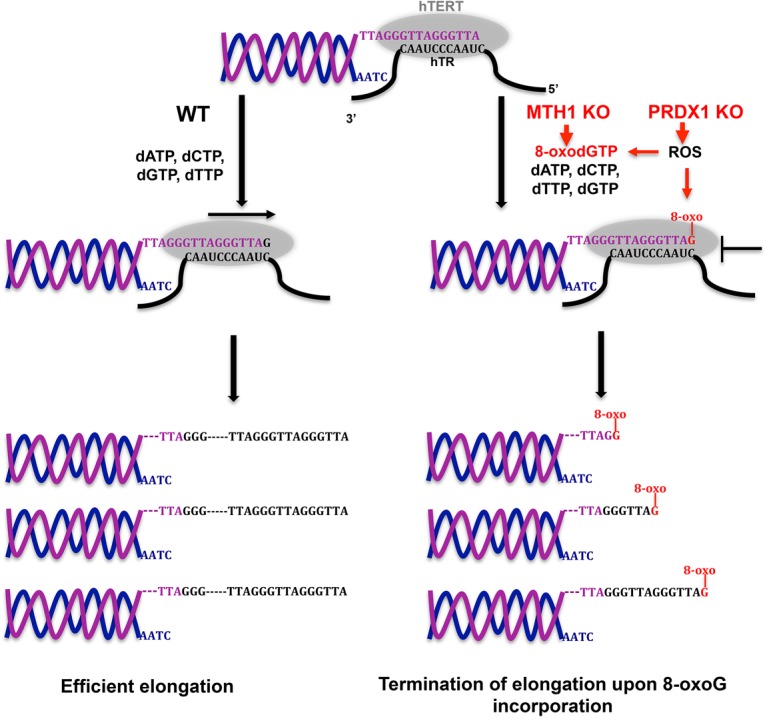
Telomerase counteracts telomere shortening and cellular senescence in germ, stem, and cancer cells by adding repetitive DNA sequences to the ends of chromosomes. Telomeres are susceptible to damage by reactive oxygen species (ROS), but the consequences of oxidation of telomeres on telomere length and the mechanisms that protect from ROS-mediated telomere damage are not well understood. In particular, 8-oxoguanine nucleotides at 3' ends of telomeric substrates inhibit telomerase in vitro, whereas, at internal positions, they suppress G-quadruplex formation and were therefore proposed to promote telomerase activity. Here, we disrupt the peroxiredoxin 1 (PRDX1) and 7,8-dihydro-8-oxoguanine triphosphatase (MTH1) genes in cancer cells and demonstrate that PRDX1 and MTH1 cooperate to prevent accumulation of oxidized guanine in the genome. Concomitant disruption of PRDX1 and MTH1 leads to ROS concentration-dependent continuous shortening of telomeres, which is due to efficient inhibition of telomere extension by telomerase. Our results identify antioxidant systems that are required to protect telomeres from oxidation and are necessary to allow telomere maintenance by telomerase conferring immortality to cancer cells.

Keywords: MTH1; PRDX1; aging; cellular senescence; oxidative stress; telomerase; telomeres.


Figures

Figure 1.
Figure 1.
MTH1 and PRDX1 cooperate for safeguarding the genome from oxidation. (A) Schematic representation of the enzymatic functions of PRDX1 and MTH1 and the putative effects of oxidation of telomere substrates on telomere maintenance by telomerase. PRDX1 reduces ROS. MTH1 hydrolyzes 8-oxo dGTP to 8-oxo dGMP, preventing its usage by telomerase and other DNA polymerases. In vitro experiments suggested that oxidation of telomeric DNA can either promote or inhibit telomerase. (B) Western blot analysis of MTH1 levels in two HCT116 wild-type cell clones (WT1 and WT2), two PRDX1 knockout clones (P KO1 and P KO2), and three PRDX1/MTH1 double-knockout clones (PM DKO1–3). The relative MTH1 levels are indicated. Tubulin was used as a loading control. (C) Western blot analysis of PRDX1 and MTH1 levels in HCT116 wild-type (WT1 and WT2), MTH1 -knockout (M KO1 and M KO2), and PRDX1/MTH1 double-knockout (PM DKO1 and PM DKO2) cells. The relative PRDX1 levels are indicated. (D) Western blot analysis of MTH1, PRDX1, and PRDX2 in HCT116 cells grown in incubators with different O2 concentrations. (E) Visualization of 8-oxo dG in DNA of wild-type, PRDX1 knockout (P KO), MTH1 knockout (M KO), and PRDX1/MTH1 double-knockout (PM DKO) cells by immunostaining with anti-8-oxoguanine (8-oxo G) antibody.
Figure 2.
Figure 2.
Oxidative stress-dependent telomere shortening in MTH1 knockout and MTH1/PRDX1 double-knockout cells. (AC) TRF length determination by Southern blot analysis of genomic DNA. Genomic DNAs from HCT116 wild-type and knockout cells grown at 20% O2 (A), 40% O2 (B), or 5% O2 (C) for the indicated PDs were purified, restriction-digested, resolved on 0.7% agarose gels, and analyzed upon in-gel hybridization using a random radiolabeled telomeric probe. Averaged rates of telomere shortening (base pairs/PD), indicated below the gels, were deduced by quantifying the changes in telomere length between the indicated consecutive PDs. (D) Comparison of telomere shortening rates between MTH1 knockout (M KO) and PRDX1/MTH1 double-knockout (PM DKO) cells exposed to different O2 concentrations. The graph represents the median TRF lengths as a function of PDs. The graphs for MTH1 knockout (5% O2), MTH1 knockout (20% O2), and PRDX1/MTH1 double knockout (20% O2) were derived from Supplemental Figure S2B, while MTH1 knockout (40% O2) data points were derived from B.
Figure 3.
Figure 3.
Inefficient elongation of telomeres in MTH1 and PRDX1 knockout cells at high O2 concentrations. (A) Western blot analysis of N-MycPOT1ΔOB in wild-type and knockout cells by immunoblotting using an anti-N-Myc antibody (9E11). Where indicated (+), wild-type and knockout HCT116 cells were stably infected with a retroviral construct expressing N-MycPOT1ΔOB. (B) Comparison of telomerase activity (RQ-TRAP assay) in the indicated knockout cells relative to wild type. (C) TRF analysis of cells expressing N-MycPOT1ΔOB grown at 5% O2. Upon completion of puromycin selection at PD10, cells were further propagated for the indicated PDs. (D) Distribution profile of telomere fragments from cells obtained in C. Cumulative fractions of telomere fragments with different sizes were plotted to generate the telomere fragment distribution profiles of wild-type and knockout cells. (E) TRF analysis of cells expressing N-MycPOT1ΔOB grown at 20% O2 and 40% O2. (F) Distribution profile of telomere fragments from cells grown at 20% O2.
Figure 4.
Figure 4.
Reduced telomerase activity at chromosome ends in MTH1 knockout and MTH1/PRDX1 double-knockout cells. (A) Schematic representation of wild-type and TSQ1 mutant telomerase specifying synthesis of TSQ1 (5′-GTTGCG-3′)n telomeric repeats. Lentiviral constructs harboring the mutant form of hTR-encoding TSQ1 repeats were introduced in wild-type and knockout cells. (B) Comparison of telomerase activity of the TSQ1–hTR–telomerase complex in wild-type and knockout cells. Cell lysates of wild-type and knockout cells expressing TSQ1–hTR (indicated by +) were analyzed in a modified RQ-TRAP assay using a substrate and a primer that specifically detects incorporated GTTGCG repeats (Materials and Methods). (C) Dot blot analysis of genomic DNA digested with restriction enzymes. At PD0 and PD21, total GTTGCG signal and Alu signal were detected upon hybridization with specific radiolabeled probes. PD0 represents the day of completion of puromycin selection. (D) Quantification of the TSQ1 signal in C. The TSQ1 signal was normalized to the Alu signal and is expressed relative to wild type. Error bars correspond to SD obtained from three independent experiments. (**) P = 0.0016; (****) P < 0.0001; (ns) P > 0.05, unpaired t-test two-tailed P-value compared with the corresponding wild type.
Figure 5.
Figure 5.
MTH1–PRDX1 double knockouts accumulate extremely short telomeres and DNA damage markers. (A) TRF analysis at PD30 and PD110. The indicated clones were grown in incubators containing 20% O2. (B) Distribution profile of telomere fragments at the indicated PDs of A. (C) Analysis of telomere signals (red) in metaphase chromosomes (blue) of clones grown for 120 PDs. White arrows indicate telomere signal-free ends, and arrowheads indicate intrachromosomal telomere fusions. (Blue) DAPI-stained chromosomal DNA; (red) TeloC probe for telomeric DNA. (D) Quantification of intrachromosomal telomere fusions. (E) Quantification of telomere-free ends across different PDs. (F) Western blot analysis of DNA damage markers in PRDX1/MTH1 double-knockout (PM DKO) cells grown for 30 and 110 PDs. For D and E, >2700 telomeres were scored for each sample.
Figure 6.
Figure 6.
Model for telomerase inhibition in MTH1 knockout and PRDX1/MTH1 double-knockout cells. ROS increase the concentration of 8-oxo dGTP. In MTH1 knockout cells, 8-oxo dGTP is not hydrolyzed to 8-oxo dGMP. Telomerase incorporates 8-oxo G, leading to premature chain termination. Loss of PRDX1 further enhances telomerase inhibition. PRDX1 loss increases ROS, promoting oxidation of dGTP to 8-oxo dGTP. PRDX1 loss may also enhance direct ROS-mediated damage of telomeres at the chromosome 3′ end, leading to inhibition of telomerase.

Comment in

References

    1. Aeby E, Ahmed W, Redon S, Simanis V, Lingner J. 2016. Peroxiredoxin 1 protects telomeres from oxidative damage and preserves telomeric DNA for extension by telomerase. Cell Rep 17: 3107–3114. - PubMed
    1. Ahmed W, Lingner J. 2018. Impact of oxidative stress on telomere biology. Differentiation 99: 21–27. - PubMed
    1. Ahmed S, Passos JF, Birket MJ, Beckmann T, Brings S, Peters H, Birch-Machin MA, von Zglinicki T, Saretzki G. 2008. Telomerase does not counteract telomere shortening but protects mitochondrial function under oxidative stress. J Cell Sci 121: 1046–1053. - PubMed
    1. Ancelin K, Brunori M, Bauwens S, Koering CE, Brun C, Ricoul M, Pommier JP, Sabatier L, Gilson E. 2002. Targeting assay to study the cis functions of human telomeric proteins: evidence for inhibition of telomerase by TRF1 and for activation of telomere degradation by TRF2. Mol Cell Biol 22: 3474–3487. - PMC - PubMed
    1. Asai A, Oshima Y, Yamamoto Y, Uochi TA, Kusaka H, Akinaga S, Yamashita Y, Pongracz K, Pruzan R, Wunder E, et al. 2003. A novel telomerase template antagonist (GRN163) as a potential anticancer agent. Cancer Res 63: 3931–3939. - PubMed

Publication types

MeSH terms

LinkOut - more resources