. 2018 Jul 6;293(27):10744-10756.
doi: 10.1074/jbc.RA118.002357. Epub 2018 May 17.

Genetic code expansion and live cell imaging reveal that Thr-308 phosphorylation is irreplaceable and sufficient for Akt1 activity

Affiliations

Genetic code expansion and live cell imaging reveal that Thr-308 phosphorylation is irreplaceable and sufficient for Akt1 activity

Nileeka Balasuriya et al. J Biol Chem. .

Abstract

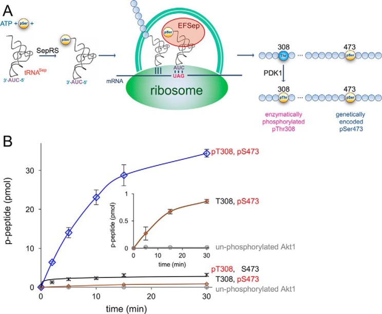
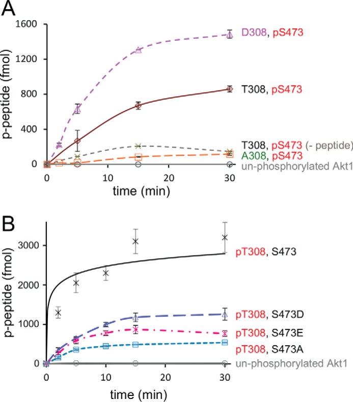
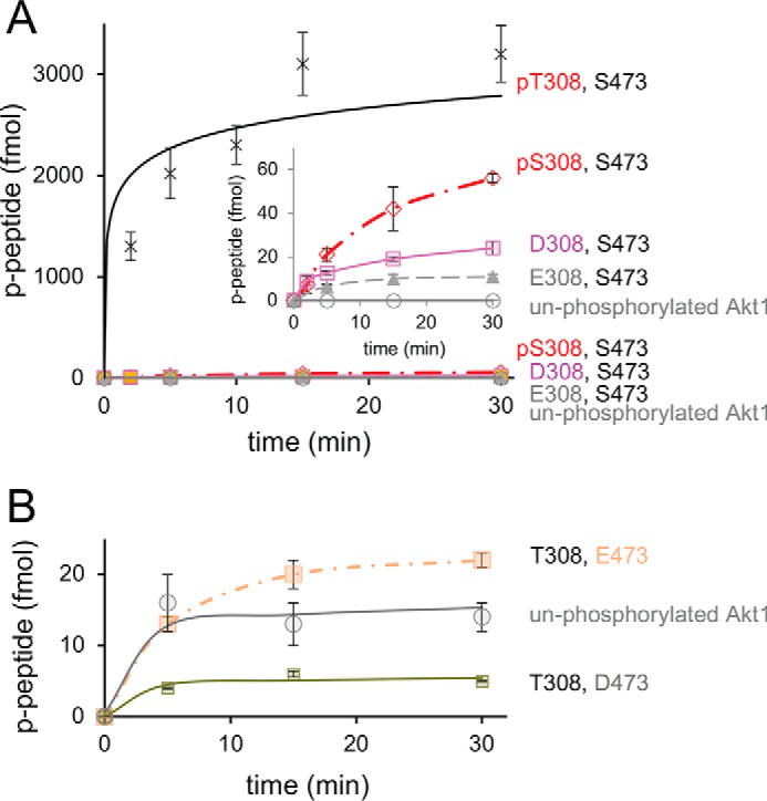
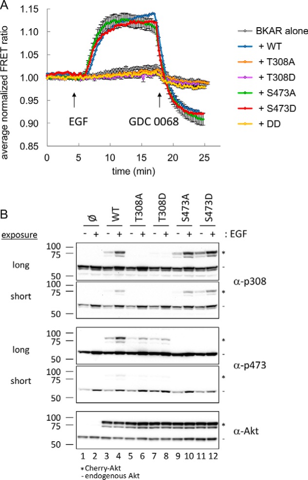
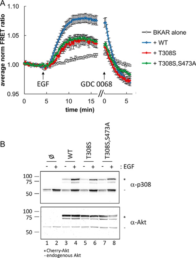
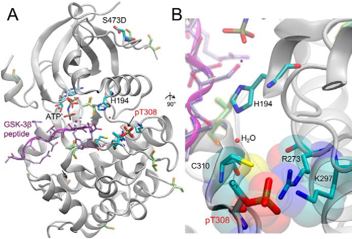
The proto-oncogene Akt/protein kinase B (PKB) is a pivotal signal transducer for growth and survival. Growth factor stimulation leads to Akt phosphorylation at two regulatory sites (Thr-308 and Ser-473), acutely activating Akt signaling. Delineating the exact role of each regulatory site is, however, technically challenging and has remained elusive. Here, we used genetic code expansion to produce site-specifically phosphorylated Akt1 to dissect the contribution of each regulatory site to Akt1 activity. We achieved recombinant production of full-length Akt1 containing site-specific pThr and pSer residues for the first time. Our analysis of Akt1 site-specifically phosphorylated at either or both sites revealed that phosphorylation at both sites increases the apparent catalytic rate 1500-fold relative to unphosphorylated Akt1, an increase attributable primarily to phosphorylation at Thr-308. Live imaging of COS-7 cells confirmed that phosphorylation of Thr-308, but not Ser-473, is required for cellular activation of Akt. We found in vitro and in the cell that pThr-308 function cannot be mimicked with acidic residues, nor could unphosphorylated Thr-308 be mimicked by an Ala mutation. An Akt1 variant with pSer-308 achieved only partial enzymatic and cellular signaling activity, revealing a critical interaction between the γ-methyl group of pThr-308 and Cys-310 in the Akt1 active site. Thus, pThr-308 is necessary and sufficient to stimulate Akt signaling in cells, and the common use of phosphomimetics is not appropriate for studying the biology of Akt signaling. Our data also indicate that pThr-308 should be regarded as the primary diagnostic marker of Akt activity.

Keywords: Akt/protein kinase B (PKB); activation loop; aminoacyl tRNA synthetase; cell biology; cell signaling; enzyme; genetic code expansion; hydrophobic motif; phosphomimetics; phosphoseryl-tRNA synthetase (SepRS); protein phosphorylation; tRNASep; transfer RNA (tRNA).


Conflict of interest statement

The authors declare that they have no conflicts of interest with the contents of this article

Figures

Figure 1.
Figure 1.
A novel route to doubly phosphorylated and active Akt1. A, schematic representation of recombinant Akt1 biosynthesis with pSer-473 genetically encoded in response to the UAG codon and pThr-308 enzymatically phosphorylated in vivo in E. coli. Genetically encoded pSer incorporation requires phosphoseryl-tRNA synthetase (SepRS), a UAG-decoding tRNASep, and the elongation factor mutant (EFSep). B, enzyme activity of differentially phosphorylated Akt1 variants with a GSK-3β substrate peptide. Akt1 quantitatively phosphorylated at both 308 and 473 (ppAktS473,T308, blue diamonds) showed maximal activity compared with the unphosphorylated Akt1 (gray circles) and singly phosphorylated Akt1 variants: pAktT308 (black cross) and pAktS473 (brown diamonds). The reported values represent the mean of triplicate experiments with error bars indicating S.D. Lower-activity variants show above-background kinase activity (inset).
Figure 2.
Figure 2.
Activity of singly phosphorylated Akt1 variants with regulatory site mutations. A, Akt1 enzyme activity is shown for variants with Ser-473 phosphorylated and Thr-308 unphosphorylated (brown diamond) or mutated: T308D (purple triangles) and T308A (orange squares). Controls include unphosphorylated Akt1 (gray circles), and pAkt1S473 in the absence of substrate peptide (−peptide, green crosses). B, compared with pAkt1T308 activity (black crosses) and in the context of Akt1 phosphorylated at Thr-308, mutations S473D (blue triangles), S473E (magenta crosses), or S473A (cyan squares) resulted in marginally reduced activity. All reported values represent the mean of triplicate experiments with error bars indicating S.D.
Figure 3.
Figure 3.
Activity of T308S and phosphomimetic Akt1 variants. A, compared with pAkt1T308 activity (black crosses), mutations of Thr-308 to either Asp (pink squares), Glu (green triangles), or pSer (red diamonds) resulted in low but above-background kinase activity (inset). B, kinase activity was also measured in the context of an unphosphorylated Akt1 (gray circles) with phosphomimetic mutations S473D (green squares) and S473E (peach squares). All reported values represent the mean of triplicate experiments with error bars indicating S.D.
Figure 4.
Figure 4.
Cellular activity of Akt1 variants. A, serum-starved COS-7 cells expressing BKAR alone (gray) or BKAR with Cherry-tagged Akt WT (blue), T308A (orange), T308D (purple), S473A (green), S473D (red), or T308D/S473D (DD, yellow) were imaged, stimulated with EGF, and then treated with the Akt inhibitor GDC 0068. Multiple cells were included from at least two independent experiments for analysis. FRET ratios from each cell were normalized, and their average was plotted over time. Error bars, S.E. B, following a 10-min treatment with EGF, lysates from serum-starved COS-7 cells expressing the indicated Cherry-tagged Akt were analyzed by Western blotting for Akt activation using phospho-specific antibodies toward the activation loop (α-p308) and hydrophobic motif (α-p473). *, migration of Cherry-Akt; −, endogenous Akt.
Figure 5.
Figure 5.
Reduced, but active, signaling from Akt T308S. A, serum-starved COS-7 cells expressing BKAR with or without minimally detectable levels of Cherry-tagged Akt were imaged during stimulation with EGF followed by treatment with the Akt inhibitor GDC 0068. Normalized average FRET ratios for WT (blue), T308S (red), and T308S/S473A (green) compared with BKAR alone (gray) are shown. Data were analyzed from cells expressing equal low levels of Cherry-Akt. Multiple cells were included from at least two independent experiments. FRET ratios from each cell were normalized, and their average was plotted over time. Error bars, S.E. B, following a 10-min treatment with EGF, lysates from serum-starved COS-7 cells expressing the indicated Cherry-tagged Akt were analyzed by Western blotting for Akt activation using phospho-specific antibodies toward the activation loop (α-p308) and hydrophobic motif (α-p473). *, migration of Cherry-Akt; −, endogenous Akt.
Figure 6.
Figure 6.
Structure of human Akt in complex with GSK-3β substrate peptide. A, structure of the active human pAktT308 (S473D) kinase domain (gray, cartoon) shown in complex with GSK-3β peptide (purple) and a nonhydrolyzable ATP analog (AMP-PNP (ATP′)). *, target of Akt1 phosphorylation on the GSK-3β substrate peptide. Key residues are labeled, and in addition to the regulatory phosphorylation sites (pThr-308 and phosphomimetic mutation S473D), other Ser/Thr phosphorylation sites are highlighted (yellow). B, a 90° rotated and close-up view of the Akt1 active site, focused on the position of pThr-308. The phosphate at position 308 makes extensive salt-bridge interactions with Arg-273 and Lys-297; pThr-308 also participates in a water-mediated hydrogen bond network with His-194. The Cγ methyl group on pThr-308 forms a hydrophobic interaction with Cys-310, as indicated in the van der Waals surface representation (transparent). The figure includes structural data from Protein Data Bank entries 3CQU (59) and 1O6K (60).

References

    1. Manning B. D., and Cantley L. C. (2007) AKT/PKB signaling: navigating downstream. Cell 129, 1261–1274 10.1016/j.cell.2007.06.009 - DOI - PMC - PubMed
    1. Hanada M., Feng J., and Hemmings B. A. (2004) Structure, regulation and function of PKB/AKT–a major therapeutic target. Biochim. Biophys. Acta 1697, 3–16 10.1016/j.bbapap.2003.11.009 - DOI - PubMed
    1. Altomare D. A., and Testa J. R. (2005) Perturbations of the AKT signaling pathway in human cancer. Oncogene 24, 7455–7464 10.1038/sj.onc.1209085 - DOI - PubMed
    1. Dai D. L., Martinka M., and Li G. (2005) Prognostic significance of activated Akt expression in melanoma: a clinicopathologic study of 292 cases. J. Clin. Oncol. 23, 1473–1482 10.1200/JCO.2005.07.168 - DOI - PubMed
    1. Wong K. K., Engelman J. A., and Cantley L. C. (2010) Targeting the PI3K signaling pathway in cancer. Curr. Opin. Genet. Dev. 20, 87–90 10.1016/j.gde.2009.11.002 - DOI - PMC - PubMed

Publication types