. 2018 Jun 8;360(6393):1126-1129.
doi: 10.1126/science.aar4142. Epub 2018 May 17.

Plants send small RNAs in extracellular vesicles to fungal pathogen to silence virulence genes

Affiliations

Plants send small RNAs in extracellular vesicles to fungal pathogen to silence virulence genes

Qiang Cai et al. Science. .

Abstract

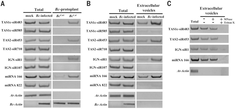
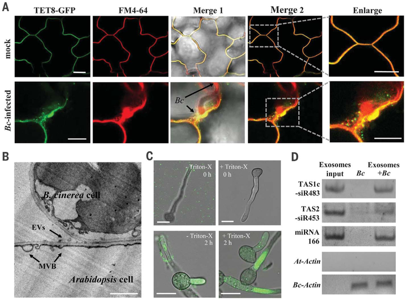
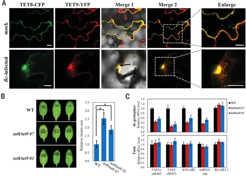
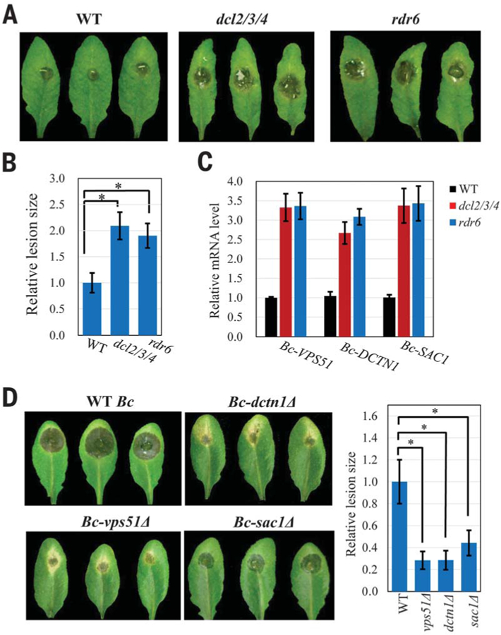
Some pathogens and pests deliver small RNAs (sRNAs) into host cells to suppress host immunity. Conversely, hosts also transfer sRNAs into pathogens and pests to inhibit their virulence. Although sRNA trafficking has been observed in a wide variety of interactions, how sRNAs are transferred, especially from hosts to pathogens and pests, is still unknown. Here, we show that host Arabidopsis cells secrete exosome-like extracellular vesicles to deliver sRNAs into fungal pathogen Botrytis cinerea These sRNA-containing vesicles accumulate at the infection sites and are taken up by the fungal cells. Transferred host sRNAs induce silencing of fungal genes critical for pathogenicity. Thus, Arabidopsis has adapted exosome-mediated cross-kingdom RNA interference as part of its immune responses during the evolutionary arms race with the pathogen.


Conflict of interest statement

Competing interests: H.J. is the inventor on a patent application (US62/573,546) held/submitted by University of California, Riverside, covering control of fungal pathogens by using RNAi-based approaches.

Figures

Fig. 1.
Fig. 1.. Plant endogenous sRNAs are transferred into fungal cells via extracellular vesicles.
(A) TAS1c-siR483, TAS2-siR453, IGN-siR1, and miRNA166 were detected by means of sRNA semiquantitative RT-PCR in B. cinerea protoplasts purified from B. cinerea–infected Col-0 Arabidopsis (BcCol). As a negative control, cultured B. cinerea mixed with uninfected leaves was subjected to the same procedure (BcCtrl). (B) These host sRNAs were also in Arabidopsis extracellular vesicles. (C) Arabidopsis sRNAs were detected in micrococcal nuclease–treated extracellular vesicles. In (A) to (C), TAS1c-siR585, TAS2-siR710, IGN-siR107, miRNA822, and Actin genes were used as controls. The “total” lane indicates total RNAs from leaves. Similar results were obtained from two biological replicates.
Fig. 2.
Fig. 2.. Tetraspanin-associated exosomes are responsible for transferring plant sRNAs to fungal cells.
(A) B. cinerea induces accumulation of TET8-GFP–labeled vesicles at the sites of infection. Short staining of FM4–64 shows extracellular membrane structures and plasma membranes. (B) Transmission electron microscopic image of Arabidopsis extracellular vesicles (EVs) near the B. cinerea infection sites. Scale bars, 1 mm. (C) TET8-GFP–labeled exosomes were taken up by B. cinerea within 2 hours of co-incubation. Scale bars, (A) and (C), 10 μm. (D) Plant sRNAs were detected in B. cinerea after uptake of the exosomes.
Fig. 3.
Fig. 3.. TET8 and TET9 coordinately regulate sRNA secretion and host immunity.
(A) TET8-CFP and TET9-YFP colocalized in vesicles that accumulated at the site of fungal infection. Scale bars, 10 μm. (B) Enhanced susceptibility to B. cinerea was observed in two independent tet8tet9 double mutant lines. Relative lesion sizes were measured at 2 days after infection. Error bars indicate the SD of more than 10 leaves. The asterisks indicate significant difference [analysis of variance (ANOVA) Dunnett’s multiple comparisons, P < 0.01]. (C)Accumulation of host transferred sRNAs was decreased in the purified fungal protoplasts isolated from the infected tet8tet9 mutants as compared with that from the wild type. In fungal protoplasts and total RNA samples, Bc-Actin and At-Actin were used as internal controls, respectively. The B. cinerea sRNA Bc-siR3.1 serves as a control.
Fig. 4.
Fig. 4.. Transferred host sRNAs silence fungal virulence genes and suppress fungal pathogenicity.
(A and B) compared with the wild type. Relative lesion sizes were measured at 2 days after infection. (C) Fungal target genes of transferred sRNAs were derepressed in B. cinerea collected from the dcl2/3/4 and rdr6 mutants. (D) Mutant B. cinerea strains with deletions in the targets of transferred host sRNAs displayed reduced virulence. Relative lesion sizes were measured at 3 days after infection. In (B) and (D), error bars indicate the SD over 10 leaves. The asterisk indicates significant difference (ANOVA Dunnett’s multiple comparisons; P < 0.01).

Comment in

  • Targeting microbial pathogens.
    Thomma BPHJ, Cook DE. Thomma BPHJ, et al. Science. 2018 Jun 8;360(6393):1070-1071. doi: 10.1126/science.aat9343. Science. 2018. PMID: 29880672 No abstract available.
  • The Trojan Horse of the Plant Kingdom.
    Castillo-González C, Zhang X. Castillo-González C, et al. Cell Host Microbe. 2018 Jul 11;24(1):1-3. doi: 10.1016/j.chom.2018.06.015. Cell Host Microbe. 2018. PMID: 30001514

References

    1. Baulcombe D, Nature 431, 356–363 (2004). - PubMed
    1. Mittelbrunn M, Sánchez-Madrid F, Nat. Rev. Mol. Cell Biol 13, 328–335 (2012). - PMC - PubMed
    1. Molnar A et al., Science 328, 872–875 (2010). - PubMed
    1. Weiberg A, Bellinger M, Jin H, Curr. Opin. Biotechnol 32, 207–215 (2015). - PMC - PubMed
    1. Wang M, Thomas N, Jin H, Curr. Opin. Plant Biol 38, 133–141 (2017). - PMC - PubMed

Publication types