. 2018 Jul;59(7):1244-1255.
doi: 10.1194/jlr.M085332. Epub 2018 May 17.

A thumbwheel mechanism for APOA1 activation of LCAT activity in HDL

Affiliations

A thumbwheel mechanism for APOA1 activation of LCAT activity in HDL

Allison L Cooke et al. J Lipid Res. 2018 Jul.

Abstract

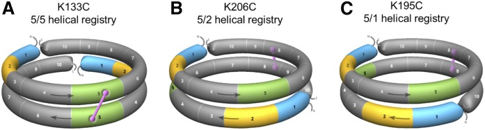
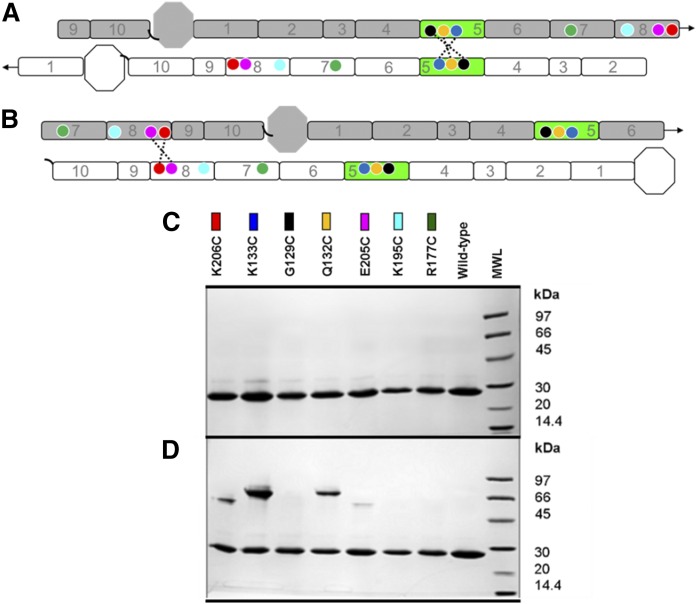
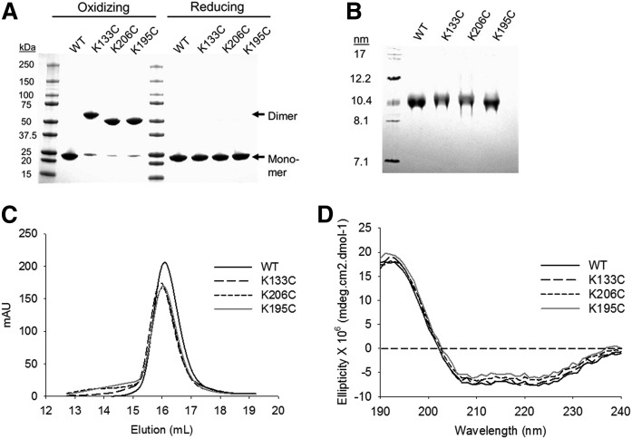
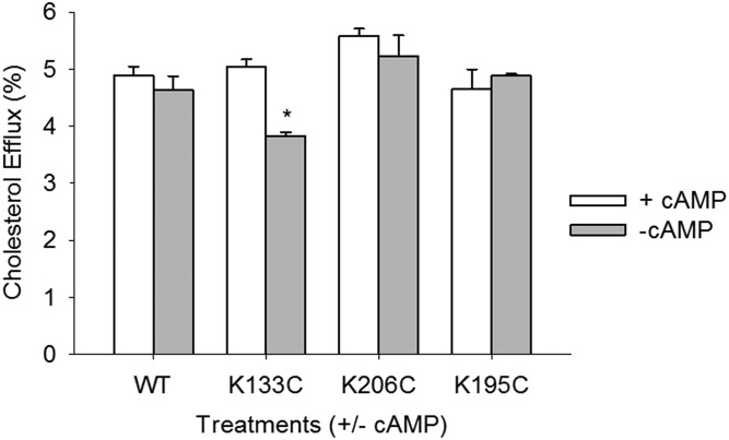
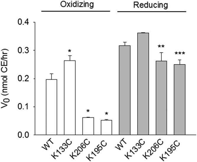
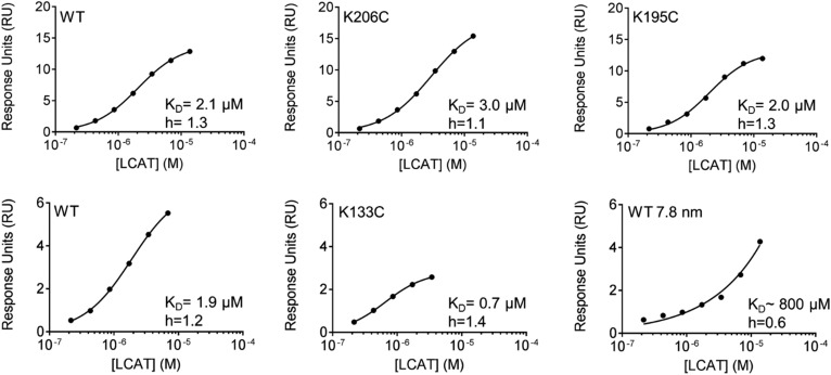
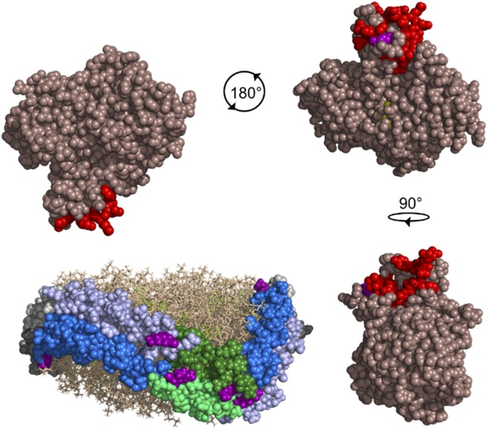
APOA1 is the most abundant protein in HDL. It modulates interactions that affect HDL's cardioprotective functions, in part via its activation of the enzyme, LCAT. On nascent discoidal HDL, APOA1 comprises 10 α-helical repeats arranged in an anti-parallel stacked-ring structure that encapsulates a lipid bilayer. Previous chemical cross-linking studies suggested that these APOA1 rings can adopt at least two different orientations, or registries, with respect to each other; however, the functional impact of these structural changes is unknown. Here, we placed cysteine residues at locations predicted to form disulfide bonds in each orientation and then measured APOA1's ability to adopt the two registries during HDL particle formation. We found that most APOA1 oriented with the fifth helix of one molecule across from fifth helix of the other (5/5 helical registry), but a fraction adopted a 5/2 registry. Engineered HDLs that were locked in 5/5 or 5/2 registries by disulfide bonds equally promoted cholesterol efflux from macrophages, indicating functional particles. However, unlike the 5/5 registry or the WT, the 5/2 registry impaired LCAT cholesteryl esterification activity (P < 0.001), despite LCAT binding equally to all particles. Chemical cross-linking studies suggest that full LCAT activity requires a hybrid epitope composed of helices 5-7 on one APOA1 molecule and helices 3-4 on the other. Thus, APOA1 may use a reciprocating thumbwheel-like mechanism to activate HDL-remodeling proteins.

Keywords: apolipoprotein A1; apolipoproteins; cholesterol metabolism; cholesterol/efflux; electron microscopy; high density lipoprotein; high density lipoprotein metabolism; lecithin:cholesterol acyltransferase; proteomics; surface plasmon resonance.


Conflict of interest statement

The authors declare no financial conflicts of interest.

Figures

Fig. 1.
Fig. 1.
Cartoons showing the 3D spatial relationships of APOA1 helix (H)5 (green), H2 (yellow), and H1 (blue) registry-locked rHDL preparations. A: In oxidizing conditions, K133C (H5) locks two antiparallel APOA1 monomers into a 5/5 helical registry through a disulfide bond linkage (pink). B: K206C (H8) locks two APOA1 monomers into a 5/2 helical registry. C: K195C (H8) locks APOA1 monomers into a 5/1 helical registry. All other helices of APOA1 are represented in gray. Arrows show the direction of the antiparallel APOA1 helices (N terminus, H1 through H10, C terminus).
Fig. 2.
Fig. 2.
Design of mutants for the postulated APOA1 registries in discoidal rHDL. A: A 2D representation of the molecular interactions between two antiparallel APOA1 monomers (imagine two APOA1 belts pulled off the edge of a particle and laid flat). Helix 5 (H5, green) of APOA1 (gray) is shown opposite to H5 of its antiparallel APOA1 partner molecule (white). Circles represent the positions of APOA1 residues K133 (dark blue), G129 (black), Q132 (yellow), K206 (red), E205 (pink), K195 (cyan), and R177 (green). The dotted line shows disulfide bonds predicted to form with APOA1 mutant K133C, G129C, and Q132C in the 5/5 registry. Arrows at the end of the molecules show the continuity of the APOA1 disc helices [N terminus (octagon), H1 through H10, C terminus (black, curved line)]. B: A 2D representation of two antiparallel APOA1 monomers with H5 (green) of APOA1 (gray) opposite to H2 of its APOA1 partner molecule (white). Circles represent the same residues indicated above. The dotted line shows the disulfide bond predicted to form in APOA1 mutant K206C and E205C in a 5/2 registry. C: SDS-PAGGE analysis of WT APOA1 and Cys-mutants reconstituted into rHDL particles under reducing conditions (3 mM DTT). Colored rectangles correspond to the positions of the residues on APOA1 diagrams in A and B. D: SDS-PAGGE (nonreducing) analysis of WT APOA1 and Cys-mutant rHDL after incubation at 21°C for 48 h in the absence of a reducing agent.
Fig. 3.
Fig. 3.
Characterization of Cys-mutant rHDL particles. A: Lipid-free WT APOA1 and Cys-mutants were predimerized in oxidizing conditions. Dimerized protein was then purified from the residual monomer by SEC. Protein purity was analyzed by SDS-PAGGE in oxidizing and reducing (3 mM β-ME) conditions. B: Sodium cholate dialysis was used to generate rHDL particles at a molar ratio of 80:8:1 POPC:cholesterol:APOA1. Native PAGGE was used to determine the purity of the particles in oxidizing conditions. C: SEC was used to determine the size and purity of the particles. Peak intensity is shown in arbitrary units (mAU) based on the elution volume of the particles. D: Far-UV CD spectra of WT and Cys-mutant rHDL particles showing the mean ellipticity of APOA1 based on the 1 nm wavelength increases in the UV spectrum (n ≥ 3 for all characterization experiments).
Fig. 4.
Fig. 4.
Cholesterol efflux capacity of WT and Cys-mutant rHDL particles. RAW mouse macrophages were exchange labeled with [3H]free cholesterol and incubated with rHDL generated with WT or Cys-mutant APOA1. Ten micrograms per milliliter (protein) of the various rHDL particles were incubated with the cells for 6 h at 37°C. The cells were treated in the presence or absence cAMP to distinguish between ABCA1-mediated (with cAMP, white bar) and aqueous diffusion (without cAMP, gray bar) cholesterol efflux. Experiments were performed in triplicate and bars represent mean ± SD. The mean was normalized to the medium alone control condition. A two-way ANOVA and Holm-Sidak post hoc test were used to determine cholesterol efflux differences between samples and within treatments with or without cAMP (K133C + cAMP vs. K133C − cAMP; *P < 0.001, t = 6.818).
Fig. 5.
Fig. 5.
LCAT activation by WT and Cys-mutant rHDL. rHDL particles (0.4 μM) generated with [3H]free cholesterol were incubated with LCAT (0.02 μM) in oxidizing (disulfide “locked,” white bars) and reducing (“unlocked,” 10 mM β-ME, gray bars) conditions as described in the Materials and Methods. The amount of labeled CE, as separated by thin-layer chromatography, was quantitated by scintillation counting. Bars represent mean ± SD of triplicate measurements for each condition. A one-way ANOVA and Holm-Sidak post hoc test were used to determine differences in the reaction velocity (V0) of CE production between WT and Cys-mutant rHDL (K133C, K206C, K195C vs. WT; *P < 0.001, F = 189.740, DF = 3; K206C reducing vs. K206C oxidizing, **P < 0.001, t = −11.823, DF = 4; K195C reducing vs. K195C oxidizing, ***P < 0.001, t = −22.023, DF = 4).
Fig. 6.
Fig. 6.
LCAT affinity for WT and Cys-mutant rHDL assessed by SPR. The 4H1, a mouse anti-APOA1 monoclonal antibody recognizing amino acids 2–8 of APOA1, was immobilized on flow cells 2–4 of a CM5 sensorchip. Mouse anti-ubiquitin monoclonal antibody was immobilized on flow cell 1 to serve as a reference channel. WT and Cys-mutant rHDL particles (2.5 mM) were coupled to 4H1 in flow cells 2–4. WT APOA1 7.8 nm rHDL particles served as a negative control. Particles were bound to the chip, washed, and then LCAT was flowed over for 120 s at concentrations from 0.2 to 55 μM in duplicate, including four buffer blanks. LCAT was washed from rHDL particles for 600 s before injecting a new concentration. Equilibrium dissociation constants (KD) were determined using GraphPad Prism 7, nonlinear regression (curve fit), specific-binding with Hill slope based on reference channel, and blank subtracted response units (RU). Binding curves are plotted on a semi-log scale. Top panel: WT, K206C, and K195C were captured on sensorchip 1. Bottom panel: WT, K133C, and WT 7.8 nm were captured on sensorchip 2.
Fig. 7.
Fig. 7.
Identification of LCAT interaction sites on rHDL containing WT and isotopically labeled APOA1 with chemical cross-linking. A 4:1 ratio of rHDL and LCAT was incubated with BS3, a lysine-to-lysine cross-linker, at a 100:1 molar ratio of cross-linker to APOA1 for 16 h at 4°C. A: NDGGE analysis of 1:1 14N/15N APOA1 rHDL prepared by sodium cholate dialysis. B: SDS-PAGGE analysis of rHDL incubated with LCAT, with (+) or without (−) BS3 cross-linking (XL). Cross-linked complexes were purified by SEC with a 50 mM ammonium bicarbonate mobile phase at 0.6 ml/min and prepared for MS analysis.
Fig. 8.
Fig. 8.
Postulated model of LCAT interacting with the 6/4 region of APOA1 molecules in the 5/5 helical registry. The Piper et al. (48) crystal structure of LCAT (dark salmon) is shown with the lid region highlighted in red and the catalytic triad in yellow. LCAT residue S108 (purple) involved in a cross-link to APOA1 is visible near the lid region. The rHDL particle is depicted with two anti-parallel molecules of APOA1 (molecule A, light gray; molecule B, dark gray) with helix (H)6 (blue), H4 (light blue), and H5 (molecule A, dark green; molecule B, light green) highlighted on both molecules. Phospholipids (tan) and cholesterol (yellow-green) are shown. Residues (purple) cross-linked in helices 4, 5, and 7 of APOA1 to LCAT are shown. In a 5/5 helical registry, there are two locations where helices 6 and 4 are opposing, meaning two possible sites for LCAT activation to occur. LCAT is depicted rotating 180° and 90° from the starting location to interact with H6 on molecule B and H4 on molecule A, and H6 on molecule A and H4 on molecule B, respectively. The figure was generated with PyMOL, version 2.0, Schrodinger, LLC.

References

    1. Gordon T., Castelli W. P., Hjortland M. C., Kannel W. B., and Dawber T. R.. 1977. High density lipoprotein as a protective factor against coronary heart disease. The Framingham Study. Am. J. Med. 62: 707–714. - PubMed
    1. Shah A. S., Tan L., Long J. L., and Davidson W. S.. 2013. Proteomic diversity of high density lipoproteins: our emerging understanding of its importance in lipid transport and beyond. J. Lipid Res. 54: 2575–2585. - PMC - PubMed
    1. Gu X., Trigatti B., Xu S., Acton S., Babitt J., and Krieger M.. 1998. The efficient cellular uptake of high density lipoprotein lipids via scavenger receptor class B type I requires not only receptor-mediated surface binding but also receptor-specific lipid transfer mediated by its extracellular domain. J. Biol. Chem. 273: 26338–26348. - PubMed
    1. Puranik R., Bao S., Nobecourt E., Nicholls S. J., Dusting G. J., Barter P. J., Celermajer D. S., and Rye K. A.. 2008. Low dose apolipoprotein A-I rescues carotid arteries from inflammation in vivo. Atherosclerosis. 196: 240–247. - PubMed
    1. Navab M., Imes S. S., Hama S. Y., Hough G. P., Ross L. A., Bork R. W., Valente A. J., Berliner J. A., Drinkwater D. C., and Laks H.. 1991. Monocyte transmigration induced by modification of low density lipoprotein in cocultures of human aortic wall cells is due to induction of monocyte chemotactic protein 1 synthesis and is abolished by high density lipoprotein. J. Clin. Invest. 88: 2039–2046. - PMC - PubMed

Publication types

MeSH terms